I. INTRODUCTION
In the past, nanotechnology has appeared as one of the very vital fields of pharmaceutical science and more specifically in drug delivery field.Reference Babaie, Ghanbarzadeh, Davaran, Kouhsoltani and Hamishehkar1 In this field, several studies were carried out and it was found that having noble carrier, most of the currently available drugs have limited potential because they are very toxic, insoluble in water, possess side effects, or highly expensive, and thus beyond the reach of the majority. At this time, over than 100 natural therapeutic molecules are in clinical progress, mostly for anticancer activities.Reference Nosrati, Sefidi, Sharafi, Danafar and Manjili2 Flavonoids are a number of ordinary anticancer agents, while the lack of water solubility limited their medical and pharmaceutical claims.Reference Zheng, Li, Pu, Lai, He and Gu3,Reference Cristescu, Visan, Socol, Surdu, Oprea, Grumezescu, Chifiriuc, Boehm, Yamaleyeva and Taylor4 Chrysin (5,7-dihydroxyflavone) is a natural flavonoid which is extracted from herbs and has been testified to have several important biological properties on the procedures of chemical defense, nitrogen fixation, inflammation, oxidation, and anti-inflammatory and anti-oxidant effects.Reference Babu, Babu, Srinivas, Kishore, Murthy and Rao5,Reference Anari, Akbarzadeh and Zarghami6 But, the lacking in solubility reduces its bioavailability and medical and pharmaceutical claims.Reference Vatten and Kvinnsland7 Chrysin also exhibits chemopreventive activity through the induction of apoptosis in a various series of cancers cells.Reference Ursin, Longnecker, Haile and Greenland8 Forasmuch as free drugs may not diffuse specifically, nanocarriers can enter into the tumor area through the lack in vessels by the enhanced permeability and retention effect.Reference Matsumura and Maeda9 Among the nanosystems that are used for drug delivery and especially cancer therapy,Reference Ghanbarzadeh, Khorrami and Arami10–Reference Nosrati, Rashidi, Danafar and Manjili13 iron oxide magnetic nanoparticles (IOMNs) are most popular as drug delivery systems for cancer therapy,Reference Aberoumandi, Mohammadhosseini, Abasi, Saghati, Nikzamir, Akbarzadeh, Panahi and Davaran14,Reference Ahmadkhani, Akbarzadeh and Abbasian15 due to their unique physical and chemical properties such as biocompatibility, low toxicity, stability, easy of surface functionalization, and magnetic direction. These properties of IOMNs enable remarkable applications, for instance catalytic processes,Reference Shaabani, Nosrati and Seyyedhamzeh16,Reference Shaabani, Boroujeni and Laeini17 magnetic resonance imaging,Reference Arami, Khandhar, Liggitt and Krishnan18 drug delivery,Reference Nosrati, Sefidi, Sharafi, Danafar and Manjili2 hyperthermia,Reference Mahmoudi, Serpooshan and Laurent19 cell labeling, and cell separation.Reference Pan, Du, Zhao and Xu20 Unfortunately, bare IOMNs are commonly unstable and the aggregation of these nanoparticles during usage is normally unavoidable.Reference Shaabani, Nosrati and Seyyedhamzeh16 Furthermore, for IOMNs application in industrial, medical, and pharmaceutical applications, the surface specific decoration and modification are main requisites.Reference Salehiabar, Nosrati, Davaran, Danafar and Manjili21 For this purpose, a diverse series of materials, such as cellulose,Reference Shaabani, Nosrati and Seyyedhamzeh16 bovine serum albumin,Reference Nosrati, Sefidi, Sharafi, Danafar and Manjili2 protein,Reference Li, Qiang, Zhong, Wang and Cui22 graphene oxide,Reference Rostami, Aghajanzadeh, Zamani, Manjili and Danafar23 dopamine,Reference Martín, Salazar, Villalonga, Campuzano, Pingarrón and González-Mora24 and amino acidsReference Nosrati, Rashidi, Danafar and Manjili13 are utilized in these stabilizing methods.
The chemical formula of phenylalanine (Phe) is C9H11NO. Phe is an α-amino acid. It can be observed as a benzyl group substituted for the methyl group of alanine. Phe due to inert and hydrophobic nature of the benzyl side chain is classified as neutral and nonpolar essential amino acid. Tyrosine, dopamine, norepinephrine, epinephrine, and the skin pigment melanin are originated from Phe.Reference Nosrati, Rashidi, Danafar and Manjili13 In recent years, various natural amino acids have been used to modify IOMNs.Reference Sousa, Rubim, Sobrinho and Tourinho25–Reference Pušnik, Peterlin, Kralj-Cigic, Marolt, Kogej, Mertelj, Gyergyek and Makovec29
Young et al. synthesized amino acid-coated magnetite nanoparticles and evaluated the stability of these nanoparticles in physiological fluids.Reference Park, Choi, Baek and Lee26 Nosrati et al. synthesized IOMNs coated with various natural amino acids, and their effects on size, morphology, and toxicity of IOMNs were evaluated.Reference Nosrati, Salehiabar, Attari, Davaran, Danafar and Manjili30 Based on our information, this strategy did not used for drug delivery by other researchers.
As part of our continuing interest in the development of amino acid-coated IOMNs for biological applications and drug delivery,Reference Salehiabar, Nosrati, Davaran, Danafar and Manjili21 we design a magnetic nanosystem for delivery of chrysin anticancer drug with the objective of improving the loading efficiency of the drug in IOMNs as well as achievement of an efficient control on the release rate of drug from nanocarriers. In this way, first, we have prepared Phe-coated IOMNs. Second, chrysin loaded as a model drug on Phe-coated IOMNs and the loading volume and release behavior of the drug from the nanoparticles were assessed. Third, the toxicity of these nanoparticles was studied on normal and cancerous cell lines.
II. MATERIALS AND METHODS
A. Materials
L-phenylalanine (Phe), FeCl3·6H2O, and FeCl2·4H2O were purchased from Merck (Darmstadt, Germany). All other chemicals and solvents were from general lab or HPLC grades and were purchased from commercial sources.
B. Synthesis of Phe-coated IOMNs
Phe-coated IOMNs were synthesized via a controlled precipitation one-pot process as described previously.Reference Nosrati, Rashidi, Danafar and Manjili13 Briefly, to 150 mL deionized water solution, 1.1 g FeCl3·6H2O, 0.4 g FeCl2·4H2O, and 0.016 mol (2.64 g Phe) Phe solution in the molar ratio of 2Fe(III):1Fe(II):4Phe were added and kept under vigorous stirring at 60 °C for 15 min. Next, under vigorous stirring and N2 gas 20 mL NH4OH (25%) solution dropwise was added till the pH reached ∼11 at which a black suspension was formed. Vigorous stirring was then continued for 6 h at 60 °C, under N2 gas atmosphere. Phe-coated IOMNs were separated from solution by using an external magnet (1.2 T) and washed with ammonium solution (1 M) (three times), then washed with deionized water and then dried in a vacuum oven overnight at 50 °C.
C. Preparation of chrysin-loaded nanocarriers (chrysin@Phe@IOMNs)
To an aqueous suspension of Phe-coated IOMNs (10 mg in 2.8 mL distilled water), 2.5 mg chrysin (in 200 μL ethanol) was added dropwise. Next, the stirring (400 rpm) continued for 24 h under the dark condition at 30 °C. Chrysin@Phe@IOMNs (chrysin-loaded nanocarrier) were separated by using an external magnet from free drug solution and then dried for 24 h at 30 °C in a vacuum oven.
D. Characterization
1. Determination of particle size
Transmission electron microscopy (TEM; Cambridge 360-1990 Stereo Scan Instrument-EDS) was used to study the morphology and size of chrysin@Phe@IOMNs.
2. Colloidal stability
Dynamic light scattering method using a nano/zetasizer (Malvern Instruments, Worcestershire, U.K., model Nano ZS) was used to measure the ζ-potential.
3. Fourier transform infrared spectroscopy
A Fourier transform infrared (FTIR) spectrometer [Bruker Tensor 27 (Biotage, Dusseldorf, Germany)] was used for the collection of FT-IR spectra.
4. Thermal analysis
Thermogravimetric analysis (TGA; Linseis Instruments model, STA PT 1000 (Robbinsville, New Jersey) was used to study the thermal stability. The TGA thermograms were documented at a heating rate of 10 °C/min, in the temperature range of 30–500 °C under N2 atmosphere.
5. Structure characterization
A Bruker AXS model D8 Advance diffractometer (Karlsruhe, Germany) was used to record X-ray diffraction (XRD) patterns with Cu Kα radiation (k = 1.542 A) with the Bragg angle ranging from 15° to 65°.
6. Magnetic properties
Magnetic properties of the samples were studied by vibrating sample magnetometer (VSM) analysis (Meghnatis Daneshpaghoh Co., Ardabil, Iran).
7. Determination of loading efficiency
The drug loading ratio (DL%) was used for the purpose of the loading efficiency of the chrysin in the chrysin@Phe@IOMNs. To calculate the DL efficiency, 2.0 mg of chrysin@Phe@IOMNs were suspended in 10 mL of ethanol. The suspension was shaken in a shaker incubator at 37 °C for overnight. Next, the samples were removed from the shaker incubator and separation of nanocarriers from free drug was done by using an external magnetic plate. The supernatant was collected and diluted, then amount of the chrysin was measured using UV-visible spectroscopy (Thermo Fisher Scientific, Madison, WI, model GENESYSTM 10S) at 348 nm. DL% was calculated as follows:
 $$\% {\rm{DL}}{{{\rm{Weight}}\;{\rm{of}}\;{\rm{drug}}\;{\rm{in}}\;{\rm{nanocarrier}}} \over {{\rm{Weight}}\;{\rm{of}}\;{\rm{nanocarrier}}}} \times 100\quad .$$
E. Drug release study
To estimate the release behavior of chrysin from chrysin@Phe@IOMNs, this study was performed. In brief, 2.0 mg of chrysin@Phe@IOMNs were dispersed in 2.0 mL PBS (pH = 5.8) containing 3% (v/v) Tween 80 and the follow-on suspension was cited within a dialysis bag (Mw: 12,000 Da) and incubated at 37 °C while dipped in 20 mL of PBS. Next, at predetermined time intervals, 2 mL of the dialysate was taken out and replaced by 2 mL new buffer/Tween solution. The content of chrysin in the dialysate was calculated using UV-Vis spectroscopy at 348 nm.
F. Biocompatibility studies
1. Hemolysis assay
The in vitro hemolysis assay was accomplished to study hemocompatibility.Reference Nosrati, Mojtahedi, Danafar and Kheiri Manjili31,Reference Rahimi, Shojaei, Safa, Ghasemi, Salehi, Yousefi and Shafiei-Irannejad32
Human blood was centrifuged at 3000 rpm for 10 min for removing serum from the blood and obtaining human red blood cells (HRBCs), then washed four times with sterile PBS solution. PBS solution was used for dilution of HRBCs (10 times), and then 0.5 mL of this mixture was mixed with 0.5 mL of sterile deionized water (positive control), 0.5 mL of PBS (negative control), 0.5 mL of Phe-coated IOMNs, and chrysin@Phe@IOMNs at a concentration of 10 mg/mL. The samples were shaken in a shaker incubator at 37 °C for 4 h. Finally, the samples were centrifuged at 13,000 rpm for 15 min, the supernatant was taken, and its absorbance was measured by using an Eppendorf BioPhotometer (Westbury, New York) (λ = 540 nm). The percentage hemolysis was calculated using the following equation:
 $${\rm{Hemolysis}}\;(\% ) = {{{A_{{\rm{sample}}}} - {A_{{\rm{negative}}}}} \over {{A_{{\rm{positive}}}} - {A_{{\rm{negative}}}}}} \times 100\quad .$$
Here, A, A negative, and A positive are the absorbance of the tested sample, negative control, and the positive control samples, respectively. The percentage of hemolysis was determined based on the average of three replicates.
2. Cell viability assay
To evaluate the biocompatibility of nanocarriers, MTT assay was performed against normal cell lines including human foreskin fibroblast (HFF-2) and human embryonic kidney cells (HEK-293) acquired from Pasteur Institute (Tehran, Iran).
RPMI-1640 (Gibco, Waltham, Massachusetts) containing 10% fetal bovine serum (Gibco, Waltham, Massachusetts), 2 mM L-glutamine, penicillin (50 IU/mL), and streptomycin (50 L g/mL) was selected as the cell cultured medium and incubated at 37 °C in a humidified incubator with 5% CO2 atmosphere.
The HFF-2 and HEK-293 cell lines’ viability was assessed in the presence of Phe-coated IOMNs compared to that of the cells in the control experiment (without IOMNs).
1 × 10⁴ cells were seeded into each well of the 96-well plate for 24 h. Then the Phe-coated IOMNs of six various concentrations ranging from 15, 30, 60, 120, 240, and 480 µM were added to wells and incubated for 72 h. The medium was removed, after the incubation time, and then washed with PBS. Next, MTT solution was added to each well followed by 4 h of incubation at 37 °C, subsequently, the medium was removed and violet formazan crystals were solubilized with DMSO (100 µL). Solubilized formazan crystals were measured at 570 nm. After mild shaking for 15 min, the absorbance ratio of the number of surviving cells in Phe-coated IOMN-treated samples was compared with the control.
G. In vitro anticancer activity
To evaluate the toxicity and antitumor activity of chrysin@Phe@IOMNs, the potential formulations were examined against human breast adenocarcinoma (MCF7) cells acquired from Pasteur Institute (Tehran, Iran). MTT assay was used for in vitro anticancer activity study. Briefly, the cells were cultured in the 96-well plate for 24 h to allow the attachment of the cell to the wells. Next, different concentrations (15, 30, 60, 120, 240, and 480 µM) of free chrysin and chrysin@Phe@IOMNs were treated to cells and incubated for 72 h. The medium was removed, after the incubation time, and then washed with PBS. Then, MTT solution was added to each well followed by 4 h of incubation at 37 °C; subsequently, the medium was removed and violet formazan crystals were solubilized with DMSO (100 µL). Solubilized formazan crystals were measured at 570 nm. After mild shaking for 15 min, the absorbance ratio of the number of surviving cells in free chrysin and chrysin@Phe@IOMN-treated samples was compared with the control.
III. RESULTS AND DISCUSSION
A. Characterization of size and morphology
IOMN surfaces were characterized with natural Phe by the available, simple, and one-pot in situ process. Therefore, chrysin was encapsulated into the nanocarrier.
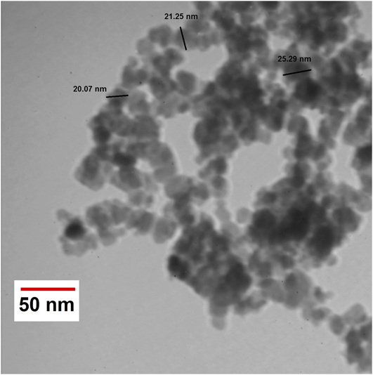
The TEM image of the chrysin@Phe@IOMNs is shown in Fig. 1. As revealed in Fig. 1, nanoparticles are discrete and have a spherical shape. Furthermore, the average size of the chrysin@Phe@IOMNs was about 19.94.20 ± 3.49 [mean ± SD (n = 40)] nm.
TEM image of chrysin@Phe@IOMNs.

B. Structure characterization
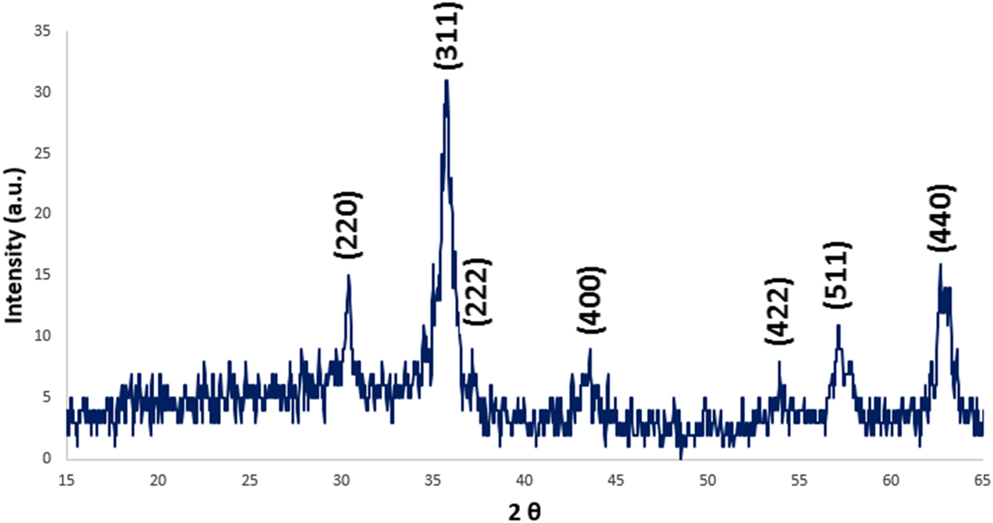
The chrysin@Phe@IOMN diffractogram achieved by XRD is shown in Fig. 2. The location and comparative intensity of diffraction peaks of the chrysin@Phe@IOMNs accorded with those of the Fe3O4 nanoparticles, which is representative of the effective synthesis of the Fe3O4 nanoparticles. The peaks shown in chrysin@Phe@IOMNs bands are at 2θ equals to 18.41°, 30.32°, 35.57°, 42.98°, 53.96°, 57.45°, and 63.03° which are accorded fine with the representative peaks and their comparative strengths of spinel structure of magnetite parallel to the (111), (220), (311), (400), (422), (511), and (440) (hkl) planes (JCPDS 019-0629).
XRD pattern of chrysin@Phe@IOMNs with the Bragg angle ranging from 15° to 65°.

C. Colloidal stability
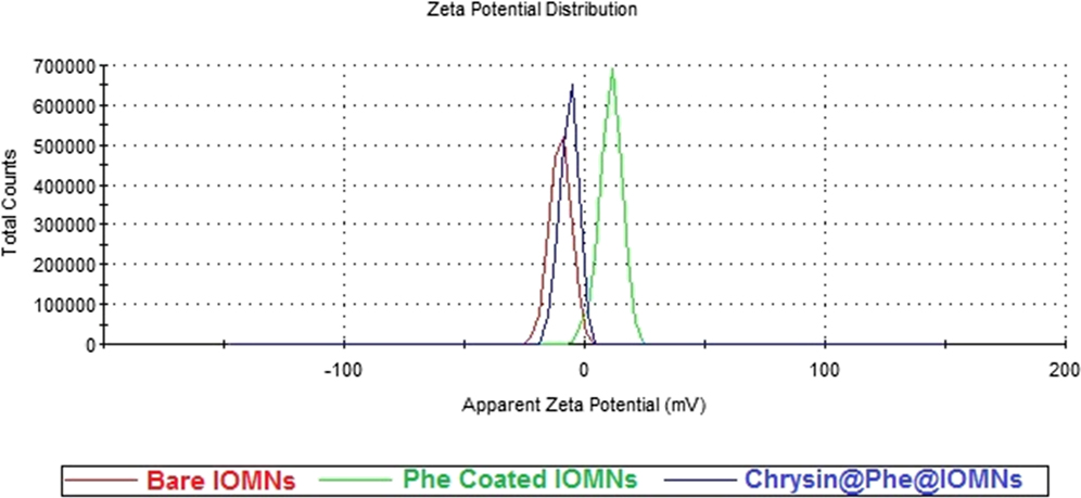
The stability of nanoparticles was affected by surface charge. Figure 3 shows the ζ-potential of pure IOMNs, Phe-coated IOMNs, and chrysin@Phe@IOMNs. The existence of Phe and chrysin on the pure IOMN surface was established with the variation of ζ-potential. As shown in Fig. 3, the ζ-potential of pure IOMNs was −10.1 mV; after reaction with Phe, the ζ-potential of Phe-coated IOMNs improved to +10.8 mV. Also, after loading of chrysin, the ζ-potential of nanoparticles was negative (−6.45 mV). The surface charge is vital in critical to the nanoparticles interact with oppositely charged cell membrane. The blood cells frequently have a negative charge; nanoparticles with negative charge can decrease the nonspecific interface with these components through electrostatic interfaces.Reference Nosrati, Rashidi, Danafar and Manjili13
ζ-potential of bare IOMNs, Phe-coated IOMNs, and chrysin@Phe@IOMNs in deionized water.

D. Magnetic properties
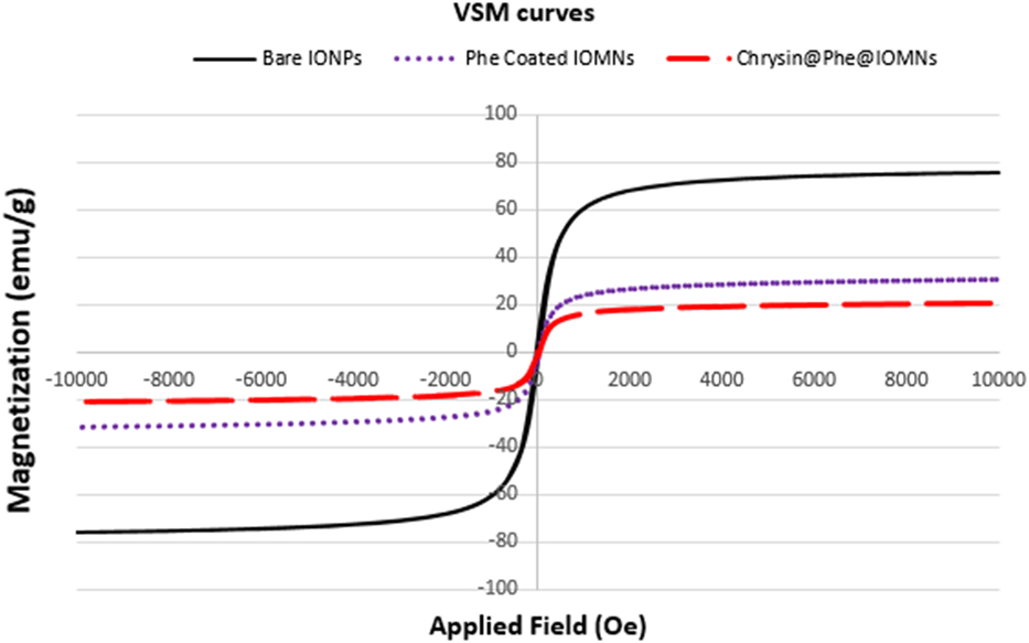
Figure 4 shows the magnetic behaviors of bare IOMNs, Phe-coated IOMNs, and chrysin@Phe@IOMNs. These properties are considered by assessing magnetization as a function of field and attained samples [saturation magnetization (Ms) of bare IOMNs, Phe-coated IOMNs, and chrysin@Phe@IOMNs]. The magnetization of results was verified in an applied magnetic field of −10,000 ≤ H (Oe) ≤ 10,000 at room temperature. The reduction of Ms for Phe-coated IOMNs (30.31 emu/g) rather than bare IOMNs (75.29 emu/g) could be explained by the coating layer on the surface of IOMNs.Reference Qu, Ma, Zhou and O’Connor33 The Ms of the chrysin@Phe@IOMNs was about 20.76 emu/g, which was lower than those of bare IOMNs and Phe-coated IOMNs. And furthermore, the decrease of Ms for chrysin@Phe@IOMNs rather than Phe-coated IOMNs can be clarified by the loading of chrysin on the Phe-coated IOMNs.
VSM curves of bare IOMNs, Phe-coated IOMNs, and chrysin@Phe@IOMNs in an applied magnetic field of −10,000 ≤ H (Oe) ≤ 10,000 at room temperature.

E. FTIR analysis
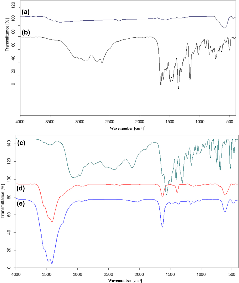
The FTIR spectra of pure Fe3O4, Phe-coated IOMNs, bare Phe, pure chrysin, and chrysin@Phe@IOMNs are compared in Fig. 5. The incidence of the amino acid covering on the IOMNs was obviously clear from the assessment of FTIR spectra of pure IOMNs with that of the Phe-coated IOMNs. Phe-coated IOMNs showed all the IR peaks characteristic to the bare Phe. In Phe-coated IOMNs, the C=O and C–O stretching vibrations can be shown at ∼1630 cm−1 and ∼1380 cm−1, respectively, that it was clearly reformed with pure Phe peaks. The peaks in the 2800–3000 cm−1 area may be fitted to the C–H stretching of methyl or methylene groups that this peak could not be seen in pure Fe3O4 and N–H stretching vibration overlays with O–H stretching at ∼3400 cm−1. Also, the absorption peaks at 1612 cm−1 which are shielded with asymmetric COO– stretching payable to the N–H bending of amine groups. Phe-coated IOMNs display strong peaks at 610 and 450 cm−1 which are related to the Fe–O vibration. These peaks confirmed a covalent bond between Phe and the surface of the IOMNs. Associated with pure Phe, shortening of the carboxyl group’s peak is due to the interaction with O–H groups on the surface of the IOMNs.Reference Park, Choi, Baek and Lee26,Reference Durmus, Kavas, Toprak, Baykal, Altınçekiç, Aslan, Bozkurt and Coşgun34
FT-IR spectra of (a) bare IOMNs, (b) free chrysin, (c) pure Phe, (d) Phe-coated IOMNs, and (e) chrysin@Phe@IOMNs from 400 to 4000 cm−1.

To the FTIR spectrum of chrysin exposed peaks at 1609 cm−1 and 1494 cm−1 due to C=C and C=O of benzene ring, respectively, 1462 cm−1 due to olefinic C–H bending vibration, 1358 cm−1 due to aromatic C–O stretching vibrations, and 1026 cm−1 due to C–O–C stretching vibrations. To the spectrum of the chrysin@Phe@IOMNs established peaks associated with chrysin, at 1490 cm−1 due to C=O of benzene ring, 1300 cm−1 due to aromatic C–O stretching vibrations, and 1020 cm−1 due to C–O–C stretching vibrations, to IOMN peaks, at a strong peak at 610 and 450 cm−1 which are given to the Fe–O vibration, which are reliable with the previous studies.Reference Li, Qiang, Zhong, Wang and Cui22
F. Thermal analysis
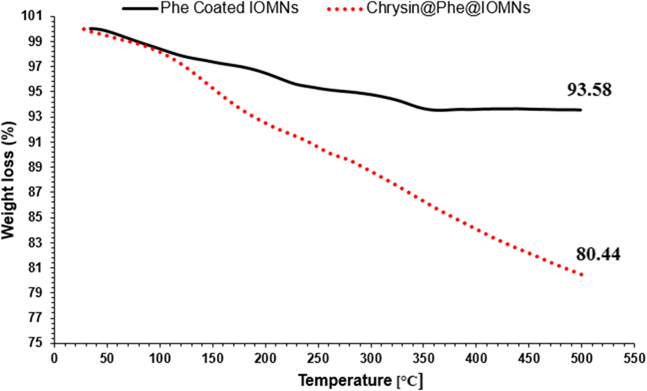
The TGA curves of the Phe-coated IOMNs and chrysin@Phe@IOMNs are shown in Fig. 6. A part from this weight loss, the coated sample demonstrates a weight loss of 6.42% relating to loss of Phe which is coated on the IOMNs. Therefore, the chrysin@Phe@IOMNs show a much higher weight loss, which is reliable with the increase in organic content of the IOMNs loaded by chrysin.
TGA curves of Phe-coated IOMNs and chrysin@Phe@IOMNs at a heating rate of 10 °C/min, in the temperature range of 30–500 °C under N2 atmosphere.

IV. CHRYSIN LOADING AND RELEASE STUDIES
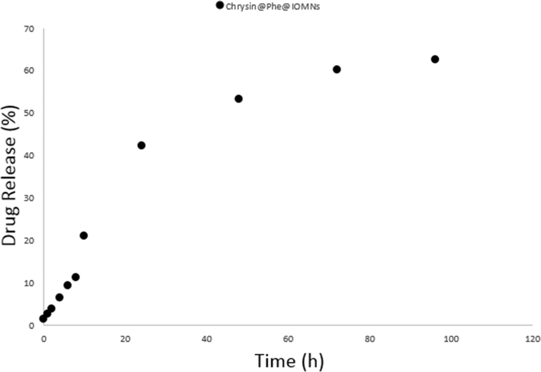
The DL of chrysin in chrysin@Phe@IOMNs was 2.4%. Figure 7 shows the release profiles of chrysin. In chrysin@Phe@IOMNs, up to 60% of chrysin was released from the chrysin@Phe@IOMNs under acidic conditions (pH 5.8) after 72 h.
Cumulative drug release profile of chrysin@Phe@IOMNs in PBS containing 3% (v/v) Tween 80 (pH = 5.8).

A. Biocompatibility studies
For assigning the biocompatibility of the Phe-coated IOMNs and chrysin@Phe@IOMNs, the nanoparticles must have blood compatibility with the negligible importance of hemolytic effect on the HRBCs. For assignment of the blood adaptability of Phe-coated IOMNs and chrysin@Phe@IOMNs, hemolysis assay was used. As predictable, the insignificant effect of Phe-coated IOMNs and chrysin@Phe@IOMNs is apparent in the experiment concentration. The hemolytic activity of the specimens was evaluated by assessing the supernatant absorbance at 540 nm (hemoglobin) with Eppendorf BioPhotometer.
Furthermore, at a high concentration, the hemolytic activity of the Phe-coated IOMNs and chrysin@Phe@IOMNs was about 2.4 and 3.89, respectively. Therefore, the negligible hemolytic activity of Phe-coated IOMNs as a nanocarrier established the excessive biocompatibility of HRBCs.
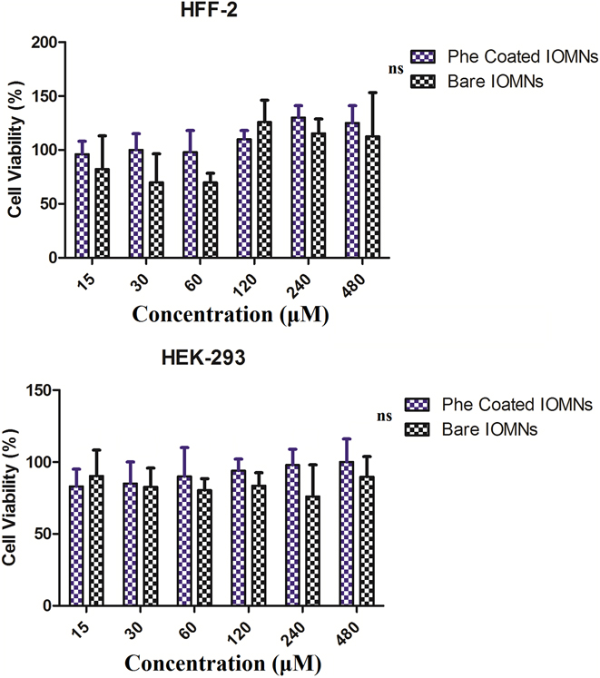
For biomedical applications, Phe-coated IOMNs are intentionally planned to interact with cells, and it is vital to confirm that these improvements are not affecting any opposing result. The cytotoxicity study of Phe-coated IOMNs was performed on HFF-2 and HEK-293 cell lines with different concentrations and the attained data are shown in Fig. 8. The cell lines were incubated with nanoparticles for 72 h with concentrations of 15, 30, 60, 120, 240, and 480 µM in a 5% CO2 atmosphere. Figure 8 compares the cell viability as a function of bare and Phe-coated IOMNs. MTT assays show no inhibitive effects of Phe-coated IOMNs against cell line growth. As a matter of fact, even slightly increased viability was observed at higher Fe concentrations, which is in agreement with other work and those of others who used different IOMN formulae.Reference Xie, Wang, Niu, Huang, Chen, Li and Chen35 The results show that the viability of HFF2 and HEK-293 cell lines is not affected by the presence of Phe-coated IOMNs.
Cytotoxicity study of bare IOMNs and Phe-coated IOMNs after incubation at 72 h on HFF-2 and HEK-293 cell lines. Each bar represents the mean of five measurements ±SD. Bars marked with ns (not significant) showed no significant difference between bare IOMNs and Phe-coated IOMNs.

B. In vitro anticancer activity
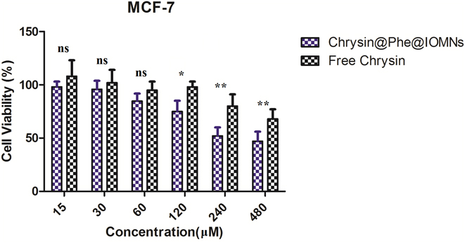
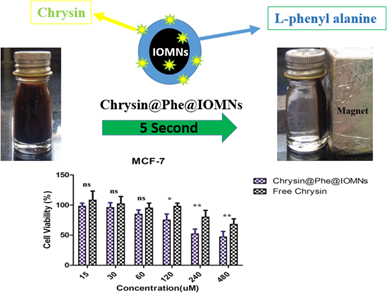
Figure 9 shows the in vitro anticancer effects of free chrysin and chrysin@Phe@IOMNs against MCF7 cell line using an MTT assay. The results presented that cell toxicity is directly commensurate with chrysin concentration. In the chrysin concentration range of 15–480 µM, the anticancer activities of the chrysin@Phe@IOMNs are higher than the activity of free chrysin (Fig. 9).
Anticancer activity study of free chrysin and chrysin@Phe@IOMNs on MCF-7 cells after incubation at 72 h. Each bar represents the mean of five measurements ±SD. Bar marked with ns (not significant) showed no significant difference between free chrysin and chrysin@Phe@IOMNs. Bars marked with * (P < 0.05) showed significant difference between free chrysin and chrysin@Phe@IOMNs. Bars marked with ** (P < 0.01) showed significant difference between free chrysin and chrysin@Phe@IOMNs.

IC50, the drug concentration at which the growth of 50% cells was inhibited, was assessed by curve fitting of the cell viability data. The IC50 value of chrysin@Phe@IOMNs at 72 h was 348.67 ± 14 µM versus free chrysin (635.33 ± 20 µM). Thus, chrysin@Phe@IOMNs had much higher cytotoxicity against MCF7 cells due to sustained drug release. The results show that using the chrysin@Phe@IOMN system is more effective over other nanotechnology-based DDS strategies. For example, Anari et al. designed chrysin-loaded PLGA-PEG nanoparticles. In this study the toxicity results show no significant difference between free chrysin and chrysin-loaded PLGA-PEG nanoparticles.Reference Anari, Akbarzadeh and Zarghami6 Our results shows that chrysin@Phe@IOMNs increased the inhibition effect of chrysin in MCF-7 cell line. Drug carriers which are produced by nanotechnology suggest the opportunity to penetrate or overcome some biological barriers to improve therapeutic efficacy of drugs.
V. CONCLUSIONS
In summary, we have established a high and simple efficient process to synthesize the Phe-functionalized IOMNs with potential claims in biomedicine. The structural, magnetic, and cytotoxic properties of Phe-coated IOMNs are elaborated. Hemolysis assay shows that these nanoparticles are hemocompatible. Also, toxicity studies on HFF-2 and HEK-293 cell lines show that Phe-coated IOMNs are biocompatible. The results show that the anticancer activities of chrysin@Phe@IOMNs are higher than the activity of free chrysin due to sustained drug release. Our study results show that the chrysin@Phe@IOMNs are appropriate system for chrysin delivery to breast cancer cells.
ACKNOWLEDGMENT
This work has been supported financially by Faculty of Pharmacy, Zanjan University of Medical Sciences, Zanjan, Iran (Grant No: A-12-430-26).