




No CrossRef data available.
Article contents
Preparation of Entamoeba histolytica antigens without enzymatic inhibitors
Published online by Cambridge University Press: 19 May 2005
Abstract
The goal of this work is to report a novel assay that preserves antigenicity of extracts with high protease content without using enzymatic inhibitors. A great reduction of proteolytic activity in the insoluble chloroform/methanol and heated amoebic fraction (IC[ratio ]MC) was obtained by this method, even in the presence of sodium dodecyl sulphate and 2-mercaptoethanol. The substrates azo-casein and the hide powder azure were used to determine the reduction of proteolytic activity of IC[ratio ]MC compared with the activity of crude extract and crude extract with iodoacetamide. The IC[ratio ]MC SDS-PAGE pattern shows a higher quantity of bands than extract with the inhibitor iodoacetamide or than crude extract. In addition, anti-Entamoeba histolytica antibodies from amoebic liver abscess patients recognized a richer antigenic Western blot pattern in the IC[ratio ]MC fraction than in crude extract alone or with inhibitor. The described method has proved to be suitable to preserve amoebic antigens for its use in diagnostic tests and it can be used for immunological response studies against E. histolytica antigens. Furthermore we propose that this method to obtain the IC[ratio ]MC fraction can be applied for the study of other microorganisms or cells with high enzymatic content.
Keywords
Information
- Type
- Research Article
- Information
- Copyright
- 2005 Cambridge University Press
INTRODUCTION
Our knowledge of Entamoeba histolytica cell biology and gene regulation has increased during the last few years (Clark et al. 2000; Freitas et al. 2004; Rawal et al. 2004).
Nevertheless, our understanding of amoebic antigen preservation methods is limited and remains with unresolved issues. The antigenic stability is important because undamaged molecules can be recognized by antibodies, allowing the development of serological tests as well as the study of protein chemistry of the parasite. A great obstacle in the development of better diagnostic tools for amoebiasis is the highly elevated enzymatic activity of the amoebic extracts. Difficulties in optimizing diagnosis methods are a result of the rapid hydrolysis of the amoebic extracts (Serrano-Luna et al. 1998). To prevent this proteolytic activity, enzymatic inhibitors have been used (Que & Reed, 2000). However, these inhibitors are not completely effective and protein degradation continues, thus extracts can only be processed for relatively short periods without the opportunity to store the same samples for further tests. (Pérez-Montfort et al. 1987).
Sodium dodecyl sulphate polyacrylamide gel electrophoresis (SDS-PAGE) and Western blot are tools for identification, purification and characterization of proteins. However E. histolytica extracts have sulfhydryl-dependent proteases which are reactivated in the presence of sodium dodecyl sulphate (SDS) and 2-mercaptoethanol (2-ME), at concentrations normally used in the SDS-PAGE (Avila et al. 1985; Araiza-Orozco et al. 1999). Proteolysis occurs despite the addition of enzymatic inhibitors. As a consequence, amoebic antigens are hydrolysed during the separation and analysis process. Furthermore, many proteases are relatively resistant to the physicochemical denaturation and maintain enzymatic activity in the detergent solution even longer than the substrate proteins (Harris & Angal, 1995).
The main goal in this work has been to preserve amoebic antigens from protease hydrolysis without using enzymatic inhibitors and demonstrate that anti-E. histolytica antibodies from amoebic liver abscess patients can recognize these antigens.
MATERIALS AND METHODS
Entamoeba histolytica
Entamoeba histolytica HK-9 trophozoites cultured in axenic conditions were harvested at the logarithmic growth phase. The trophozoites were kindly provided by Dr Salvador Said (Instituto Mexicano del Seguro Social, Monterrey Nuevo León México).
Trophozoites were washed with 0·15 M phosphate buffered 0·85% saline (pH 7·4) centrifuged at 510 g, and then the pellet was freeze-dried and stored at room temperature until use. The protein content from each trophozoites' batch was determined according to the method of Lowry et al. (1951).
Crude extract
The crude extracts were prepared by suspending the freeze-dried trophozoite pellets in a suitable assay buffer to a final concentration required for each enzymatic assay. The mixture was stirred and kept on ice, ultrasonicated at 20 kHz in bursts of 30 sec until complete disruption, then centrifuged at 400 g for 5 minutes. The pellet was discarded and the supernatant of the crude extract was immediately analysed and used as a positive control in the enzymatic assays.
Preparation of the amoebic fractions
Insoluble chloroform-methanol (IC[ratio ]M) fraction
The freeze-dried trophozoite pellet was suspended in chloroform: methanol (vol/vol), mixed and centrifuged at 400 g for 5 min, 4 °C. The supernatant was discarded and the resultant pellet was dried (IC[ratio ]M fraction). The protein concentration was determined and the IC[ratio ]M fraction was stored at room temperature (Flores-Castañeda, 1999).
Insoluble chloroform-methanol fraction heated (IC[ratio ]MC)
The IC[ratio ]M fraction was suspended in the buffer required for each assay and heated for 10 min in a boiling water bath to obtain the IC[ratio ]MC fraction (Flores-Castañeda, 1995).
Proteolytic activity assays
Two colorimetric enzyme assays were used to determine the proteolytic activity in the amoebic fractions. In the first assay we used the water-soluble substrate azo-casein as substrate according to McLaughlin & Faubert (1977). In the second method we used the water-insoluble substrate hide powder azure (partially denatured collagen) according to the method of Rinderknecht et al. (1968). The azo-casein and hide powder azure were obtained from Fluka, Sigma, St Louis MO, USA.
Protease activity assays using azo-casein
The method followed that described by McLaughlin & Faubert (1977). The crude extract and the IC[ratio ]MC fraction were suspended in 0·1 M Tris-HCl buffer (pH 7·4), 0·5% Triton X-100 at a final protein concentration of 25 mg/ml equivalent to 31·11×106 trophozoites/ml. The samples were incubated 1 h at 4 °C, then centrifuged at 400 g for 2 min, at 4 °C. The supernatant fractions were recovered and diluted in 1 ml of 0·1 M Tris-HCl (pH 7·4), 0·5% Triton X-100 to obtain samples with increasing protein concentrations, from 0 to 1·5 mg. Additional tubes containing 2% SDS and 5% 2-ME were mixed with increasing protein concentrations of IC[ratio ]MC to simulate the conditions used in SDS-PAGE. In this assay, we used serial dilutions of trypsin as an internal positive control.
Then 100 μl of 1% azo-casein were added to serial dilutions of extracts in 150 μl of 0·1 M Tris-HCl (pH 7·4), 0·5% Triton X-100, incubated for 90 min at 37 °C. To stop the reaction 250 μl of 10% trichloroacetic acid were added. The mixtures were centrifuged at 7000 g for 6 min and 200 μl of each supernatant were diluted in 1·8 ml of 0·5 M NaOH and the absorbance was measured at 420 nm on a PMQ spectrophotometer (Zeiss, Germany). The enzymatic activity of 0·5 mg of crude extract (absorbance at 420 nm=0·320) was used as reference for determining the relative proteolytic activity. Six replicates were made.
Protease activity assays using hide powder azure
The method used was according to that of Rinderknecht et al. (1968). The activities of crude amoebic extracts, IC[ratio ]M and IC[ratio ]MC fractions corresponding to 6×10⁶ trophozoites were tested. The samples were suspended in 1·5 ml of 0·05 M Tris-HCl (pH 7·0), 0·5% Triton X-100, incubated for 1 h at 4 °C. Later the supernatant fractions were recovered by cold centrifugation at 11000 g for 15 min. The enzymatic assay was performed by mixing 1 ml of each supernatant sample with 1 ml of 0·05 M Tris-HCl (pH 7·0), 0·5% Triton X-100 containing 5 mg hide powder azure and 100 μl of 0·2 M cysteine. The reactions were incubated at 37 °C for 1 h, and then the reactions were stopped by placing the tubes in an iced water-bath. The assay tubes were cold-centrifuged at 450 g, for 5 min. The optical density of the supernatants was measured at 600 nm. Additional samples were treated with 2% SDS and 5% 2-ME or 4 mM iodoacetamide. As a positive control in this assay, we measured the activity of 0·23 μg papain activated by a 10-min incubation with 0·7 mM 2-ME in 0·05 M Tris-HCl (pH 8·0) at 4 °C, followed by a second incubation for 15 min at 37 °C with frequently stirrings. As negative controls, the reactants without amoebic extracts were used. The relative proteolytic activity was determined by using the proteolytic activity from a crude extract of 6×10⁶ trophozoites as a reference (0·750±0·09 optical density units at 600 nm). Nine replicates were made.
Sodium dodecyl sulphate polyacrylamide gel electrophoresis (SDS-PAGE)
The amoebic extracts were electrophoretically analysed on polyacrylamide 12–18% T gradient separating slab gels under denaturing conditions. Five percent stacking gels were used. The protein concentration used for each sample was 22 μg of protein/cm. Protein measurement was determined according to the method of Lowry et al. (1951). Commercial molecular weight markers were included (SDS-Dalton mark VII-L and SDS-GH, Sigma, St Louis MO, USA). A part of the gel was silver stained by minor modifications of Merril's technique (Merril et al. 1981). Briefly, the gels were fixed in 50% methanol[ratio ]12% acetic acid solution for 20 min. To eliminate the SDS excess, the gels were washed in a 10% ethanol[ratio ]5% acetic acid solution, followed by 3 distilled water washings of 5 min each. The gels were incubated in 0·02 M silver nitrate solution for 30 min. After washing the gel twice for 5 min in water, the bands were developed in a reducing solution containing 0·28 M sodium carbonate, 0·05% formalin. The stain development was stopped with 1% acetic acid. Molecular weight standard proteins were plotted versus their migration coefficient (Rf) and relative molecular weights of the amoebic bands were estimated over this plot.
Western blot
The crude extract, IC[ratio ]M and IC[ratio ]MC fractions were subjected to a gradient SDS-PAGE. The proteins were electro-transferred to nitrocellulose sheets, using a Trans-blot cell and a power supply model 250/2.5 (Bio-Rad Laboratories, Richmond, CA, USA). The nitrocellulose membranes were blocked with 3% gelatine in 0·15 M phosphate-buffered 0·85% saline (pH 7·4) (Hypure liquid gelatine, Norland, New Brunswick, NJ, USA). The sera to be analysed were diluted in 0·15 M phosphate-buffered saline, pH 7·4 containing Tween 20 1[ratio ]1000 (PBS/Tween), 0·3% gelatin, and incubated at 4 °C overnight. The membranes were washed extensively with PBS/Tween and incubated with a horseradish peroxidase-conjugated mouse anti-human immunoglobulin (Sigma, St Louis, MO, USA), for 2 h at room temperature. The immunoreactive bands were developed by using H2O2 and 3′,3′-diaminobenzidine-tetra-hydrochloride.
Sera
The amoebic liver abscess (ALA) patients were admitted at the Dr José Eleuterio González University Hospital, in Monterrey Nuevo León, México, with acute symptoms, ALA echography compatible diagnosis, and anti-E. histolytica antibodies (1[ratio ]1024 titration value), as detected by the indirect haemagglutination (IHA) method (IHA Cellognost-Amebiasis Kit Hoechst, Behring, Germany). Immediately after obtaining the sera, aliquots were stored at −20 °C until use.
Chemicals
The products whose origin was not indicated above were reagent grade from Sigma, St Louis, MO, USA, and/or Bio-Rad Laboratories, Richmond, CA, USA.
RESULTS
Proteolytic activity
In a quantitative analysis using the azocasein method, the proteolytic activity detected in the crude extract showed an increased linearity vs protein content. In contrast, equivalent amounts of IC[ratio ]MC fraction did not show proteolytic activity even in the presence of 2% SDS and 5% 2-ME (Fig. 1).

Fig. 1. Reduction of amoebic proteolytic activity on azo-casein. ([bull ]) Crude extract as control (□) IC[ratio ]MC alone, ([utri ]) IC[ratio ]MC+2% SDS and 5% 2-mercaptoethanol. Relative proteolytic activity based on 0·5 mg of crude extract. Bars correspond to standard errors, n=6.
In addition, the substrate hide powder azure method was assessed to increase the proteolytic reaction sensitivity (Table 1). The proteolytic activity of the crude extract on hide powder azure was 67% reduced by using 4 mM iodoacetamide, meanwhile the IC[ratio ]M fraction showed 92% reduction vs the crude extract enzymatic activity. Some sulfhydryl-dependent amoebic proteases are reactivated with SDS and 2-ME even if enzymatic inhibitors are added. We investigated whether the chloroform/methanol/heated treatment can prevent the sulfhydryl-dependent proteases reactivation. The IC[ratio ]M fraction showed a slight proteolytic activity increase when 2% SDS and 5% 2-ME were added. Nevertheless such activity increase was quantitatively similar to the activity obtained by the crude extract plus 4 mM iodoacetamide (0·32 vs 0·33 units). In contrast, the IC[ratio ]MC fraction showed more than 90% activity reduction, even in the presence of SDS and 2-ME (Table 1).

SDS-PAGE analysis
The amoebic extracts were electrophoretically analysed on polyacrylamide slab gels under denaturing conditions. The protein concentration used for each sample was 22 μg of protein/cm. Silver staining of the gels (Fig. 2 I) revealed that the crude extract (lane B) shows only a small number of protein bands and a dark-stained smear starting around 24 kDa and extending down to the migration front. Also the crude extract lane became wider than the other lanes. The crude extract prepared with iodoacetamide (Fig. 2-I, lane C) did not show the dark-stained smear at the migration front. This fraction exhibited fewer bands than the IC[ratio ]MC fraction (Fig. 2 I, lane A). In agreement with the results of the azocasein and hide powder azure proteolytic assays, the IC[ratio ]MC fraction exhibited the best resolution, with a great range of different molecular weight bands.

Fig. 2. SDS-PAGE and Western blot patterns from amoebic fractions. I – SDS-PAGE silver stained. II – Western blot patterns using serum from an amoebic liver abscess patient. IC[ratio ]MC (A), Crude extract (B), Crude extract+4 mM iodoacetamide (C).
Western blot patterns
Western blots were performed to verify and compare the antigenic preservation in each extract. Fig. 2 II, shows the Western blot pattern obtained from an ALA-patient serum. As expected, the IC[ratio ]MC fraction showed more well-defined antigenic bands corresponding to antigen-antibody reactions (Fig. 2 II, lane A) than those observed in the crude extract (lane B) and extract treated with iodoacetamide (Fig. 2 II, lane C). The antigenic structure of amoebic molecules was best preserved in IC[ratio ]MC fraction even when compared to antigens from extract treated with the protease inhibitor iodoacetamide (Fig. 2 II, lane C). There were many bands of low molecular weight in IC[ratio ]MC that were not present in the iodoacetamide-treated extract.
The IC[ratio ]MC fraction showed antigenic bands whose molecular weights ranged from 6 kDa to greater than 155 kDa (Fig. 2 II, lane A). Only few antigen-antibody bands were observed in the crude extract lane (Fig. 2 II, lane B), implying that amoebic enzymes had degraded part of the antigens. Treatment of amoebic crude extracts with iodoacetamide partially prevents the proteolytic activity leading to the degradation of many antigens.
DISCUSSION
We describe here a method to preserve the antigens from amoebic extracts without using enzymatic inhibitors. The reduction of the proteolytic activity in the IC[ratio ]MC fraction in the absence of enzymatic inhibitors was demonstrated by two different colorimetric enzyme methods and it was compared with a conventional procedure using the inhibitor iodoacetamide to reduce the proteolytic activity of crude amoebic extracts. The amoebic IC[ratio ]MC fraction showed undetectable enzymatic activity on the water-soluble azo-casein substrate, even in the presence of 2% SDS and 5% 2-ME. Noteworthy, was a dramatic percentage reduction in activity observed on the skin-insoluble substrate hide powder azure with IC[ratio ]MC preparations when compared with the activity of the crude amoebic extract plus iodoacetamide inhibitor. Frequently, data obtained through the proteolytic activity on azo-casein and hide powder azure does not reveal any correlation (Cantera et al. 1999), but IC[ratio ]MC showed the least activity on both substrates.
Protease inhibitors are not totally effective for long-term preservation of amoebic extracts, because they only allow working for short periods of time. The method reported herein allows us to preserve the amoebic IC[ratio ]M fraction at room temperature for several years. Moreover, the IC[ratio ]MC fraction may be frozen and preserved for periods up to several months (Flores-Castañeda, 2002, 1999, 1995).
The chloroform-methanol treatment was performed on freeze-dried trophozoites because we wanted to eliminate lipids and maintain the trophozoite pellet as far away as possible from aqueous solutions to avoid protease activation. The treatment performed on freeze-dried trophozoites preserves the antigenicity of amoebic molecules with a broad molecular weight range. The method herein described to prevent the proteolytic activity has not been applied to the purification of biologically active molecules; it is possible that solvent treatment could damage the biological activity of molecules.
The proteolytic inactivation of the IC[ratio ]MC fraction could have been be due to enzyme denaturation and/or protease inactivation by the chloroform-methanol and heat treatment. In addition, hydrophobic proteases could be discarded in the chloroform-methanol phase. Despite these problems, an important quantity of antigenic molecules was preserved in the IC[ratio ]MC fraction. The amoebic crude extracts analysed by SDS-PAGE contained dark smears at the migration front of the electrophoresis and significantly, the IC[ratio ]MC fraction did not show these dark smears. It could be assumed that these dark smears are lipids which are removed from the IC[ratio ]MC fraction by chloroform-methanol mixture. However, in disagreement with this idea, it is clear that the amoebic extracts containing iodoacetamide did not present the dark smear even though these extracts were not treated with organic solvents. We propose that the dark smear present in the crude extract is formed by low molecular weight peptides produced by protein degradation during the analysis process.
In agreement with the results of the azo-casein and hide powder azure proteolytic assays, the IC[ratio ]MC fraction exhibited the best resolution with a great range of different molecular weight bands, even though we analysed the same protein concentration in all samples. The IC[ratio ]MC fraction preserved an extensive variety of molecules with low, medium and high molecular weights as detected by the silver stain in SDS-PAGE analysis. This led us to explore the antigenic stability of the E. histolytica molecules on the different extracts. Using antibodies from the same ALA patient serum, we compared the Western blot patterns of each fraction. According to our proposition that small peptides produced by protein degradation cause the dark smear at the electrophoretic front of the crude extract, its Western blot showed a marked lack of immunoreactivity with the anti-E. histolytica antibodies from ALA patients. Here we showed that the protease inhibitor iodoacetamide preserves the amoebic antigenic structures less well than IC[ratio ]MC. As expected the IC[ratio ]MC fraction showed more highly preserved antigenic patterns determined by Western blot than the crude extract with or without iodoacetamide.
We have reproduced and compared the Western blot patterns by using sera from different ALA patients and negative controls. In addition, using this antigenic preservation method we have designed immunoassay diagnostic tests for invasive amoebiasis (Flores-Castañeda, 2002, 1999, 1995). Furthermore, we have demonstrated IC[ratio ]MC can be used for immunological response studies against E. histolytica antigens in mice models (Tamez-Treviño et al. 2000), analysis and identification of antigenic amoebic molecules (Flores, MS unpublished observations) and we propose that the described method could be as well applied for the study of other microorganisms or cells with high enzymatic contents.
We conclude that the longer shelf-life of amoebic antigens and the broad pattern of antigenic molecules preserved in IC[ratio ]MC provide advantages of this method over the methods using protease inhibitors.
We thank Dr Salvador Said-Fernández who kindly provided Entamoeba histolytica HK-9 trophozoites and Dr Antonio Luna de la Rosa for the artwork. We also thank Dr Carlos Medina, Jennifer C. Sims-Mourtada and Morgan McKeller for the critical reading of the manuscript. This project was partially supported with the following grants: PAICYT SA-255-99, PAICYT CN921-04 and SIREYES 19980602010. Tamez E. and Rangel R. were recipients of a fellowship from the Consejo Nacional de Ciencia y Tecnología (CONACYT).
Footnotes
U.S.A. Patents 5,459,042 and 5,861,263, Mexican Patents 209646 and 209648.
References
REFERENCES
ARAIZA-OROZCO, L. M.,
AVILA, E.,
MUNOZ, M. L. &
ARIAS-NEGRETE, S. (
1999).
Entamoeba histolytica: surface proteolytic activity and its relationship with in vitro virulence.
Folia Parasitologica 46,
161–167.Google Scholar
AVILA, E.,
SÁNCHEZ-GARZA, M. &
CALDERÓN, J. (
1985).
Entamoeba histolytica and E. invadens: Sulfhydril-dependent proteolytic activity.
Journal of Protozoology 32,
163–166.Google Scholar
CANTERA, C. S.,
GOYA, L.,
GARCIA, R. &
SOFIA, A. (
1999).
Proteolytic activity assessment: use of the chromogenic substrate hide powder azure.
Journal of the Society of Leather Technologists and Chemists 83,
101–106.Google Scholar
CLARK, C. G.,
ESPINOSA-CANTELLANO, M. &
BHATTACHARYA, A. (
2000).
Entamoeba histolytica: an overview of the biology of the organism. In
Amebiasis
(ed. Ravdin, J. I.), pp.
1–45.
Imperial College Press,
London.CrossRef
FLORES DE CASTAÑEDA, M. S. (
2002).
Procedimiento para la preservación de moléculas antigénicas, sin el uso de inhibidores enzimáticos. Mexican Patent 209646.
FLORES DE CASTAÑEDA, M. S. (
2002).
Procedimiento para la preservación de moléculas antigénicas, sin el uso de inhibidores enzimáticos y su aplicación en métodos inmunológicos. Mexican Patent 209648.
FLORES-CASTAÑEDA, M. S. (
1999).
Procedure to preserve antigens of Entamoeba histolytica without enzymatic inhibitors and their use in immunological methods. United States Patent 5 861 263.
FLORES DE CASTAÑEDA, M. S. (
1995).
Procedure to preserve antigens of E. histolytica without enzymatic inhibitors. United States Patent 5459042.
FREITAS, M. A.,
VIANNA, E. N.,
MARTINS, A. S.,
SILVA, E. F.,
PESQUERO, J. L. &
GOMES, M. A. (
2004).
A single step duplex PCR to distinguish Entamoeba histolytica from Entamoeba dispar.
Parasitology 128,
625–628.CrossRefGoogle Scholar
HARRIS, E. L. V. &
ANGAL, S. (
1995).
Protein Purification Methods: A Practical Approach.
IRL Press,
Oxford.
LOWRY, O. H.,
ROSEBROUGH, N. J.,
FARR, A. L. &
RANDALL, R. J. (
1951).
Protein measurement with the Folin phenol reagent.
Journal of Biological Chemistry 193,
265–275.Google Scholar
McLAUGHLIN, J. &
FAUBERT, G. (
1977).
Partial purification and some properties of a neutral sulphydril and an acid proteinase from Entamoeba histolytica.
Canadian Journal of Microbiology 23,
420–425.CrossRefGoogle Scholar
MERRIL, C. R.,
GOLDMAN, D.,
SEDMAN, S. A. &
EBERB, M. (
1981).
Ultrasensitive stain for proteins in polyacrylamide gels, regional variations in cerebrospinal fluid proteins.
Science 211,
1434–1438.CrossRefGoogle Scholar
PÉREZ-MONFORT, R.,
OSTOA-SALOMA, P.,
VELAZQUEZ-MEDINA, L.,
MONTFORT, I. &
BECKER, I. (
1987).
Catalytic classes of proteinases of Entamoeba histolytica.
Molecular and Biochemical Parasitology 26,
87–97.CrossRefGoogle Scholar
QUE, X. &
REED, S. L. (
2000).
Cysteine proteinases and the pathogenesis of amebiasis.
Clinical Microbiology Reviews 13,
196–206.CrossRefGoogle Scholar
RAWAL, S.,
MAJUMDAR, S.,
DHAWAN, V. &
VOHRA, H. (
2004).
Entamoeba histolytica Gal/GalNAc lectin depletes antioxidant defences of target epithelial cells.
Parasitology 128,
617–624.CrossRefGoogle Scholar
RINDERKNECHT, H.,
GEOKAS, M. C.,
SILVERMAN, P. &
HAVERBACK, B. J. (
1968).
A new ultrasensitivity method for the determination of proteolytic activity.
Clinica Chimica Acta 21,
197–203.CrossRefGoogle Scholar
SAÍD-FERNÁNDEZ, S. &
FLORES-CASTAÑEDA, M. S. (
1996).
Diagnóstico molecular de amibiasis.
Gaceta Médica de México 132,
314–318.Google Scholar
SERRANO-LUNA, J. J.,
NEGRETE, E.,
REYES, M. &
DE LA GARZA, M. (
1998).
Entamoeba histolytica HM1:IMSS: hemoglobin-degrading neutral cysteine proteases.
Experimental Parasitology 89,
71–77.CrossRefGoogle Scholar
TAMEZ-TREVIÑO, E.,
SEPÚLVEDA-SAAVEDRA, J.,
TIJERINA-MENCHACA, R. &
FLORES-CASTAÑEDA, M. S. (
2000).
Systemic anti-amoeba antibodies induced orally in mice: detection by an ELISA test using preserved Entamoeba histolytica antigens without enzymatic inhibitors.
Archives of Medical Research 31 (Suppl.),
S28–S29.Google Scholar
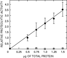
Fig. 1. Reduction of amoebic proteolytic activity on azo-casein. ([bull ]) Crude extract as control (□) IC[ratio ]MC alone, ([utri ]) IC[ratio ]MC+2% SDS and 5% 2-mercaptoethanol. Relative proteolytic activity based on 0·5 mg of crude extract. Bars correspond to standard errors, n=6.
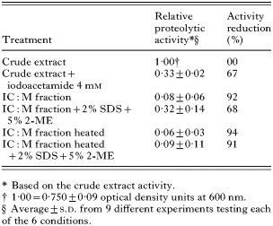
Table 1. Decrease of proteolytic activity in Entamoeba histolytica extracts using hide powder azure as substrate
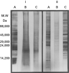
Fig. 2. SDS-PAGE and Western blot patterns from amoebic fractions. I – SDS-PAGE silver stained. II – Western blot patterns using serum from an amoebic liver abscess patient. IC[ratio ]MC (A), Crude extract (B), Crude extract+4 mM iodoacetamide (C).
 You have
Access
- 8
- Cited by