Introduction
Pneumatisation of the temporal bone begins in utero and continues into adolescence. It occurs as a result of epithelial out-pouching from the eustachian tube and middle ear, with subsequent subepithelial bone resorption.Reference Virapongse, Sarwar, Bhimani, Sasaki and Shapiro1 This physiological process is thought to serve several functions, namely, reducing the mass of the skull, protecting against trauma, providing acoustic resonance, and acting as an air reservoir to counteract negative middle-ear pressure.Reference Tumarkin2 Although there are certain characteristic patterns of pneumatisation, great variation has been demonstrated between normal, healthy individuals.Reference Virapongse, Sarwar, Bhimani, Sasaki and Shapiro1
Hyperpneumatisation of the craniocervical junction and cervical vertebrae is an extremely rare phenomenon hitherto lacking a convincing aetiological and pathophysiological explanation.
Case report
A fit, healthy, 40-year-old man presented with a 2-year history of left ear discomfort and autophony preceded by a short spell of vomiting. His symptoms were exacerbated by sneezing and coughing and relieved by performing the Valsalva manoeuvre against closed nostrils. He described an exacerbation of his symptoms following a minor road traffic accident, although he did not seek medical attention at the time. He also complained of a short history of worsening pain in the left shoulder and neck. He denied any hearing loss, vertigo or otorrhoea.
On examination, there was significant bulging of both the pars tensa and pars flaccida of both tympanic membranes (Figure 1). Examination of the post-nasal space and eustachian tube orifices was unremarkable, and there were no positive neurological findings in the upper limbs.
Endoscopic view of the left tympanic membrane showing bulging pars flaccida and pars tensa.

Audiometry was normal, and tympanography showed an immittance of 0 and a volume of 1.7 cm3 on the left.
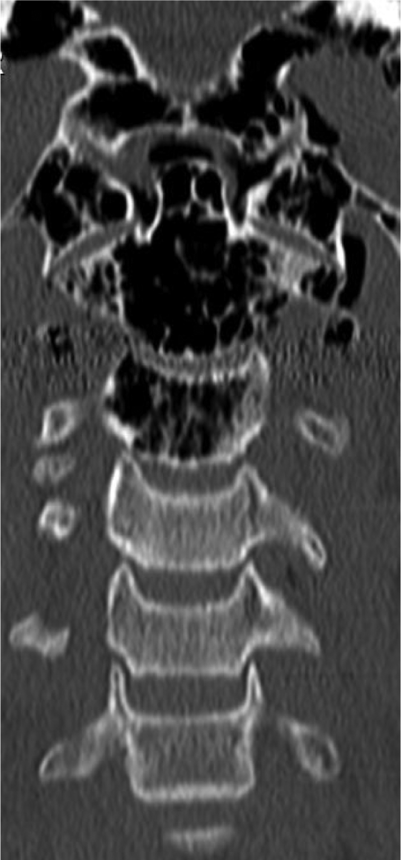
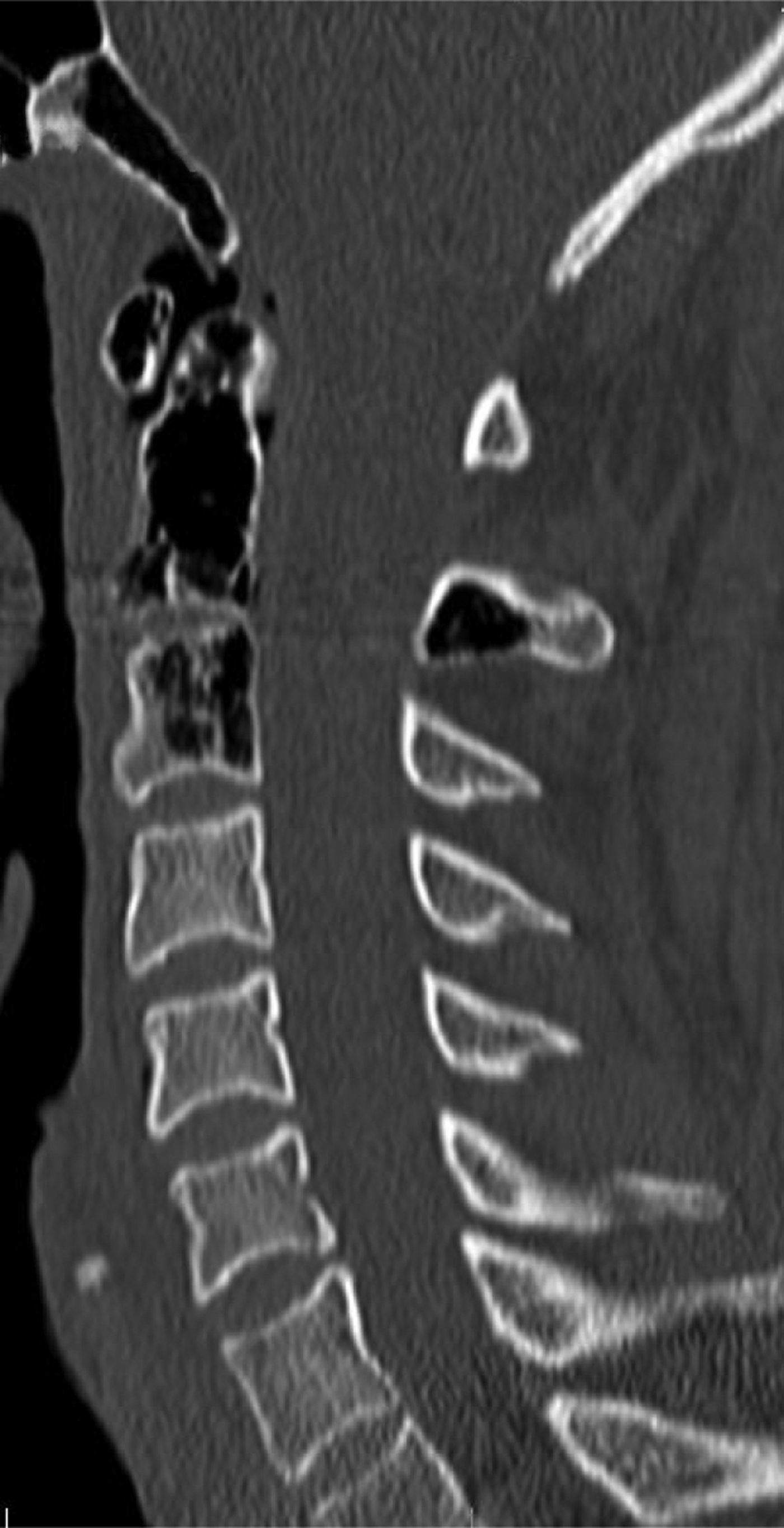
Computed tomography (CT) imaging revealed extensive hyperpneumatisation of the left mastoid, occipital bone and upper three cervical vertebrae, with extension into the surrounding soft tissues (Figures 2 and 3).
Initial coronal computed tomography scan showing pneumatisation of the occipital condyles and upper three cervical vertebrae.
Initial sagittal computed tomography scan showing extensive craniocervical pneumatisation, including free air in the soft tissues.
Initially, the patient was advised to stop performing the Valsalva manoeuvre. At follow up two weeks after the initial presentation, he reported a significant improvement in his symptoms. At that point, it was noted that his left tympanic membrane moved with respiration, suggesting a patulous eustachian tube.
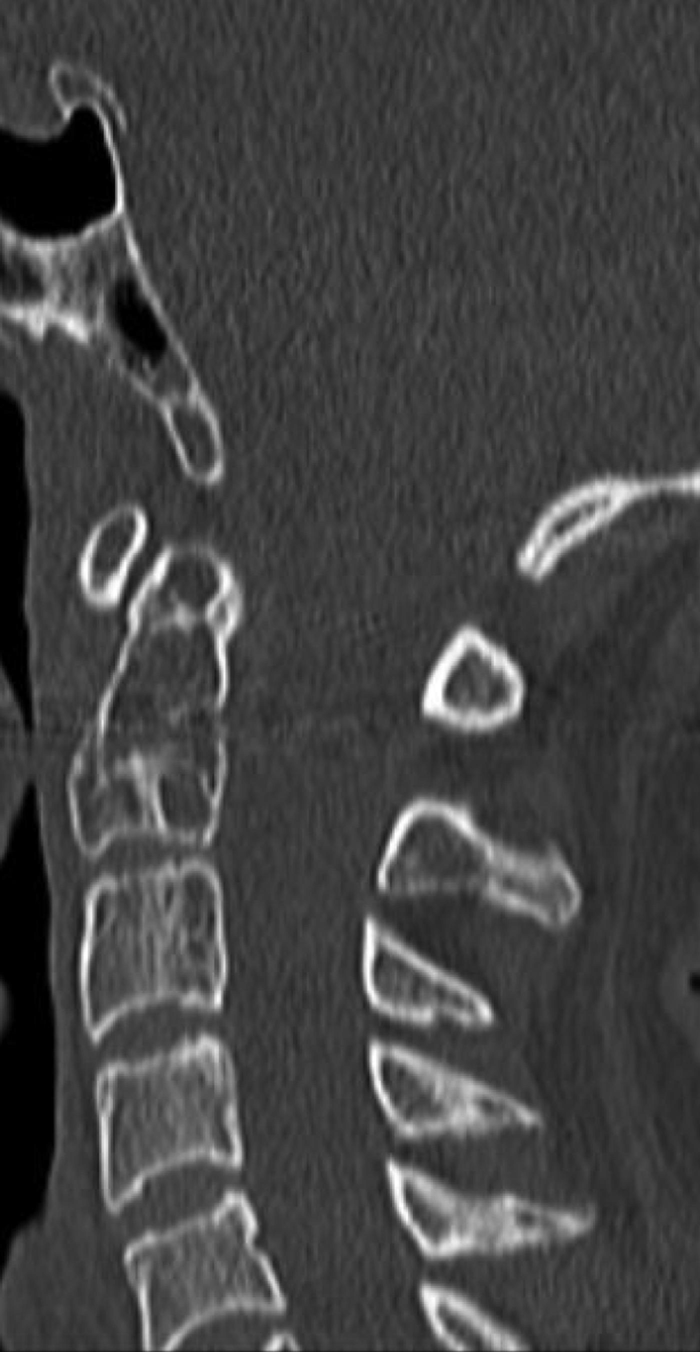
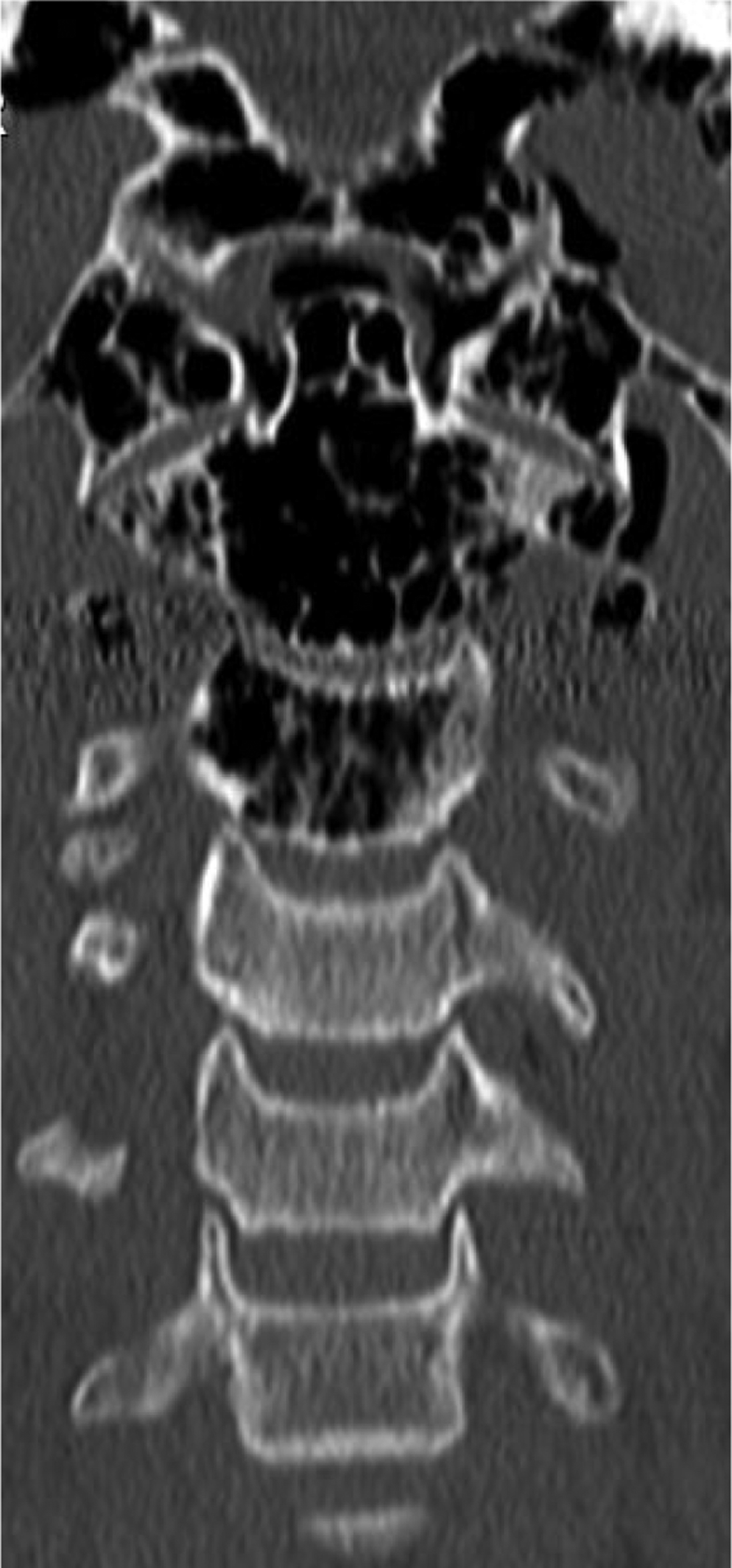
A repeated CT scan at three weeks confirmed almost complete resolution of the previously observed hyperpneumatisation (Figures 4 and 5). By this point, the patient was almost completely symptom-free.
Coronal computed tomography scan after three weeks of abstaining from performing the Valsalva manoeuvre, showing opacification of the previously pneumatised bones.
Sagittal computed tomography scan three weeks after presentation, showing significant improvement.
Discussion
This case represents an advanced form of the very rare phenomenon of hyperpneumatisation of the craniocervical junction and upper cervical vertebrae. It is likely that our patient's habit of repeatedly performing the Valsalva manoeuvre resulted in increased pressure in the middle ear and mastoid antrum, causing bony erosion and pneumatisation. There are very few other cases in the literature of skull base hyperpneumatisation involving the upper cervical vertebrae; however, all have in common a history of raised middle-ear pressure, minor trauma or both.Reference Martin, Bhargava, Ashforth and Russell3–Reference Sadler, Doyle, Hall and Crawford8
The most intriguing aspect of this condition is the fact that the pneumatisation process is able to cross seemingly intact synchondroses and joints. Both Turowski et al.Reference Turowski, Rettig, Raab and Zanella4 and Littrell et al.Reference Littrell, Leutmer, Lane and Driscoll5 described cases of craniocervical pneumatisation in patients who performed habitual Valsalva manoeuvres. However, these authors' pathophysiological explanations differed, with Turowski et al. favouring a theory of spread through soft tissues, and Littrell et al. postulating a defective eustachian tube acting as a ball-valve and raising air pressure in the mastoid air cells. Al Felasi et al.Reference Al Felasi, Venail and Lonjon7 and Martin et al.Reference Martin, Bhargava, Ashforth and Russell3 both described less extensive cases in a wind-instrument player and a patient with a chronic cough, respectively. Whilst the former authors proposed a mechanism of micro-fractures caused by pressure changes in the mastoid air cells, the latter supported the eustachian tube ball-valve theory, and suggested that persistently raised middle-ear pressure could lead to an erosive process of bony destruction capable of crossing the temporo-occipital synchrondosis and atlanto-occipital joints.Reference Martin, Bhargava, Ashforth and Russell3
• Pneumatisation of the craniocervical junction is rare and its aetiology poorly understood
• A case of advanced craniocervical hyperpneumatisation is presented
• All reported cases have a history of raised middle-ear pressure, minor trauma or both
• Bony destruction appears to result from increased middle-ear pressure
• Hyperpneumatisation crosses synovial joints via either micro-fractures or congenital assimilation of bones
• Conservative management and repeated imaging is recommended
The patient described in the current report had evidence of a patulous eustachian tube. By performing the Valsalva manoeuvre and producing positive pressure in the middle ear, the patient was able to tense the tympanic membrane, thus reducing his autophony symptoms. It is plausible that his repeated Valsalva manoeuvres eventually produced a eustachian tube mucosal fold which acted as a ball-valve, preventing dissipation of raised middle-ear pressure; over time, this positive pressure led to the development of hyperpneumatisation. The exacerbation of symptoms following minor trauma may be explained by micro-fractures at the atlanto-occipital joint allowing communication between the pneumatised and fragile occipital bone and the cervical vertebrae.
An interesting consideration is the possibility of congenital abnormalities resulting in bony continuity at the craniocervical junction.Reference Smoker9 Atlanto-occipital assimilation may result in a direct path for pneumatisation to cross seemingly intact joints, and was the mechanism proposed by Saddler et al.Reference Sadler, Doyle, Hall and Crawford8 in their report of atlanto-occipital pneumatisation in a patient with a rudimental atlanto-occipital joint. Atlanto-occipital assimilation is associated with increased prevalence of fusion of the axis and third cervical vertebra;Reference Smoker9 this is relevant, as in both our case and the similarly advanced case reported by Turowski et al.Reference Turowski, Rettig, Raab and Zanella4 pneumatisation did not spread beyond the third cervical vertebra, and there are no reports of the process involving the fourth.
Hyperpneumatisation of the craniocervical bones has the potential for serious complications. Pneumatised and weakened cervical vertebrae are more susceptible to traumatic injury, and the spread of air across the synovial joints of the craniocervical junction raises the possibility of spread of pathogens from the nasopharynx and middle ear. Treatment reported by other authors has ranged from advising against performing the Valsalva manoeuvre,Reference Massmann, Garcia, Pizanis, Roth, Nieder and Schneider6 through steroid nasal spays,Reference Littrell, Leutmer, Lane and Driscoll5 to myringotomy and grommet insertion,Reference Martin, Bhargava, Ashforth and Russell3, Reference Turowski, Rettig, Raab and Zanella4 and in one case surgical exploration and obliteration of airspaces using bone and fat grafts.Reference Al Felasi, Venail and Lonjon7 As our case demonstrates, addressing the cause of raised middle-ear pressure may result in both a rapid improvement in symptoms and a reversal of the pathological process.
Whilst the mechanism of pneumatisation of the bones of the craniocervical junction remains unclear, the present case adds to the evidence in favour of a theory of positive pressure in the middle ear, with micro-fractures facilitating spread across joints. As more experience of the condition is being reported, it seems that a pattern of raised middle-ear pressure and physical trauma or barotrauma is emerging. The present case also highlights the effectiveness of conservative treatment, with the emphasis being placed on behaviour modification.
Conclusion
Pneumatisation of the craniocervical junction is rare, and the aetiology of this condition is poorly understood. This paper presents an advanced case of craniocervical hyperpneumatisation, and examines possible aetiological factors in previously reported cases. All cases have in common a history of raised middle-ear pressure, minor trauma or both. Bony destruction appears to result from increased middle-ear pressure. The process of hyperpneumatisation crosses synovial joints either via micro-fractures or congenital assimilation of bones.
We recommend that this condition be managed conservatively, with repeated imaging.