





Non-technical Summary
Signs of feeding on fossils of extinct species are key to understanding how ancient animals interacted and interpreting the environments they lived in. A recently discovered skull of a tyrannosaur, Albertosaurus, aids in our interpretation of the prehistoric past. The fossil is the smallest skull of Albertosaurus discovered to date. The animal was at least eight years old when it died. Two sets of tooth marks are preserved on the lower jaw of the tyrannosaurid. These marks are caused by the teeth of a predator/scavenger scraping against the bone during feeding. The spaces between the marks left on the bone were measured and used to identify a potential animal that fed on this Albertosaurus. With the application of a new method to better estimate tooth spacing from tooth marks, the most likely predator/scavenger is a raptor (Dromaeosauridae). Tyrannosaurs likely ate raptors, but this fossil represents the first evidence of raptors feeding on a tyrannosaur.
Introduction
Tyrannosaurids were the dominant carnivorous dinosaurs of the Late Cretaceous in North America and Asia (Brusatte et al., Reference Brusatte, Norell, Carr, Erickson, Hutchinson, Balanoff, Bever, Choiniere, Makovicky and Xu2010). The Upper Cretaceous rocks of Alberta, Canada preserve representatives of both major tyrannosaurid clades; Albertosaurinae (Gorgosaurus, Albertosaurus) and Tyrannosaurinae (Daspletosaurus, Thanatotheristes, Tyrannosaurus). Of the two currently recognized albertosaurines, Gorgosaurus libratus Lambe, Reference Lambe1914, from the Dinosaur Park Formation (Upper Cretaceous: upper Campanian) of the Belly River Group has been the subject of a greater number of scientific studies. A large number of specimens, including several nearly complete skeletons of juvenile through adult individuals of Gorgosaurus have been studied in detail, providing invaluable information about the anatomy and ontogeny of the taxon (Carr, Reference Carr1999; Bradley, Reference Bradley2015; Voris, Reference Voris2018; Voris et al., Reference Voris, Zelenitsky, Therrien, Ridgely, Currie and Witmer2021; Therrien et al., Reference Therrien, Zelenitsky, Tanaka, Voris, Erickson, Currie, DeBuhr and Kobayashi2023). Conversely, Albertosaurus sarcophagus Osborn, Reference Osborn1905, from the Horseshoe Canyon Formation (Upper Cretaceous: Campanian–Maastrichtian) of the Edmonton Group (Eberth and Braman, Reference Eberth and Braman2012; Eberth et al., Reference Eberth, Evans, Brinkman, Therrien, Tanke and Russel2013, Reference Eberth, Evans, Ramezani, Kamo, Brown, Currie and Braman2023) is known from a more limited number of specimens (Tanke and Currie, Reference Tanke and Currie2010). Juvenile and sub-adult Albertosaurus individuals are rare and poorly known, most consisting of isolated elements (Currie, Reference Currie1998).
Over the past few decades, studies have begun to improve our understanding of prehistoric organismal relationships, including dinosaurs, using tooth marks left on bones (Currie and Jacobsen, Reference Currie and Jacobsen1995; Jacobsen, Reference Jacobsen1998; Rogers et al., Reference Rogers, Krause and Curry Rogers2003; Hone et al., Reference Hone, Choiniere, Sullivan, Xu, Pittman and Tan2010; Hone and Tanke, Reference Hone and Tanke2015; Drumheller et al., Reference Drumheller, McHugh, Kane, Riedel and D’Amore2020; Brown et al., Reference Brown, Tanke and Hone2021; Coppock and Currie, Reference Coppock and Currie2024, Wyenberg-Henzler and Scannella, Reference Wyenberg-Henzler and Scannella2026). Most recognized dinosaur feeding traces are preserved on herbivorous dinosaur bones, such as hadrosaurs and ceratopsians, and comparatively few are recorded on tyrannosaurid bones (e.g., Jacobsen, Reference Jacobsen1998). Among tyrannosaurids, most tooth marks are observed on maxillae and dentaries, but signs of healing and bone remodeling indicate they are healing/healed injuries and interpreted as non-lethal, intraspecific aggression rather than feeding (Tanke and Currie, Reference Tanke and Currie1998; Peterson et al., Reference Peterson, Henderson, Scherer and Vittore2009; Bell and Currie, Reference Bell and Currie2010; Hone and Tanke, Reference Hone and Tanke2015; Brown et al., Reference Brown, Currie and Therrien2022). Unhealed tyrannosaurid tooth marks on tyrannosaurid bones also evinces intra-familiar feeding, and in some cases cannibalism (Longrich et al., Reference Longrich, Horner, Erickson and Currie2010; Hone and Tanke, Reference Hone and Tanke2015; McLain et al., Reference McLain, Nelsen, Snyder, Griffin, Siviero, Brand and Chadwick2018; Coppock and Currie, Reference Coppock and Currie2024). In some cases, it is possible to associate tooth marks identified as feeding traces with the dentition of theropod dinosaurs as the trace maker, but in other cases this is prone to uncertainty (D’Amore and Blumenschine, Reference D’Amore and Blumenschine2009; Hone and Chure, Reference Hone and Chure2018; Lei et al., Reference Lei, Tschopp, Hendrickx, Wedel, Norell and Hone2023).
Based on size, spacing, and denticle rakes, most theropod tooth marks from the Late Cretaceous have been interpreted as having been produced by tyrannosaurids (Fiorillo, Reference Fiorillo1991; Jacobsen, Reference Jacobsen1998; Hone and Watabe, Reference Hone and Watabe2010; Bell et al., Reference Bell, Currie and Lee2012). Other contemporaneous carnivorous theropods (i.e., dromaeosaurids and troodontids; Eberth et al., Reference Eberth, Evans, Brinkman, Therrien, Tanke and Russel2013, Reference Eberth, Evans, Ramezani, Kamo, Brown, Currie and Braman2023), have few feeding traces attributed to them (see Currie and Jacobsen, Reference Currie and Jacobsen1995; Jacobsen and Bromley, Reference Jacobsen and Bromley2009). Here, we describe cranial remains of an immature Albertosaurus sarcophagus (TMP 2022.007.0040) from the Horseshoe Canyon Formation (HCFm) of Alberta that preserves unhealed tooth marks and develop a novel method of accounting for tooth mark and denticle mark rake angle to identify the trace maker as a dromaeosaurid.
Materials and methods
TMP 2022.007.0040 consists of an associated, disarticulated skull, including a left maxilla, right dentary, right surangular, and left splenial, and a portion of limb bone. The specimen was primarily found as ex-situ bones at the surface, although some elements were found in-situ in the rock layer. The stratigraphic position of the specimen relative to regional stratigraphic marker beds (i.e., coal beds) was determined using a 1.5-m Jacob’s staff (pogo stick) with an Abney level.
Paraloid B-72 in acetone was applied to the specimen as a consolidant and adhesive during fieldwork and preparation. Matrix surrounding the jacketed elements was removed with an air scribe and hand tools (scalpel, brush). The final surface of some of the elements was prepared with an air abrasive on low pressure with sodium bicarbonate powder. TMP 2022.007.0040 is stored and accessible for researchers in the collections of the Royal Tyrrell Museum of Palaeontology, Drumheller, Alberta, Canada.
A Canon EOS 80D DSLR camera and a Canon 100 mm macro lens with electronic focus were used to photograph the specimen. Raw images were processed and modified in Adobe Photoshop version 25.4.0. Some elements were coated with an ammonium chloride powder prior to photography to enhance surface textures following the dry method of Parsley et al. (Reference Parsley, Lawson and Pojeta2018). Illustrations of the specimen were created in ArtRage version 5.0.8, and final figures were compiled in Adobe Illustrator version 30.0.
Morphometric measurements of the specimen were taken with calipers or a measuring tape if greater than 100 millimeters. All measurements were taken in millimeters (mm). Tooth denticles were observed under a microscope at 112.5× magnification and measured through the microscope using the ocular lens graticule calibrated with a mm scale bar. Because preserved teeth were often broken at the basal end (near the root), denticle numbering started at the apical tip of the tooth and increased along the carina towards the root. Denticles with the observed largest base width (BW) were used for measurements, typically in the mid-portion of both the anterior and posterior carinae.
Tooth mark and denticle mark length, width, and angle were measured following the methods outlined in Wyenberg-Henzler et al. (Reference Wyenberg-Henzler, Fowler, Currie and Sullivan2024) (by the best fit line). To avoid artificially increasing tooth mark length, cracks running through the tooth marks were subtracted from measurements. Tooth mark depth was measured on silicone peels of the tooth marks. The spacing between tooth marks or between denticle marks (henceforth “mark spacing”) was measured between midpoints (center of the marks or the deepest point in the “V”-shaped indentation). Raw spacing was measured perpendicular to the long axis of the mark. Denticle mark spacing of the tooth mark preserved on the surangular was quantified by dividing the number of denticles by the linear distance over which the denticles occur (perpendicular to the long axis). All spacing data were log10 transformed prior to analysis.
For the true tooth spacing of the trace maker to be accurately represented by the tooth mark spacing on the bone, the teeth must contact the bone surface and rake perpendicularly (parallel with the bone surface and rake 90° from the long axis of the tooth row). Any divergence from a 90° angle would result in an underestimation of the true mark spacing and consequently an underestimation of the true tooth spacing of the trace maker (D’Amore and Blumenschine, Reference D’Amore and Blumenschine2012; Hone and Chure, Reference Hone and Chure2018; Lei et al., Reference Lei, Tschopp, Hendrickx, Wedel, Norell and Hone2023). As the raking angle decreases from 90°, the apparent mark spacing will decrease following the sine of the angle between the tooth row orientation and the rake direction (θ). At a certain angle threshold, the tooth marks will likely blur together and not be distinctly visible (Fig. 1). This same underestimation applies to denticle mark spacing (φ) (Fig. 2). Overestimation due to angle is not possible because the angle cannot create a spacing wider than what is truly present in the trace maker, however, teeth that do not occlude due to absence or incomplete replacement can still result in overestimation (Hone and Chure, Reference Hone and Chure2018). Denticle spacing is largely free of overestimation.
Adjusting for underestimation in tooth mark spacing measurements. (1) Demonstration of effect of trace maker tooth row orientation and rake direction on resulting mark raw spacing, black dots indicate spacing of trace maker’s teeth upon contact to bone; (2) hypothetical scenario of a theropod dinosaur creating tooth marks on bone; (3) enlarged view of tooth marks calculation of the corrected spacing (Cs) by using the raw spacing (Rs) and the angle between the tooth marks and a hypothesized tooth row orientation (θ).

Figure 1. Long description
Panel 1 is a table with two columns. The left column, Tooth row orientation and rake direction, shows three scenarios of a tooth row moving against a surface. At 90 degrees, sin(90) equals 1. At 45 degrees, sin(45) equals 0.71. At 20 degrees, sin(20) equals 0.34. The right column, Resulting mark spacing, shows the horizontal distance between marks decreasing from 1 unit to 0.71 unit to 0.34 unit as the angle narrows.
Panel 2 shows a green theropod dinosaur skull biting down on a tan-colored bone. A dashed purple line indicates the tooth row orientation, and a blue dotted arrow indicates the rake direction. A rectangular box zooms into the contact point.
Panel 3 is the enlarged view of two parallel tooth marks. A dashed pink line represents the tooth row orientation. A blue dotted line follows the path of the rake. The distance between the starting points of the two marks along the tooth row is labeled C s (Corrected spacing). The perpendicular distance between the two rake paths is labeled R s (Raw spacing). The angle between the tooth row and the rake path is labeled with the Greek letter theta. A legend on the right defines these terms: C s equals Corrected spacing and R s equals Raw spacing.
Adjusting for underestimation in denticle mark spacing measurements. (1) Demonstration of effect of trace maker tooth carina orientation and rake direction on resulting mark raw spacing, black dots indicate spacing of trace maker’s tooth denticles upon contact to bone; (2) hypothetical scenarios of a theropod dinosaur tooth creating three potential tooth marks with denticle marks on bone; (3) enlarged view of a scenario where two possible tooth carina orientations hypotheses can be applied, creating two possible angles (φ1 and φ2); (4) enlarged view of a scenario where denticle marks are bordered by tooth mark scores, but a single tooth carina orientation hypothesis can be applied, resulting in a single angle (φ); (5) enlarged view of a scenario with only one bordering tooth mark score and a single angle (φ); (6) enlarged view of the denticle marks in (5) applying the calculation of the corrected spacing (Cs) by using the raw spacing (Rs) and the angle between the denticle marks and a hypothesized tooth carina orientation (φ).

Figure 2. Long description
Panel 1 is a table with two columns. The left column shows carina orientation and rake direction at 90, 45, and 20 degrees. The right column shows resulting mark spacing, where 90 degrees equals 1 unit, 45 degrees equals 0.71 unit, and 20 degrees equals 0.34 unit.
Panel 2 shows a grey theropod tooth contacting a tan bone surface. A rectangular inset highlights three sets of parallel striations on the bone, each with blue dashed arrows indicating rake direction. A legend at the top right defines dashed pink lines as Carina orientation, blue dotted arrows as Rake direction, C s as Corrected spacing, and R s as Raw spacing.
Panel 3 shows a set of parallel marks between two hypothesized carina orientations, labeled i and ii, forming angles phi 1 and phi 2.
Panel 4 shows marks bordered by two vertical tooth scores, with a single carina orientation i forming angle phi at the top and bottom.
Panel 5 shows marks with only one bordering tooth score on the right and a single carina orientation i forming angle phi.
Panel 6 is a close-up of panel 5, showing a right-angled triangle geometry. The hypotenuse is the corrected spacing C s along the vertical carina line. The side adjacent to angle phi is the raw spacing R s, which is perpendicular to the parallel marks. The blue dotted arrows indicate the rake direction perpendicular to the carina.
To correct for the underestimation of tooth and denticle spacing caused by the rake angle, we developed a novel method where a hypothesized tooth row/tooth carina orientation (best fit line) is placed along the origination or termination of the marks. The hypothesized tooth row/tooth carina orientation attempts to encompass the end point of each mark, under the assumption that for serially spaced tooth or denticle marks, common origin/termination points are a reasonable proxy for tooth row/tooth carina orientation. This corrective method can only be used when an estimation of tooth row or carinae angle is possible, either due to common origin/termination points or other indications. Corrected spacing was calculated with the following equation:

Where: Raw spacing = measured spacing (mm) between tooth/denticle marks perpendicular to rake direction; θ = mean angle between tooth marks and hypothesized tooth row orientation; φ = mean angle between denticle marks and hypothesized tooth carina orientation.
For tooth and denticle marks preserved on TMP 2022.007.0040, hypothesized corrected spacings were generated by applying two hypothesized tooth row or tooth carinae orientations. The tooth mark spacing was compared to the alveolar spacing of potential trace makers present in the HCFm. Alveolar and denticle spacing measurements of potential trace makers were obtained from specimens housed at the University of Alberta Laboratory for Vertebrate Paleontology (UALVP) and Royal Tyrrell Museum of Palaeontology (TMP) collections. Because complete tooth bearing elements of taxa from the HCFm are limited, closely related taxa of similar proportions are also included from the better sampled Dinosaur Park Formation (Brown et al., Reference Brown, Tanke and Hone2021, Reference Brown, Currie and Therrien2022). If alveolar spacing data were not available for a given taxon, alveolar length was used as a proxy instead, because the amount of bone separating alveoli has a negligible influence on spacing. Alveolar spacing measurements for Dromaeosauridae (N = 132), Troodontidae (N = 51), and Tyrannosauridae (N = 365) were augmented with data from Brown et al. (Reference Brown, Tanke and Hone2021, Reference Brown, Currie and Therrien2022) and Funston et al. (Reference Funston, Powers, Amber Whitebone, Brusatte, Scannella, Horner and Currie2021). Alveolar spacing for Richardoestesia (N = 17) was measured based on the figured holotype R. gilmorei Currie, Rigby, and Sloan, Reference Currie, Rigby, Sloan, Carpenter and Currie1990 (CMN 343) from Currie et al. (Reference Currie, Rigby, Sloan, Carpenter and Currie1990, fig. 8.4), thus some error in spacing for Richardoestesia is expected. Denticle mark spacing (using denticle base width [BW] as a reliable proxy) was compared to the denticle spacing of potential trace makers from the HCFm. Denticle characteristics have been noted as being reliable indicators of species (Chandler, Reference Chandler1990; Smith et al., Reference Smith, Vann and Dodson2005; Hendrickx and Mateus, Reference Hendrickx and Mateus2014; Hendrickx et al., Reference Hendrickx, Mateus, Araújo and Choiniere2019) and striated tooth marks are recognized as being a reliable indicator of body size for the trace maker (D’Amore and Blumenschine, Reference D’Amore and Blumenschine2012). Denticle spacing measurements for Richardoestesia (N = 15), Dromaeosauridae (N = 71), Troodontidae (N = 53), and Tyrannosauridae (N = 243) were augmented with data from Larson et al. (Reference Larson, Brown and Evans2016), Torices et al. (Reference Torices, Reichel and Currie2014), and Buckley et al. (Reference Buckley, Larson, Reichel and Samman2010). Anterior and posterior carinae data were pooled because it is not known if the denticle marks on TMP 2022.007.0040 were caused by anterior or posterior serrations.
Because estimates of both denticle spacing and tooth spacing can be obtained from traces on the specimen (TMP 2022.007.0040), these two metrics are able to be visualized as bivariate data on a biplot with potential trace makers. For specimens of potential trace making taxa with in-situ teeth, mean alveolar and denticle spacing were calculated and plotted. Because some potential trace making taxa have very limited samples of jaw material with in-situ teeth (e.g., Albertavenator, Richardoestesia), the jaw-based data were augmented with data from isolated teeth. For isolated teeth, fore-aft base length (FABL) was used as a proxy to alveolar spacing and denticle spacing was calculated as for in-situ teeth.
The isolated tooth data from Torices et al. (Reference Torices, Reichel and Currie2014) and Buckley et al. (Reference Buckley, Larson, Reichel and Samman2010) provide poor sampling of juvenile tyrannosaurs. As a result, we augmented these datasets with additional measurements of the smallest HCFm Tyrannosauridae teeth from the TMP collection to attempt to capture the full variability and ontogeny of denticle spacings in tyrannosaurids. Specimens of potential trace making taxa were plotted in 2-dimensional morphospace and compared to mark spacing seen in TMP 2022.007.0040. Due to low sampling in the HCFm, these jaw-based metrics were also augmented with closely related taxa from the Dinosaur Park Formation (e.g., Gorgosaurus for Albertosaurus).
Histological sampling of an associated limb bone fragment of TMP 2022.007.0040 was performed following a method similar to that outlined by Funston et al. (Reference Funston, Wilkinson, Simon, Leblanc, Wosik and Currie2020). Imaging of the thin section was performed on a Nikon Eclipse E600POL trinocular polarizing microscope using Nikon NIS Elements Document software.
Statistical tests were conducted in the R statistical program (R version 4.2.2) using RStudio (Version 2022.12.0+353). Tooth mark/denticle mark spacing and taxon tooth/denticle spacing were compared using a Monte Carlo permutation tail-probability test (using base R functions) to evaluate the probability that tooth mark/denticle mark spacing could be caused by the different taxa. Probability is evaluated as the distance from a taxon’s median over 10,000 bootstrap replicates. Taxa were rejected if p-value < 0.05 (α = 0.05). Tooth mark/denticle mark spacing was plotted against the distribution of alveolar/denticle spacing in HCFm and closely related taxa through density histograms (function: hist).
Repositories and institutional abbreviations
Types, figures, and other specimens examined in this study are deposited in the following institutions: American Museum of Natural History (AMNH), New York, USA; Canadian Museum of Nature (CMN), Ottawa, Canada; Museum of the Rockies (MOR), Bozeman, USA; Royal Tyrrell Museum of Palaeontology (TMP), Drumheller, Canada; University of Alberta Laboratory for Vertebrate Paleontology (UALVP), Edmonton, Canada.
Systematic paleontology
Dinosauria Owen, Reference Owen1842
Saurischia Seeley, Reference Seeley1888
Theropoda Marsh, Reference Marsh1881
Coelurosauria von Huene, Reference von Huene1914
Family Tyrannosauridae Osborn, Reference Osborn1906
Subfamily Albertosaurinae Currie et al., Reference Currie, Hurum and Sabath2003
Genus Albertosaurus Osborn, Reference Osborn1905
Type species
Albertosaurus sarcophagus Osborn, Reference Osborn1905 (CMN 5600) from the Horseshoe Canyon Formation, Edmonton Group, at the Knee Hills Creek of the Red Deer River, Alberta, Canada.
Albertosaurus sarcophagus Osborn, Reference Osborn1905
Stratigraphic position of TMP 2022.007.0040 within the Horseshoe Canyon Formation (HCFm). (1) Map of TMP 2022.007.0040 quarry locality along the Red Deer River valley in Alberta, Canada, west of the town of Morrin, approximately 5 km north of the Morrin Bridge (HWY 27) crossing the Red Deer River; (2) photograph of the quarry from the coulee bottom facing east, white arrow indicates specimen in-situ level, geological hammer for scale (~30 cm long), the two coals above the specimen level are the top of the Coal 9 swarm, with the upper coal being the boundary between the Horsethief and Morrin members of the HCFm, Coal 10 is visible to the east along the valley wall; (3) stratigraphic column displaying the upper Horsethief Member above the specimen, numbers indicate height in m; abbreviations: MM, Morrin Member; grain size: Cl, clay; Si, silt; f, fine-grained sandstone; m, medium-grained sandstone; c, coarse-grained sandstone.

Figure 3. Long description
Panel 1 contains a map of Alberta, Canada, showing Edmonton and Calgary, with an inset satellite map of the Red Deer River valley. A star marks the TMP 2022.007. 0040 quarry North of Highway 27 and West of the town of Morrin. A 1 km scale bar is provided.
Panel 2 is a photograph of the excavation site. A white arrow points to a geological hammer resting on the specimen in-situ level in the foreground. Dotted lines lead to the valley wall in the background, identifying the Top of coal 9 and Coal 10 layers.
Panel 3 is a stratigraphic column of the Horsethief Member within the Horseshoe Canyon Formation. The vertical axis measures height from 0 to 6 m. The horizontal axis indicates grain size from right to left: Cl for clay, Si for silt, f for fine sandstone, m for medium sandstone, and c for coarse sandstone.
* At 0.5 m: A yellow sandstone layer contains the TM P 2022.007. 0040 specimen, indicated by a dinosaur skull icon.
* 1.0 to 1.5 m: Green shale or mudstone with a thin black coal seam.
* 1.5 to 5.5 m: Alternating thick green shale or mudstone and thin red ironstone bands.
* 5.5 to 6.0 m: A yellow sandstone layer capped by a black coal seam, marking the boundary with the M M (Morrin Member).
A legend at the bottom right defines the colors: yellow for Sandstone, green for Shale or Mudstone, black for Coal, and red for Ironstone.
TMP 2022.007.0040 left maxilla. (1) Maxilla in lateral view; (2) reconstruction of maxilla in lateral view, light-gray shading indicates missing sections of element, ascending ramus repositioned to align ventral end of interfenestral strut with the remainder of the element, solid line indicates fresh break between the repositioned contacting surfaces; (3) maxilla in medial view.

Figure 4. Long description
The figure consists of three vertically stacked panels showing the TMP 2022.007. 0040 specimen.
Panel 1: A photograph of the maxilla in lateral view. The bone is dark brown with a triangular profile. It features a large circular opening, the antorbital fenestra, in the center. The ascending ramus rises steeply at the front, while the posterior end tapers into a long, thin process. A 10 cm scale bar with alternating 1 cm black and white segments is located at the bottom of the figure.
Panel 2: A line drawing reconstruction of the lateral view. Stippled texture represents the preserved bone surface. Light-gray shaded areas indicate missing sections at the anterior tip, the dorsal edge of the ascending ramus, and the posterior process. A solid line marks a fresh break where the ascending ramus was repositioned to align the interfenestral strut with the main body of the bone.
Panel 3: A photograph of the maxilla in medial view, showing the inner surface. The bone texture is more irregular here, with visible ridges and recesses. The orientation is reversed compared to the lateral views, with the tapering posterior process pointing toward the left side of the frame.
TMP 2022.007.0040 right dentary. (1) Dentary in lateral view; (2) dentary in medial view.

TMP 2022.007.0040 right surangular. (1) Surangular in lateral view; (2) surangular in medial view, black arrow indicates bulbous pathology on ventral margin; (3) enlarged view of tooth mark on anterior end of surangular; (4) augmentation of (3) showing preservation of denticle marks associated with the tooth mark, identifiable denticle marks (solid light-blue lines), best fit line and angle of denticle marks (dotted dark-blue line), two hypothesized carina orientations (dashed pink lines) (i) and (ii), and the angle between the hypothesized carina orientation and best fit line of denticle marks (φ); specimen dusted in ammonium-chloride powder in (3) and (4).

Figure 6. Long description
A multi-panel figure containing four numbered photographs of specimen TMP 2022.007. 0040.
* Panel 1 at the top-left shows the surangular in lateral view. It is a long, fragmented bone with a 10 centimeter scale bar below it.
* Panel 2 at the bottom-left shows the surangular in medial view. A black arrow at the bottom center points to a rounded, bulbous pathology on the ventral margin.
* Panel 3 at the top-right is a close-up of a rectangular area from the anterior end of the bone, dusted in white ammonium-chloride powder. It shows a deep, oval-shaped tooth mark with a 2 millimeter scale bar.
* Panel 4 at the bottom-right is an augmentation of panel 3 with analytical overlays. Solid light-blue lines trace individual denticle marks within the indentation. A dotted dark-blue line represents the best fit line of these marks. Two dashed pink lines, labeled i and ii, indicate hypothesized carina orientations. The Greek letter phi marks the angle between the best fit line and the hypothesized carina orientation.
TMP 2022.007.0040 left splenial. (1) Splenial in medial (lingual) view; (2) splenial in lateral (labial) view; (3) enlarged image of tooth marks on posterior process of splenial; (4) Augmentation of (3) showing tooth mark spacing measurements, tooth marks numbered (1, 2, 3), inter-mark spacing (a and b, dotted black lines) measured between midpoint of tooth marks (solid light-blue lines), best fit lines and angles of each tooth mark (dotted dark-blue lines), two hypothesized tooth row orientations (dashed pink lines) (i) and (ii), and the angle between the hypothesized tooth row orientation and best fit lines of tooth marks (θ); specimen dusted in ammonium-chloride powder in (3) and (4).

Figure 7. Long description
A four-panel anatomical study of a fossilized splenial bone.
Panel 1 (top left): A photo of the splenial in medial lingual view. The bone is elongated and dark brown with a rectangular box highlighting the posterior process. A 5 centimeter scale bar is positioned between panels 1 and 2.
Panel 2 (bottom left): A photo of the splenial in lateral labial view, showing the opposite side of the bone with a white specimen label attached.
Panel 3 (top right): An enlarged photo of the posterior process from panel 1, dusted in white ammonium-chloride powder to highlight three diagonal tooth marks. A 1 centimeter scale bar is at the bottom left.
Panel 4 (bottom right): An augmented version of panel 3 with a 5 millimeter scale bar. It includes technical overlays:
- Three diagonal tooth marks are numbered 1, 2, and 3.
- Solid light-blue lines mark the midpoints of the tooth marks, with inter-mark spacing labeled ‘a’ (between 1 and 2) and ‘b’ (between 2 and 3).
- Dotted dark-blue lines indicate the best fit lines and angles of each mark.
- Two dashed pink lines labeled (i) and (ii) represent hypothesized tooth row orientations.
- The Greek letter theta marks the angle between the hypothesized tooth row orientation and the best fit lines of the tooth marks.
Histological thin section of TMP 2022.007.0040 limb fragment demonstrating lines of arrested growth (LAGs) (black arrows, N = 6) and annuli (red arrows, N = 2), white arrow indicates example of a Haversian canal near the endosteum; abbreviations: E, endosteum; P, periosteum.

Skull element measurements/counts for TMP 2022.007.0040 (mm), asterisk (*) indicates less certain measurements because the element may be incomplete, length is antero-posterior, height is dorso-ventral; abbreviations: Spec, specimen(s)

Table 1. Long description
The table is divided into three primary sections.
Section 1: Maxilla (left)
- Length: 408 mm.
- Height at 6th alveolus: 170 mm.
- Maximum height at back of lacrimal process: 178* mm.
- Tooth row length: 295 mm.
- Tooth count: 13* (14 if including a possible small tooth at the tip).
- Antorbital fossa length: 270* mm (estimated based on similar-sized specimens).
- Maxillary fenestra maximum length: 52.5 mm.
- Antorbital fenestra length: 175* mm (estimated).
- Antorbital fenestra height: 110* mm (estimated).
- Promaxillary fenestra height: 29.98 mm.
Section 2: Dentary (right)
- Length: 360* mm.
- Minimum height at 8th alveolus: 70.5 mm.
- Maximum height: 120* mm.
- Tooth row length: 288 mm.
- Tooth count: 15.
Section 3: Surangular (right)
- Length: 293* mm.
- Maximum depth: 91* mm.
Note: Asterisks indicate less certain measurements due to incomplete elements.
Alveoli length measurements for the left maxilla and right dentary of TMP 2022.007.0040; asterisk (*) indicates alveoli that are excluded in the means (anteriormost 2 and posteriormost 5), double asterisk (**) indicates alveoli excluded in the mean of the maxilla only

Table 2. Long description
The table consists of three columns: Tooth position, Left maxilla alveolus length (m m), and Right dentary alveolus length (m m).
* Position 1 (anterior): Left maxilla 22, Right dentary 13.8.
* Position 2: Left maxilla 25.5, Right dentary 16.8.
* Position 3: Left maxilla 32.2, Right dentary 21.6.
* Position 4: Left maxilla 26.2, Right dentary 22.5.
* Position 5: Left maxilla 25, Right dentary 21.3.
* Position 6: Left maxilla 25.2, Right dentary 22.7.
* Position 7: Left maxilla 21.1, Right dentary 24.3.
* Position 8: Left maxilla 20.8, Right dentary 20.5.
* Position 9: Left maxilla 24.1, Right dentary 20.1.
* Position 10: Left maxilla 16.7, Right dentary 19.5.
* Position 11: Left maxilla Incomplete, Right dentary 20.6.
* Position 12: Left maxilla Incomplete, Right dentary 19.2.
* Position 13: Left maxilla 12.5, Right dentary 15.9.
* Position 14: Left maxilla blank, Right dentary 14.4.
* Position 15 (posterior): Left maxilla blank, Right dentary 13.
* Mean: Left maxilla 25.1, Right dentary 21.6.
Asterisks indicate exclusions from the mean: positions 1, 2, 11, 12, 13, 14, and 15 are excluded for both; positions 9 and 10 are excluded for the maxilla only.
Holotype
Partial skull (CMN 5600) from the Horseshoe Canyon Formation, Red Deer River, Alberta, Canada.
Occurrence
TMP 2022.007.0040 was found at the base of a mudstone slump on the eastern slope of the Red Deer River valley, five kilometers north of the Morrin Bridge (precise GPS coordinates on file at TMP). Two coal seams were identified above the specimen, 1.0 m and 5.3 m above the specimen (Fig. 3), representing the top of the Horsethief Member (Coal 9 swarm) of the HCFm (sensu Eberth and Braman, Reference Eberth and Braman2012). Coal 10, situated near the top of the overlying Morrin Member, is not visible on the hill directly above the specimen but is visible to the east along the valley wall (Fig. 3.2). This stratigraphic location is consistent with the known stratigraphic range of Albertosaurus sarcophagus in the HCFm (Eberth et al., Reference Eberth, Evans, Brinkman, Therrien, Tanke and Russel2013, Reference Eberth, Evans, Ramezani, Kamo, Brown, Currie and Braman2023).
Description
TMP 2022.007.0040 preserves a left maxilla, right dentary, right surangular (preserving a single tooth mark), and left splenial (preserving three tooth marks), nine associated ex-situ teeth, and a portion of a shattered limb bone. A small amount of additional material has not yet been prepared, including one additional ex-situ tooth. Given the limited amount of in-situ material, the site was not mapped in detail. However, a rough quarry map (available on file at the TMP) confirms the skull was disarticulated. Autapomorphic characters for Albertosaurus sarcophagus have not been clearly assigned, thus attribution of TMP 2022.007.0040 to A. sarcophagus is challenging. Omitting stratigraphic position as a valid diagnosis of the taxon, TMP 2022.007.0040 remains most likely referable to A. sarcophagus by the presence of a broad anterior angle of the maxilla as well as a narrow maxillary interfenestral strut (Carr, Reference Carr2010; Voris et al., Reference Voris, Zelenitsky, Therrien, Ridgely, Currie and Witmer2021).
Maxilla
The left maxilla has a broad anterior angle (83°), consistent with the characteristic differentiating Albertosaurus and Gorgosaurus (Voris et al., Reference Voris, Zelenitsky, Therrien, Ridgely, Currie and Witmer2021). The interfenestral strut is 13.2 mm anteroposteriorly wide at the narrowest point. It was displaced medially postmortem, causing the ventral end to not contact the remainder of the antorbital fossa (Fig. 4). Following realignment of the interfenestral strut (Fig. 4.), the ascending ramus projects at a 35° to the jugal ramus. The maxillary fenestra does not meet the ventral margin of the antorbital fossa, with a 5.7 mm separation at the nearest point. The maxillary depth (anterior end of antorbital fenestra) and length (tooth row length) provide a depth-to-length ratio of 0.58. The medial surface of the maxilla is heavily eroded and fragmented due to weathering (Fig. 4.3). The medial wall and shelf, as well as the anteromedial process, are almost completely eroded. Most of the interdental plates are absent, with only portions remaining to support a few teeth. The alveoli are fully exposed with only the lateral wall visible and half of the interdental bone (septa). The tooth row is complete and 295 mm long, preserving 13 identifiable alveoli (with possibly a 14th anterior alveolus) (Table 1). Although there is variability in the number of alveoli, TMP 2022.007.0040 falls within the expected range for Albertosaurus (Currie, Reference Currie2003b). The mean alveolus length is 22.8 mm, or 25.1 mm when excluding the anterior two and posterior five alveoli, as done for the size metric by Brown et al. (Reference Brown, Currie and Therrien2022) (Table 2). A fragment of the palatine ramus was recovered detached from the maxilla. Additional maxillary fragments were found surrounding the in-situ element but had no clear contact with the larger element.
Dentary
The right dentary preserves a complete tooth row with 15 alveoli (Fig. 5). The posterior end of the dentary is incomplete and therefore the full length of the element is unknown (Table 1). The tooth row is 288 mm long, and the mean alveolar length is 17.7 mm, or 21.6 mm when excluding the anterior two and posterior five alveoli (Table 2). The anterior end of the dentary adjacent to the first two alveoli is fragmented and the rugosity of the mandibular symphysis cannot be determined. As with the maxilla, much of the medial surface of the dentary is eroded, exposing the lateral wall and interdental bone. Nevertheless, a portion of the medial surface of the dentary is preserved up to the Meckelian groove between alveoli eight and eleven. Additionally, the ventral margin of the Meckelian foramen is preserved at alveolus four. The dentary groove is well defined on the lateral side along the length of the tooth row. The groove is moderately deep and there is an abundance of nutrient foramina that perforates the surface of the bone.
Surangular
The dorsal margin of the right surangular is complete, with the surangular shelf clearly defined. The remainder of the body of the surangular is a mosaic of fragments with an incomplete ventral margin (Fig. 6). Only the dorsal margin of the posterior surangular fenestra is preserved, but it is estimated to be 26 mm antero-posteriorly long. A separate fragment, on which a tooth mark is preserved (see subsequent section on “Surangular tooth mark”), contains a portion of the anterior surangular fenestra and the beginning of the dentary contact. These two fragments do connect, but the smaller anterior fragment was left unconnected to the main block of the surangular to facilitate analysis. A pathologic bulb three times thicker than the surrounding bone is preserved on the main body of the surangular (Fig. 6.1, 6.2). The bulb surrounds a foramen-like pit, most easily seen in medial view on the most ventral fragment (Fig. 6.2). Remodeled bone texture on the medial surface surrounds the pit, extending 64 mm in anteroposterior length. A similar remodeled bone texture extends 20 mm around the pit on the lateral surface. Trichomonas-like lesions have been identified previously on tyrannosaur jaws (Wolff et al., Reference Wolff, Salisbury, Horner and Varricchio2009), but the morphology of these lesions typically entails complete perforations of the bone, whereas the pathology on TMP 2022.007.0040 is a deep pit, not completely passing through the bone.
Splenial
The preserved portion of the left splenial is approximately 210 mm antero-posteriorly long and 35 mm dorso-ventrally wide. Only the posterior process of the splenial is well preserved, where the dentary contact surface is visible. The remainder of the splenial is poorly preserved and shattered (Fig 7). The shattered anterior end projects at nearly 90° from the posterior process, likely due to postdepositional processes. The mylohyoid foramen is not preserved. Three tooth marks are preserved on the tip of the posterior process (see subsequent section on “Splenial tooth marks”).
Additional elements
Other elements collected with the specimen include 10 associated teeth (Supplementary Data, Fig. S1), and a shattered partial limb element preserving a handful of unconnected fragments (possibly a tibia, which was sectioned to examine histology) (Fig. 8). The 10 teeth belong to either the maxilla or dentary of the specimen, but their original location is unknown. The mean tooth crown height is 34 mm and a mean denticle base width of 0.43 mm corresponds well with the denticle base width for the maxilla (0.41 mm) and dentary (0.38 mm) (Supplementary Data, Tables S1 and S2).
Surangular tooth mark
A single tooth mark containing approximately 28 denticle marks is preserved on the lateral surface of the anterior end of the surangular (Fig. 6.3, 6.4). The tooth mark runs perpendicular to the long axis of the bone and is located slightly below the origin of the anterior surangular foramen and the dentary contact surface of the surangular. The tooth mark morphology is defined as a linear, non-elongate, shallow pit, with a rough-edged external and raked internal morphology (sensu Wyenberg-Henzler et al., Reference Wyenberg-Henzler, Fowler, Currie and Sullivan2024). The raked internal morphology (or “striations” as defined by Drumheller et al., Reference Drumheller, D’Amore, Njau, Woodward and Farlow2023) are sub scores that are formed by tooth denticles raking across the bone surface. Similar marks have been described previously as the ichnospecies Knethichnus parallelum Jacobsen and Bromley, Reference Jacobsen and Bromley2009.
The denticle marks rake across the indentation, with some striae visible outside the indented groove on the otherwise unaltered surface of the bone. The mark is 8.2 mm long dorso-ventrally, 3.8 mm wide anteroposteriorly, and has a maximum depth of 0.57 mm. The denticle marks rake at 47° from the long axis of the bone and are regularly spaced and parallel. Raw mean denticle mark spacing is 0.167 mm (Supplementary Data, Table S3). D’Amore and Blumenschine (Reference D’Amore and Blumenschine2012) used the raw maximum denticle mark spacing as the best estimate for true minimum denticle mark spacing to infer the identity of the trace maker. However, when possible, adjusting for the rake angle reaches a closer approximation of the true denticle mark spacing.
Two hypothesized orientations of the tooth carinae ((i) and (ii)) are identified based on a combination of correlated origin/termination of striae and furrows running sub-perpendicular to striae, and result in two potential corrected spacings (Fig. 6.4). Hypothesized carina orientation (i) extends along the termination of at least nine denticle marks, and is the preferred hypothesis, because it is likely the beginning or end of the tooth denticles raking across the bone (corrected spacing = 0.191 mm). Hypothesized carina orientation (ii) is along the deepest furrow of the mark and the longest portion of the mark (corrected spacing = 0.236 mm) (Fig. 6.4).
Splenial tooth marks
Three sub-parallel tooth marks are preserved on the medial (lingual) surface (the surface facing the midline of the body, away from the dentary) of the posterior process of the splenial (Fig. 7.3). The marks rake at a mean angle of 39.3° from the long axis of the splenial. Distances between the three tooth marks are regular, except for the dorsal convergence of tooth marks 2 and 3 (Fig. 7.4). The mean raw tooth mark spacing is 2.35 mm (Supplementary Data, Table S4), and each mark has an approximate depth of 0.55 mm. Following Wyenberg-Henzler et al. (Reference Wyenberg-Henzler, Fowler, Currie and Sullivan2024), the tooth marks are defined as curved, elongate, shallow scores, with a rough-edged external and chattered internal morphology. These marks have a deep “V”-shaped profile due to the apical tip of a tooth being dragged across the surface of the bone (Drumheller et al., Reference Drumheller, D’Amore, Njau, Woodward and Farlow2023). Tooth contact directionality can be inferred from redirection of the bone fibers (Currie and Jacobsen, Reference Currie and Jacobsen1995), but the bone fibers in TMP 2022.007.0040 do not have any visible change in directionality at the boundary of the marks. Consequently, directionality of the tooth contact on the surface cannot be inferred by this method.
Two hypothesized tooth row orientations ((i) and (ii)) produce two corrected tooth mark spacings. The first hypothesized tooth row orientation (i) extends along the dorsal side of the origination/termination of the tooth marks, and the area where two curved traces converge, with the convergence of the curved marks suggesting a rake angle approaching parallel to tooth row angle. Horizontal spacing between the dorsal tips of each tooth mark are regular (between mark 1–2 = 7.16 mm; between mark 2–3 = 7.88 mm), suggesting the three teeth contacted or released from the bone at the same time. Hypothesized tooth row orientation (ii) extends along the ventral tip of the three tooth marks. Horizontal spacing between the marks is less consistent (between mark 1–2 = 3.87 mm; between mark 2–3 = 5.83 mm), suggesting the teeth contacted or released from the bone at slightly different times, likely due to the ventral curvature of the splenial at the tip of the marks, or teeth that are inconsistently spaced. The tooth marks rake at a mean angle of 16° relative to hypothesized tooth row orientation (i) (corrected spacing = 8.53 mm) and 32° relative to hypothesized tooth row orientation (ii) (corrected spacing = 4.44 mm) (Fig. 7.4; Supplementary Data, Table S5). Due to the regularity of the initial spacing, hypothesized tooth row orientation (i) is the preferred hypothesis.
Bone histology
The thin section of the limb bone fragment associated with TMP 2022.007.0040 only provides a small portion of the entire cross-section of the long bone but does span the entire radial endosteal to periosteal transect (26 mm thick). Six lines of arrested growth (LAGs) and two annuli (nearest the endosteum) are visible in the limb bone cross-section (Fig. 8). The LAGs are consistent with the interpretation of these features as reduced periods of growth (Reid, Reference Reid, Brett-Surmann, Holtz and Farlow2012), known to be annual in living tetrapods and likely extinct dinosaurs (Horner et al., Reference Horner, De Ricqlès and Padian1999, for an alternate view see Chinsamy and Pereyra, Reference Chinsamy and Pereyra2026).
Remarks
Age and growth
The restricted view of the limb bone cross-section provided by the histological thin section of TMP 2022.007.0040 limits our ability to determine the exact age of the specimen but provides a plausible estimate. The bone is primarily woven bone, typical of young animals (e.g., Chinsamy-Turan, Reference Chinsamy-Turan2005; Woodward et al., Reference Woodward, Tremaine, Williams, Zanno, Horner and Myhrvold2020). The six LAGs and two annuli indicate the individual was at least eight years old at time of death. Although it is difficult to determine which limb element the fragment is from, it is likely a weight-bearing element (tibia?), which has been identified to be more reliable in the uniformity and consistency of LAGs (e.g., Chinsamy-Turan, Reference Chinsamy-Turan2005; Cullen et al., Reference Cullen, Canale, Apesteguía, Smith, Hu and Makovicky2020). Asymmetric expansion of the medullary cavity and bone remodeling during growth, as indicated by the presence of Haversian canals near the endosteum, possibly could have erased (at least partially) early LAGs (Fig. 8). Had the entire cross-section of the limb element been preserved, it is possible that additional LAGs (possibly 1–2 more) may have been visible near the medullary cavity. The LAGs increase slightly in spacing towards the periosteal surface, suggesting that the animal was still in a period of rapid growth. Additionally, the absence of external fundamental systems (EFS) near the periosteal surface indicates the animal was not fully grown (Horner et al., Reference Horner, De Ricqlès and Padian1999). Beyond the histological data, the presence of striated bone texture in the antorbital fossa of the maxilla may be another size-independent indication of the immature status of TMP 2022.007.0040 (Bradley, Reference Bradley2015). Based on mean alveolar length, TMP 2022.007.0040 is the smallest associated Albertosaurus skull described to date, adding to the limited sample of immature Albertosaurus individuals.
Body size estimations also indicate that TMP 2022.007.0040 was an immature individual. Following Albertosaurinae allometric regressions reported in Currie (Reference Currie2003a, table 3), we estimate that TMP 2022.007.0040 possessed a basal skull length of 63.25 cm (R 2 = 0.93) and femoral length of 66 cm (R 2 = 0.98), based on maxillary tooth row length. Skull length was subsequently used to estimate body length and body mass following the regressions of Therrien and Henderson (Reference Therrien and Henderson2007), resulting in an estimated 6 m long and 533 kg Albertosaurus. Based on relationships established between femur length and biological age in Albertosaurus, TMP 2022.007.0040 is estimated to have been ~10 or 11 years old following the methods of Eberth and Currie (Reference Eberth and Currie2010) and Erickson et al. (Reference Erickson, Currie, Inouye and Winn2006), respectively.
Although caution is warranted due to the application of stepwise regressions, the estimated skull length, femur length, body length, body mass, and age for TMP 2022.007.0040 are slightly smaller than those of a subadult Gorgosaurus individual, TMP 1991.036.0500, (maxillary tooth row length = 30.25 cm, skull length = 63.9 cm, femur length = 65 cm, body length = 6.29 m, estimated body mass = 595.5 kg (see Therrien et al., Reference Therrien, Zelenitsky, Tanaka, Voris, Erickson, Currie, DeBuhr and Kobayashi2023), estimated age = 11 years following Eberth and Currie’s (Reference Eberth and Currie2010) method). Although an age estimate of 10 or 11 years is slightly above the estimated age based on visible LAGs in TMP 2022.007.0040 (8 years), the fact that additional LAGS would likely have been present near the medullary cavity makes an age of ~10 or 11 years old reasonable.
Taxonomic identity of tooth mark maker
To identify the trace maker of the tooth marks left on TMP 2022.007.0040, damage caused by sullegic (fossil extraction) and trephic (fossil preparation) processes (sensu Marin-Monfort et al., Reference Marin-Monfort, Suñer and Fernández-Jalvo2018) need to be ruled out. No known damage to the bone surface occurred during specimen extraction and preparation, and tool marks were ruled out following the methods of Blumenschine et al. (Reference Blumenschine, Marean and Capaldo1996) and through comparison with known preparation marks (Wiest et al., Reference Wiest, Ferraro, Binetti, Forman, Esker, Kibunjia, Brugal and Zechmann2018). As such, the marks can be confidently identified as tooth marks.
The denticle marks on the surangular are regularly spaced, reminiscent of ziphodont dentition characteristic of theropods (D’Amore and Blumenschine, Reference D’Amore and Blumenschine2012), thus non-dinosaurian carnivores from the HCFm are ruled out as the potential trace maker. Crocodiles, champsosaurs, and mammals can be excluded because they would have left different mark morphologies than those seen on the splenial of TMP 2022.007.0040. Actualistic work on crocodylian feeding traces shows these bite marks are characterized by high rates of bisected pits, hook scores, fractures, and low rates of furrows (Njau and Blumenschine, Reference Njau and Blumenschine2006; Baquedano et al., Reference Baquedano, Domínguez-Rodrigo and Musiba2012; Drumheller and Brochu, Reference Drumheller and Brochu2014). Champsosaurs, with their needle-like teeth, may be expected to leave similar type marks to crocodiles, though finer and possibly with higher fracture rates, but actualistic analyses are impossible. Most mammal feeding traces from assemblages similar to those seen in the HCFm are characterized by ‘gnaw’ marks leaving pairs of grooves in the bone (Longrich and Ryan, Reference Longrich and Ryan2010; Hone et al., Reference Hone, Tanke and Brown2018) that are quite distinct from the marks observed on TMP 2022.007.0040.
Given the HCFm fauna, dinosaurian candidates with dentition capable of causing the tooth marks on TMP 2022.007.0040 are limited to dromaeosaurids (Atrociraptor marshalli Currie and Varricchio, Reference Currie, Varricchio, Currie, Koppelhus, Shugar and Wright2004), troodontids (Albertavenator curriei Evans et al., Reference Evans, Cullens, Larson and Rego2017), tyrannosaurids (Albertosaurus sarcophagus), and the maniraptoran Richardoestesia gilmorei (Larson et al., Reference Larson, Brinkman and Bell2010; Eberth et al., Reference Eberth, Evans, Brinkman, Therrien, Tanke and Russel2013, Reference Eberth, Evans, Ramezani, Kamo, Brown, Currie and Braman2023). Tyrannosaurids are, by far, the most commonly recognized producers of tooth marks in the Late Cretaceous of North America and Asia, leaving marks generally similar to those seen on TMP 2022.007.0040 (Erickson and Olson, Reference Erickson and Olson1996; Jacobsen, Reference Jacobsen1998; Hone and Watabe, Reference Hone and Watabe2010; Bell et al., Reference Bell, Currie and Lee2012; Rivera-Sylva et al., Reference Rivera-Sylva, Hone and Dodson2012; Robinson et al., Reference Robinson, Jasinski and Sullivan2015). In contrast, fewer dromaeosaurid feeding traces have been recognized in the fossil record. Feeding traces from the dromaeosaurid Saurornitholestes have been identified on a tibia of the giant azhdarchid Cryodrakon (Currie and Jacobsen, Reference Currie and Jacobsen1995), and a pterosaur bone preserved in the chest cavity of a Velociraptor specimen provides additional support for dromaeosaurs feeding on pterosaurs (Hone et al., Reference Hone, Tsuihiji, Watabe and Tsogtbaatr2012). Furthermore, velociraptorine feeding traces have been reported on the protoceratopsid Protoceratops (Hone et al., Reference Hone, Choiniere, Sullivan, Xu, Pittman and Tan2010), and tooth marks reported on bones of the iguanodontian Tenontosaurus have been attributed to Deinonychus (Gignac et al., Reference Gignac, Makovicky, Erickson and Walsh2010). Finally, feeding traces attributable to troodontids are even scarcer. Jacobsen and Bromley (Reference Jacobsen and Bromley2009) attributed tooth marks of the ichnotaxon Linichnus serratus Jacobsen and Bromley, Reference Jacobsen and Bromley2009, on a ceratopsid bone to Troodon sp., but no other troodontid tooth marks have been recognized.
Identity of surangular tooth mark maker
The denticle marks preserved on the surangular of TMP 2022.007.0040 were used to eliminate less probable trace makers and provide a narrower list of potential trace makers. Comparisons between the raw (0.167 mm) and corrected ((i) 0.191 mm and (ii) 0.236 mm) denticle mark spacing on TMP 2022.007.0040 and the denticle spacing on theropod teeth from the HCFm reveal that the denticle mark spacings are incompatible with the denticle spacings of Tyrannosauridae and Troodontidae (Table 3; Fig. 9.3, 9.4), consistent with the previous observation that tyrannosaurs produce mark spacing of 0.3 mm or larger (Jacobsen, Reference Jacobsen1995). Similarly, despite the small body size of troodontids, their large denticles likely resulted in a denticle mark spacing similar to, or larger than, the larger tyrannosaurids. Additionally, the number of preserved denticle marks (approximately 28) surpasses the known number of denticles on the anterior or posterior carina of troodontid teeth (personal observation). Alternatively, the data for both raw and corrected denticle mark spacings fall within the expected variation in denticle spacing of Dromaeosauridae and Richardoestesia (Table 3; Fig. 9.1, 9.2).
Results from a Monte Carlo permutation tail-probability test with 10,000 bootstrap replicates, comparing raw and corrected denticle mark spacings preserved on the surangular of TMP 2022.007.0040 with potential trace maker taxa denticle spacing in the Horseshoe Canyon Formation (HCFm)

Table 3. Long description
The table consists of five columns: Taxon, N (number of teeth), and three columns of p-values for TMP 2022.007.0040 denticle mark spacing: Raw spacing (0.167 mm), Corrected spacing i (0.191 mm), and Corrected spacing ii (0.236 mm).
* Richardoestesia: N = 15. Raw p = 0.4006. Corrected i p = 0.197. Corrected ii p = 0.1998.
* Dromaeosauridae: N = 71. Raw p = 0.0957. Corrected i p = 0.2854. Corrected ii p = 0.956.
* Troodontidae: N = 53. Raw p < 0.0001. Corrected i p < 0.0001. Corrected ii p < 0.0001.
* Tyrannosauridae: N = 243. Raw p = 0.0081. Corrected i p = 0.008. Corrected ii p = 0.0119.
Corrections for rake angle did not significantly affect the results, but the two proposed carinae orientations provide slightly different likelihoods for the trace maker. Corrected spacing (i) at 0.191 mm is more consistent with Dromaeosauridae (p = 0.2958) over Richardoestesia (p = 0.197). Correct spacing (ii) at 0.236 mm strongly favors Dromaeosauridae (p = 0.956) as the trace maker over Richardoestesia (p = 0.1998), with Dromaeosauridae nearly a perfect match.
Identity of splenial tooth marks maker
The spacing of the three tooth marks on the splenial of TMP 2022.007.0040 was compared to alveolar spacing of potential trace maker taxa. Comparisons were made for raw tooth mark spacing (2.35 mm) and corrected spacings from the two hypothesized tooth row orientations ((i) 8.53 mm and (ii) 4.44 mm) (Table 4; Fig. 10). The results reveal that the raw spacing does not differ significantly only from the alveolar spacing in Troodontidae (p = 0.4172). Corrected spacing (ii) at 4.44 mm is inconsistent with all taxa, likely due to its position along the tail end of the distributions for all four groups. Corrected spacing (i) at 8.53 mm is weakly consistent with the distribution of alveolar spacing in Tyrannosauridae (p = 0.0527), and just below the threshold of 0.05 for Dromaeosauridae (p = 0.0459). However, when comparing to individual taxa, corrected spacing (i) is significantly more consistent with Dromaeosaurus specifically (p = 0.3779).
Results from a Monte Carlo permutation tail-probability test with 10,000 bootstrap replicates, comparing raw and corrected tooth mark spacings preserved on the splenial of TMP 2022.007.0040 with potential trace maker taxa alveolar spacing; Tyrannosauridae N total includes the single specimen of ?Daspletosaurus (MOR 268; specimen N =1, alveoli N= 9)

Table 4. Long description
The table consists of six columns: Taxon, N specimens, N alveoli, and three columns of p-values for TMP 2022.007.0040 tooth mark spacing: Raw spacing (2.35 mm), Corrected spacing ii (4.44 mm), and Corrected spacing i (8.53 mm).
* Richardoestesia: 1 specimen, 17 alveoli. p-values are less than 0.0001 for all three spacing categories.
* Atrociraptor: 1 specimen, 30 alveoli. p-values are less than 0.0001 for all three spacing categories.
* Dromaeosaurus: 1 specimen, 18 alveoli. p-values are less than 0.0001 for Raw and Corrected ii, and 0.3799 for Corrected i.
* Saurornitholestes: 6 specimens, 84 alveoli. p-values are less than 0.0001 for Raw and Corrected i, and 0.0375 for Corrected ii.
* Dromaeosauridae: 8 specimens, 132 alveoli. p-values are less than 0.0001 for Raw, 0.0217 for Corrected ii, and 0.0459 for Corrected i.
* Albertavenator: 1 specimen, 25 alveoli. p-values are 0.367 for Raw, 0.0402 for Corrected ii, and less than 0.0001 for Corrected i.
* Stenonychosaurus: 3 specimens, 26 alveoli. p-values are 0.4637 for Raw, 0.0397 for Corrected ii, and less than 0.0001 for Corrected i.
* Troodontidae: 4 specimens, 51 alveoli. p-values are 0.4172 for Raw, 0.0366 for Corrected ii, and less than 0.0001 for Corrected i.
* Albertosaurus: 33 specimens, 192 alveoli. p-values are less than 0.0001 for Raw and Corrected ii, and 0.0167 for Corrected i.
* Gorgosaurus: 9 specimens, 164 alveoli. p-values are 0.0069 for Raw, 0.005 for Corrected ii, and 0.0719 for Corrected i.
* Tyrannosauridae: 43 specimens, 365 alveoli. p-values are 0.0261 for Raw, 0.0264 for Corrected ii, and 0.0527 for Corrected i.
Comparison between denticle mark spacing on the surangular of TMP 2022.007.0040 (vertical lines) with the denticle spacing of teeth from potential trace maker taxa in the Horseshoe Canyon Formation (HCFm) (histograms 1–4); N = number of teeth, dashed line (0.167 mm) represents raw denticle mark spacing, solid lines indicate denticle mark spacing corrected for rake angle with two hypothesized carinae orientations (0.191 mm) and (0.236 mm), (i) and (ii) from Figure 6.4, respectively; spacing measured in mm, all data log10 transformed prior to analysis, scale bar for tooth silhouettes = 2 mm, scale bar for enlarged view of tooth denticles = 1 mm (all enlarged denticles to scale).

Figure 9. Long description
A multi-panel plot with four stacked histograms labeled 1 through 4. The X-axis represents Denticle Spacing in millimeters log 10 transformed, ranging from negative 1.5 to 1.0. The Y-axis represents Frequency, with varying scales for each panel. Three vertical reference lines intersect all panels: a dashed line at 0.167 representing raw spacing, and two solid lines at 0.191 and 0.236 representing corrected spacing.
* Panel 1: Purple histogram for Richardoestesia (N equals 15). The distribution is concentrated between negative 1.2 and negative 0.7, mostly to the left of the reference lines.
* Panel 2: Blue histogram for Dromaeosauridae Atrociraptor (N equals 71). The distribution is centered around negative 0.6, overlapping the reference lines.
* Panel 3: Green histogram for Troodontidae Albertavenator (N equals 53). The distribution is shifted right, peaking between negative 0.3 and 0.0.
* Panel 4: Orange histogram for Tyrannosauridae Albertosaurus (N equals 243). The distribution is the most frequent, peaking sharply between negative 0.5 and negative 0.3.
To the right of each histogram is a black silhouette of a tooth with an inset magnifying the denticles. Scale bars indicate 2 mm for the teeth and 1 mm for the enlarged denticles. The top of the chart is titled Denticle Mark Spacing on TMP 2022.007. 0040.
Comparison between tooth mark spacing on the splenial of TMP 2022.007.0040 (vertical lines) with the alveolar spacing of potential trace maker taxa (histograms 1–4); N = number of alveoli, dashed line (2.35 mm) represents raw tooth mark spacing, solid lines indicate tooth mark spacing corrected for rake angle with two hypothesized tooth row orientations (8.53 mm) and (4.44 mm), (i) and (ii) from Figure 7.4, respectively, Daspletosaurus is a single embryonic dentary ?Daspletosaurus (MOR 268) from Funston et al. (Reference Funston, Powers, Amber Whitebone, Brusatte, Scannella, Horner and Currie2021), all data log10 transformed prior to analysis, scale bar for skull silhouettes = 5 cm.

Figure 10. Long description
A multi-panel histogram with a shared X-axis labeled Alveolar Spacing (mm) Log sub 10, ranging from -0.5 to 2.5. The Y-axis for each panel represents Frequency. Three vertical reference lines intersect all panels: a dashed line at 0.37 (raw spacing of 2.35 mm), a solid line labeled (ii) at 0.65 (corrected spacing of 4.44 mm), and a solid line labeled (i) at 0.93 (corrected spacing of 8.53 mm).
* Panel 1: Features Richardoestesia (N=17) with a pink histogram centered between 0.7 and 0.9. A silhouette of a slender jaw is on the right.
* Panel 2: Features Dromaeosauridae including Atrociraptor (N=30), Dromaeosaurus (N=18), and Saurornitholestes (N=84). The blue-shaded histograms are clustered between 0.6 and 1.0. A skull silhouette with a large orbital opening is on the right.
* Panel 3: Features Troodontidae including Albertavenator (N=25) and Stenonychosaurus (N=26). The green-shaded histograms are clustered between 0.3 and 0.7, overlapping the dashed raw spacing line. A long, low skull silhouette is on the right.
* Panel 4: Features Tyrannosauridae including Albertosaurus (N=192), Gorgosaurus (N=164), and Daspletosaurus (N=9). The orange and yellow histograms are spread widely from 0.0 to 1.7, with the highest frequency peaks between 1.0 and 1.6. A robust, deep skull silhouette is on the right.
A scale bar of 5 cm is provided next to the skull silhouettes.
Atrociraptor marshalli is the only confidently identified dromaeosaur from the HCFm, yet the only Atrociraptor marshalli specimen with preserved alveoli, TMP 1995.166.0001, does not overlap with either hypothesized corrected spacing; the range of alveolar spacing for Atrociraptor (mean = 6.24 mm) falls directly between the two hypothesized corrected tooth mark spacings preserved on TMP 2022.007.0040 (Fig. 10.3). Atrociraptor should not be omitted as a possibility, despite this difference. The continuous range of hypothesized tooth spacing means that the true tooth spacing would more likely fall between the two tooth row orientation hypotheses than outside of them. As for Tyrannosauridae, p-values are consistently higher for the distribution of smaller tyrannosaurids (Gorgosaurus) than larger ones (Albertosaurus) (Table 4). This is due to the presence of more juvenile and sub-adult specimens of Gorgosaurus than Albertosaurus in our database of measured specimens. Smaller Albertosaurus individuals with alveolar spacing sizes similar to the known subadult and juvenile Gorgosaurus sizes are expected to be present in the HCFm. TMP 2022.007.0040 extends the Albertosaurus alveolar spacing distribution to smaller sizes. Yet, there is still a lack of small Albertosaurus specimens between adults/sub-adults and very young juveniles. Young juvenile Albertosaurus specimens, TMP 1998.123.0028 and UALVP 62347, show the current smallest alveolar spacing range for Albertosaurus. The embryonic ?Daspletosaurus dentary (Funston et al., Reference Funston, Powers, Amber Whitebone, Brusatte, Scannella, Horner and Currie2021) preserves what we would expect to be near the lowest alveolar spacing limit for tyrannosaurids (Fig. 10.4). In addition to tooth mark spacing, the relatively narrow width of the tooth marks (1.14 mm, Supplementary Data, Table S5) may also support a narrower/gracile tooth of a dromaeosaurid contacting the bone rather than the more robust teeth of tyrannosaurids. Tooth marks that are possibly attributed to small tyrannosaurids have larger widths than those on TMP 2022.007.0040 (Brown et al., Reference Brown, Tanke and Hone2021; Nielsen et al., Reference Nielsen, Fowler, Wyenberg-Henzler, Jacobsen and Pearce2026). However, tooth mark width is influenced by multiple variables, including the applied bite force, and may not strictly reflect tooth morphology (Wyenberg-Henzler et al., Reference Wyenberg-Henzler, Fowler, Currie and Sullivan2024). Thus, the width of the marks on TMP 2022.007.0040 may not be a reliable indicator for the tooth morphology of the trace maker.
Overall tooth mark analysis
Trace maker size is estimated following the regression data of D’Amore and Blumenschine (Reference D’Amore and Blumenschine2012; see Supplementary Data, Table S6). Three potential size estimates of FABL, skull length (SL), and body length (BL) were estimated based on the denticle mark spacing on TMP 2022.007.0040 (Table 5) and compared with known sizes from Atrociraptor marshalli Currie and Varricchio, Reference Currie, Varricchio, Currie, Koppelhus, Shugar and Wright2004 (TMP 1995.166.0001), Saurornitholestes langstoni Sues, Reference Sues1978, (UALVP 55700, Currie and Evans, Reference Currie and Evans2019), and Dromaeosaurus albertensis Matthew and Brown, Reference Matthew and Brown1922 (AMNH 5356, Currie, Reference Currie1995). The size estimates for the theropod that produced tooth marks on TMP 2022.007.0040 are consistent with known late Campanian-Maastichtian dromaeosaurids from Alberta.
Size estimates for the trace maker of the denticle marks on the surangular of TMP 2022.007.0040 compared to known sizes from relatively complete specimens of dromaeosaurids; size estimates use raw and corrected denticle spacings based on the regression data of D’Amore and Blumenschine (Reference D’Amore and Blumenschine2012); regression data and calculations provided in Supplementary Data, Table S6; abbreviations: FABL, fore–aft base length; SL, skull length; BL, body length (tip of snout to tip of tail)

Table 5. Long description
The table consists of four columns: Taxon, Tooth fore-aft base length (FABL) in millimeters, skull length (SL) in centimeters, and body length (BL) in meters.
* Row 1: Raw denticle spacing 0.167 mm. Tooth F A B L 1.98. S L 15.5. B L 1.94.
* Row 2: Corrected denticle spacing i 0.191 mm. Tooth F A B L 2.41. S L 18.3. B L 2.22.
* Row 3: Corrected denticle spacing ii 0.236 mm. Tooth F A B L 3.51. S L 25.1. B L 2.89.
* Row 4: Atrociraptor marshalli T M P 1995.166.0001. Tooth F A B L 5.41. S L approximately 20. B L approximately 2.
* Row 5: Saurornitholestes langstoni U A L V P 55700. Tooth F A B L 5.63. S L 21.3. B L approximately 2.
* Row 6: Dromaeosaurus albertensis A M N H 5356. Tooth F A B L 8.1. S L 22.5. B L approximately 2.
Assuming the tooth marks on the surangular and on the splenial of TMP 2022.007.0040 were left by the same individual, or individuals of the same taxon, the relationship between denticle marks and tooth marks spacing can be compared to the relationship of denticle spacing and alveolar spacing in potential trace maker taxa. This is shown using a Denticle/Alveolar Spacing Biplot (Fig. 11). FABL is shown to be a reliable proxy for alveolar spacing, FABL ≈ alveolar length ≈ alveolar spacing, as is shown by isolated teeth plotting in the same morphospace as the skulls (Fig. 11). Plotted in 2D space, the denticle mark and tooth mark bivariate data of TMP 2022.007.0040 (raw and corrected) plot nearly entirely within the morphospace occupied by Dromaeosauridae (both jaws and isolated teeth) and the mean for isolated dromaeosaurid teeth (Fig. 11).
Denticle/Alveolar Spacing biplot; paired denticle spacing and tooth fore–aft base length/alveolar spacing data demonstrating morphospace occupied by potential trace makers for the tooth and denticle marks on TMP 2022.007.0040; axes log10 transformed; Dromaeosauridae overlaps with the raw tooth mark spacing (small light-gray square), Dromaeosauridae and Tyrannosauridae overlap with the corrected tooth mark spacings (dark rectangle), left most Tyrannosauridae point with an inset white dot is TMP 1996.005.0011, an isolated young juvenile premaxillary tooth.

Figure 11. Long description
A line graph biplot with log10-transformed axes. The y-axis represents Denticle Spacing in millimeters, ranging from 0.04 to 8. The x-axis represents Alveolar Spacing in millimeters, ranging from 1 to 100.
Data clusters are categorized by color-coded polygons and icons:
* Purple (bottom): Richardoestesia, associated with a jaw bone icon.
* Blue (center-left): Dromaeosauridae, associated with a skull icon.
* Green (top-left): Troodontidae, associated with a skull icon.
* Orange (center-right): Tyrannosauridae, associated with a large skull icon. This cluster is divided by vertical dashed lines into Juvenile, Subadult, and Adult stages.
Specific data points for TM P 2022.007.0040 include:
* A small light-gray square representing raw tooth marks, located within the Dromaeosauridae blue polygon.
* A dark gray rectangle representing corrected tooth marks, overlapping both the Dromaeosauridae and Tyrannosauridae polygons.
Symbols in the legend indicate:
* Solid circles: Isolated teeth fore-aft base length.
* Diamonds: Cranial elements mean alveolar spacing.
* Triangles: Taxon mean.
* A specific Tyrannosauridae point with a white dot represents TMP 1996.005.0011, a juvenile premaxillary tooth.
The morphospace occupied by Tyrannosauridae extends into the space occupied by the corrected spacing hypotheses as well, but is due to the presence of a single isolated premaxillary tooth (TMP 1996.005.0011; indicated by the white dot inside the left-most tyrannosaurid point, Fig. 11), which is the smallest tyrannosaurid tooth from the HCFm with denticles from the TMP collection, and belongs to a young juvenile with an estimated skull length of 25 cm (Funston et al., Reference Funston, Powers, Amber Whitebone, Brusatte, Scannella, Horner and Currie2021). However, the denticles are not present on the apical end of the tooth (which would be more likely to occlude with bone during feeding), and only present on the basal half of the crown. Other small tyrannosaur teeth with a similar FABL to TMP 1996.005.0011 (e.g., TMP 2022.007.0053 and TMP 1996.039.0027) lack denticles completely, suggesting teeth from the youngest Albertosaurus specimens may lack denticles, as seen in Tyrannosaurines (Funston et al., Reference Funston, Powers, Amber Whitebone, Brusatte, Scannella, Horner and Currie2021). The two next tyrannosaurid teeth with the smallest FABL (TMP 1996.039.0027 and TMP 1990.082.0016), which are considered young juveniles as well, still fall on the larger side of the corrected spacing area.
In tyrannosaurids, there is a significant positive relationship between alveolar spacing/tooth FABL and denticle spacing (p <0.001 and <0.001, R 2 = 0.542 and 0.619, respectively), part of which is due to allometry (Fig. 11). As a result, younger smaller individuals with subsequently smaller denticle and alveolar spacing (between TMP 1996.005.0011 and the next largest tyrannosaur data point) that would overlap with the denticle marks on TMP 2022.007.0040 are expected to be present in the HCFm. The two smallest Albertosaurus cranial elements (TMP 1997.058.0001 and UALVP 62347) are still larger than the largest hypothesized corrected alveolar spacing/denticle spacing estimate for the tooth marks on TMP 2022.007.0040. The embryonic ?Daspletosaurus, which lacks denticles and thus cannot be included in the biplot, is smaller than the required size for the tooth marks (Fig. 10). A tyrannosaur with an alveolar spacing between this embryonic tyrannosaur and young Albertosaurus juveniles would be appropriately sized to create the tooth marks on TMP 2022.007.0040. If the trace maker is presumed to be a young Albertosaurus, a 2.89-m-long individual (as predicted from corrected denticle spacing (ii); see Table 5) would have been less than two years of age (based on regression age estimates from Erickson et al., Reference Erickson, Currie, Inouye and Winn2006). This would indicate that cannibalism was a feeding behavior that began at a very young age in tyrannosaurids (see Nielson et al., Reference Nielsen, Fowler, Wyenberg-Henzler, Jacobsen and Pearce2026). Although a dromaeosaurid (i.e., Atrociraptor) is the best supported tracemaker, both Richardoestesia and a very young tyrannosaurid (Albertosaurus) cannot fully be eliminated.
Trace maker behavior
The tooth marks present on TMP 2022.007.0040 show no evidence of healing or bone remodeling and are therefore either peri- or post-mortem marks attributable to predation or scavenging (Hone and Tanke Reference Hone and Tanke2015). Differentiation of tooth marks inflicted during predation and subsequent feeding (both peri-mortem to post-mortem) from those inflicted during scavenging (post-mortem only) is difficult (Blumenschine, Reference Blumenschine1986; Shipman, Reference Shipman1986; Hone and Watabe, Reference Hone and Watabe2010; Hone and Chure, Reference Hone and Chure2018; Drumheller et al., Reference Drumheller, McHugh, Kane, Riedel and D’Amore2020). In this case, however, given the predicted much smaller relative body size of the trace maker, a scavenging scenario is more likely (see below).
The position of the tooth traces on the lateral surangular and medial splenial would suggest bites occurred to both sides of the lower jaw. This location is an area close to jaw closing musculature and oral tissues, likely high-economy consumption targets (but lower priority than the hindquarters and viscera). If the feeding occurred at, or soon after, the time of death, this location would represent a vulnerable area ideal for a predator to subdue prey, commonly targeted among modern mammalian carnivores (Emerson and Radinsky, Reference Emerson and Radinsky1980; Černe et al., Reference Černe, Krofel, Jonozovič, Sila, Marenče, Molinari, Kusak, Berce and Bartol2019; Pollock et al., Reference Pollock, Hocking and Evans2022). The location of the splenial on the medial side of the mandible makes it a difficult target to reach and may have required the skull to be partially or fully disarticulated for the trace maker to access. This suggests that TMP 2022.007.0040 may alternatively have been at least partly disarticulated when feeding occurred, or that the actions of the trace maker contributed to the dismemberment of the carcass. Such a tooth mark could have been produced while feeding on high-economy areas (e.g., tongue) or be an indicator of feeding after the high-economy areas are exhausted, as an example of late-stage carcass consumption (Hone and Watabe, Reference Hone and Watabe2010; Drumheller et al., Reference Drumheller, McHugh, Kane, Riedel and D’Amore2020).
Fresh versus dry/weathered bones behave differently in response to mechanical stress (e.g., a tooth contacting the surface). A dry bone becomes dehydrated and more brittle, whereas a wet bone will resist fracture but is still subject to damage (Wescott, Reference Wescott2019). The tooth marks on TMP 2022.007.0040 are preserved in high detail with clean edges and the surrounding outer cortex of the bone does not show any signs of flaking from desiccation. Thus, the feeding behavior that resulted in the tooth marks is interpreted to have happened before the bone could fully desiccate and weather (Behrensmeyer, Reference Behrensmeyer1978). This supports a feeding scenario in which the trace maker is feeding on meat around the jaw. The tooth marks are more likely caused by accidental contact of the trace maker’s teeth with the bone while feeding, rather than direct evidence of osteophagy by a small theropod (Hone and Rauhut, Reference Hone and Rauhut2010).
It is impossible to determine with certainty if the tooth marks on the splenial and surangular were produced over the course of a single or multiple feeding events, and by one or several individuals of a single (i.e., Atrociraptor) or different species (i.e., Atrociraptor and juvenile Albertosaurus). Feeding on the same carcass by multiple species at various times is common among extant animals (Shardlow, Reference Shardlow2013; Gallagher et al., Reference Gallagher, Papastamatiou and Barnett2018; Morris et al., Reference Morris, Smith, Chakrabarti, Lala, Nyaga and Bump2023) and is also recorded in the fossil record (Paik et al., Reference Paik, Kim, Lim, Huh and Lee2011; Benites-Palomino et al., Reference Benites-Palomino, Aguirre-Fernández, Velez-Juarbe, Carrillo-Briceño, Sánchez and Sánchez-Villagra2023).
In the instance where the trace maker was Albertosaurus sarcophagus, the tooth marks would be a sign of cannibalism, as only a single tyrannosaurid taxon (Albertosaurus) is known to occur in the HCFm despite extensive sampling (Tanke and Currie, Reference Tanke and Currie2010; Eberth et al., Reference Eberth, Evans, Brinkman, Therrien, Tanke and Russel2013; Mallon et al., Reference Mallon, Bura, Schumann and Currie2020). Cannibalism has frequently been reported in tyrannosaurs (Longrich et al., Reference Longrich, Horner, Erickson and Currie2010; Hone and Tanke, Reference Hone and Tanke2015; McLain et al., Reference McLain, Nelsen, Snyder, Griffin, Siviero, Brand and Chadwick2018; Drumheller et al., Reference Drumheller, McHugh, Kane, Riedel and D’Amore2020) and has been identified in Albertosaurus (Coppock and Currie, Reference Coppock and Currie2024). Thus, the tooth marks on TMP 2022.007.0040 could represent another occurrence of cannibalism in the species. Interestingly, it would also represent an instance of conspecific scavenging of a subadult individual (~ 6 m in length, 8–10 years old) by a very young juvenile individual (~ 2.9 m in length, < 2 years old). Nielsen et al. (Reference Nielsen, Fowler, Wyenberg-Henzler, Jacobsen and Pearce2026) recently documented a case in which a smaller tyrannosaurid individual fed upon the skeleton of a larger one. In this case, the ‘small’ tyrannosaur was of an estimated 5.8 m in length, twice the estimated length of the trace maker for TMP 2022.007.0040 (and similar in size to TMP 2022.007.0040), which fed on the metatarsal of a larger adult individual of an estimated 10–12 m in length.
If the tooth marks on TMP 2022.007.0040 were caused by dromaeosaurids, the addition of tyrannosaurids to the known feeding evidence on pterosaurs (Currie and Jacobsen, Reference Currie and Jacobsen1995; Hone et al., Reference Hone, Tsuihiji, Watabe and Tsogtbaatr2012), protoceratopsids (Hone et al., Reference Hone, Choiniere, Sullivan, Xu, Pittman and Tan2010), iguanodontians (Gignac et al., Reference Gignac, Makovicky, Erickson and Walsh2010), birds, lizards, fish, and small mammals (Hone et al., Reference Hone, Dececchi, Sullivan, Xing and Larsson2022) by dromaeosaurs supports the notion of a generalized or opportunistic diet for these predators. The addition of scavenging tyrannosaurids would contribute to their suggested possible ecological role of cleaning up large carcasses (Hone et al., Reference Hone, Tsuihiji, Watabe and Tsogtbaatr2012). The estimated body masses of the dromaeosaurid tracemaker (~16–18 kg, based on Dromaeosaurus albertensis and Saurornitholestes langstoni: Therrien and Henderson, Reference Therrien and Henderson2007; Benson et al., Reference Benson, Campione, Carrano, Mannion, Sullivan, Upchurch and Evans2014) and Albertosaurus individual TMP 2022.007.0040 (533 kg), suggest a very large body size disparity of 30:1 between the two theropods. Although some modern carnivores (e.g., lions, wolves, wild dogs, Komodo dragons, crocodiles) routinely take prey larger than themselves (e.g., giraffe, moose, water buffalo), often via cooperative hunting, the predator–prey body mass ratios are lower than that seen here (e.g., 650 kg giraffe:125 kg lion = 26:5; Caraco and Wolf, Reference Caraco and Wolf1975; Nudds, Reference Nudds1978; Creel and Creel, Reference Creel and Creel1995; Owen-Smith and Mills, Reference Owen‐Smith and Mills2008; Hansen et al., Reference Hansen, Domenici, Bartashevich, Burns and Krause2023). One major exception is the wolverine (Gulo gulo Linnaeus, Reference Linnaeus1758, 25 kg), which can successfully hunt several species of large ungulate, including moose (Alces alces [Linnaeus, Reference Linnaeus1758], 700 kg; 28:1) and caribou (Rangifer tarandus, 210 kg; 42:5) (Haglund, Reference Haglund1974; Magoun et al., Reference Magoun, Laird, Keech, Valkenburg, Parrett and Robards2018). Within Archosauria, crocodyliforms can consume prey significantly larger than themselves (e.g., 800 kg Bos gaurus Hamilton-Smith, Reference Hamilton-Smith and Cuvier1827, 281 kg; Crocodylus palustris Lesson, Reference Lesson1831; ~20:7), although the taxa that are known to consume large prey all have a specific snout morphology that facilitates it (Drumheller and Wilberg, Reference Drumheller and Wilberg2020). In all these extant cases, however, the predation is on an herbivore, rather than feeding on a carnivore, as is seen on TMP 2022.007.0040.
The possibility of cooperative hunting in dromaeosaurids may have made predation on a larger tyrannosaurid plausible, especially if attacking a sick/injured prey individual (see Roach and Brinkman, Reference Roach and Brinkman2007, for a critical evaluation of the possibility of pack hunting behavior in dromaeosaurids). The predatory toolkit of a dromaeosaur (sickle claws and jaws) may suggest the group was optimized for specialized predation rather than scavenging or a generalized diet (Manning et al., Reference Manning, Payne, Pennicott, Barrett and Ennos2005; Fowler et al., Reference Fowler, Freedman, Scannella and Kambic2011, Tse et al., Reference Tse, Miller and Pittman2024). However, there was likely interspecific and intraspecific/ontogenetic variability within dromaeosaurid diets and feeding habits (Frederickson et al., Reference Frederickson, Engel and Cifelli2020). Although dromaeosaurids may have been better suited for specialized diets, they were still unlikely to pass up an opportunity to scavenge on an accessible carcass. We think dromaeosaurid scavenging of a tyrannosaurid carcass is the most likely scenario (Fig. 12). Interestingly, Jacobsen (Reference Jacobsen, Tanke and Carpenter2001) described a nearly identical, but opposite situation from the Dinosaur Park Formation—specifically a dromaeosaurid (Saurornitholestes) lower jaw with feeding traces attributed to a small tyrannosaur. Together, with the specimen described herein, this suggests that dromaeosaurids and immature tyrannosaurids engaged in reciprocal intra-guild predation/scavenging between theropod dinosaurs in the Late Cretaceous of North America.
Reconstruction of potential scenario for the cause of tooth marks on TMP 2022.007.0040: Atrociraptor scavenge the carcass of Albertosaurus along the edge of a river floodplain; artwork by JAD.

Caveats and application of tooth/denticle mark spacing corrections
Variation in trace maker dentition, including gaps in the tooth row caused by missing teeth, heterodonty, curvature of the tooth row, curvature of the bitten bone surface, and variation in biting trajectories and feeding behaviors, can all affect confidence in attributing traces to a particular taxon (D’Amore and Blumenschine, Reference D’Amore and Blumenschine2012; Hone and Chure, Reference Hone and Chure2018; Lei et al., Reference Lei, Tschopp, Hendrickx, Wedel, Norell and Hone2023). Although the correction method applied here cannot fully account for these sources in variation, it allows for a closer approximation to the true denticle or tooth spacing of the trace maker.
For tooth marks, the inclusion of all available tooth spacing data (i.e., all alveoli from all tooth-bearing elements) for potential trace makers helps minimize any uncertainty introduced by heterodonty. Theropods had frequent tooth replacement, resulting in temporary gaps in the tooth row where replacement teeth had not fully erupted (D’Emic et al., Reference D’Emic, O’Connor, Pascucci, Gavras, Mardakhayava and Lund2019). Consequently, measured tooth mark spacing may be overrepresented (e.g., the tooth mark spacing is double the true tooth spacing if a single tooth is missing in between). However, when three or more tooth marks are preserved, and spacing is consistent between adjacent marks (as in TMP 2022.007.0040; see Supplementary Data, Table S4), it is more parsimonious to interpret the mark spacing as representative of tooth spacing rather than assuming a missing tooth between each mark. In cases where only two tooth marks are preserved, it is not possible to determine whether gaps in erupted dentition influenced the mark spacing. The identity of a trace maker can generally be inferred with greater confidence from tooth marks with raking denticle marks. Nevertheless, curvature of the tooth and variation in denticle size along the carina still introduce uncertainty when attributing traces to a specific taxon. Including all denticle sizes (from the base to the apical tip of the carina) for potential trace makers in the analysis may help account for variation in denticle spacing.
Applying the tooth/denticle mark spacing correction method to examples in the literature demonstrates that these corrections can influence the interpretation of trace maker identity. For example, when correction for rake angle is applied to tooth marks on a diplodocoid femur from the Morrison Formation described by Hone and Chure (Reference Hone and Chure2018), the mean tooth mark spacing increases from 17 mm to 20–26.4 mm (Fig. 13.1; Supplementary Data, Table S7). Comparing the corrected spacing to the potential theropod trace makers of the Morrison Formation (Allosaurus, Ceratosaurus, Marshosaurus, Torvosaurus; Lei et al., Reference Lei, Tschopp, Hendrickx, Wedel, Norell and Hone2023), the mark spacing is most consistent with the dental spacing of Ceratosaurus (Supplementary Data, Figure S2). Although isolating a specific trace maker remains difficult due to the abundance of similar-sized theropods in the Morrison Formation, correcting for rake angle may still help narrow down trace maker identifications. Similarly, applying the correction method to the tooth marks on a tyrannosaur metatarsal described by Nielsen et al. (Reference Nielsen, Fowler, Wyenberg-Henzler, Jacobsen and Pearce2026) increases the maximum inferred trace maker tooth spacing from 13 mm to 15 mm (Supplementary Data, Table S8). This revised maximum includes the tooth spacing range of adult specimens in their sample, suggesting the trace maker may have been a larger individual than initially proposed.
Tooth/denticle mark spacing correction method applied to examples in literature. (1) Tooth mark spacing correction applied to tooth marks on a diplodocoid femur from Hone and Chure (Reference Hone and Chure2018); two hypothesized tooth row orientations (dashed pink lines) (i) and (ii) are applied to seven tooth marks (dotted blue lines; marks A–G in Hone and Chure, Reference Hone and Chure2018) resulting in two potential corrected spacings; measurements for raw spacings (Rs1–7) provided in Supplementary Data, Table S7; image modified from Hone and Chure (Reference Hone and Chure2018, fig. 4) used and licensed under CC-BY 4.0; (2) denticle mark spacing correction applied to a tooth mark on an abelisaurid tooth from Boschetto et al. (Reference Boschetto, Maggia, De Brito, Borgese, Clavel, Chenet, Roques, Sèbe, Veyssières and Goedert2026); a single hypothesized carina orientation (dashed pink line) is applied to eight denticle marks (dotted blue lines) resulting in one corrected spacing, raw spacing is measured by the number of marks over 3 mm (3 mm/7 marks = 0.43 mm); image modified from Boschetto et al. (Reference Boschetto, Maggia, De Brito, Borgese, Clavel, Chenet, Roques, Sèbe, Veyssières and Goedert2026, fig. 2G) used and licensed under CC-BY 4.0; abbreviations: Cs, corrected spacing; Rs, raw spacing.

Figure 13. Long description
Panel 1 shows a large fossilized diplodocoid femur with a 5 centimeter scale bar. Seven tooth marks are highlighted with blue dotted lines. Two hypothesized tooth row orientations, labeled i and ii, are shown as dashed pink lines intersecting the marks. Orientation i forms a 58 degree angle with the marks, while orientation ii forms a 40 degree angle. Raw spacing measurements R s 1 through R s 6 are indicated between the blue lines. To the right, calculations show: R s / Sin theta = C s. With a Mean R s of 17 millimeters, the corrected spacing C s i is 20 millimeters and C s ii is 26.4 millimeters.
Panel 2 shows a close-up of an abelisaurid tooth with a 5 millimeter scale bar. Eight parallel denticle marks are traced with blue dotted lines. A single pink dashed line representing the carina orientation intersects them at a 74 degree angle. A measurement of 0.43 millimeters is labeled between two marks. To the right, the calculations show: R s / Sin phi = C s. The R s is calculated as 3 millimeters over 7 marks, equaling 0.43 millimeters. The final corrected spacing C s is 0.45 millimeters.
Denticle mark examples in the literature similarly demonstrate the applicability of the correction method. The least desirable scenario, in which denticle marks lack clear bounding tooth mark scores (Fig. 2.3), can be more challenging to apply a tooth carina orientation, particularly when the marks do not appear to originate or terminate at a common point (see Drumheller et al., Reference Drumheller, McHugh, Kane, Riedel and D’Amore2020, fig. 2A, as an example). In contrast, applying a tooth carina orientation to denticle marks with bounding tooth mark scores (Fig. 2.4, 2.5) can be done with more confidence. Denticle marks left on an abelisaurid tooth described by Boschetto et al. (Reference Boschetto, Maggia, De Brito, Borgese, Clavel, Chenet, Roques, Sèbe, Veyssières and Goedert2026) closely resemble the theoretical scenario illustrated in Figure 2.4. Applying the correction method increases the denticle spacing from 0.43 mm to 0.45 mm. In this case, the correction has little effect because the denticle marks rake nearly perpendicular to the hypothesized carina orientation (Fig. 13.2). Consequently, the corrected spacing remains most consistent with the largest ziphodont teeth known from the region (Abelisauridae indet.; Boschetto et al., Reference Boschetto, Maggia, De Brito, Borgese, Clavel, Chenet, Roques, Sèbe, Veyssières and Goedert2026). The case of denticle marks left on the chevron of Majungasaurus described by Rogers et al. (Reference Rogers, Krause, Curry Rogers, Rasoamiaramanana and Rahantarisoa2007, fig. 7C) provides support for both the reliability of the correction method and the placement of hypothesized carina orientations based on the origination or termination of the marks. The denticle marks on the chevron change direction as they rake across the bone surface, such that the left side of the trace is nearly perpendicular to the left hypothesized carina orientation and the right side of the trace intersects the right hypothesized carina orientation at a steeper angle. When both sides of the trace are corrected using their respective hypothesized carina orientations, the resulting denticle spacings are identical (Supplementary Data, Table S9). Both corrected denticle spacings closely approximate what is likely the true denticle spacing despite substantial variation in rake angle. Unfortunately, the original figure lacks a scale bar, preventing any reassessment of trace maker identity based on the corrected measurements. Ultimately, the novel tooth/denticle mark spacing correction method demonstrates clear utility in identifying trace makers. Application of the method to TMP 2022.007.0040 and to examples from the literature displays how the correction for rake angle can alter tooth or denticle spacing estimates to either strengthen support for trace makers proposed by raw spacing or expand the range of plausible trace maker candidates.
Conclusion
The Albertosaurus specimen TMP 2022.007.0040 provides a better understanding of the Late Cretaceous Horseshoe Canyon ecosystem. The smallest associated Albertosaurus skull described to date (TMP 2022.007.0040) is an immature individual estimated to be ~ 6 m in length and 8–10 years old. The morphology and spacing of the tooth marks preserved on the splenial and surangular of TMP 2022.007.0040 suggest the trace maker was most likely a dromaeosaurid, thereby expanding the known diet of dromaeosaurids and supporting their role as generalist or opportunistic predators/scavengers. Although less likely, it is also possible that the tooth marks could have been produced by a very young tyrannosaurid, which would imply that scavenging and cannibalism was a feeding behavior that began at an early age (< 2 years old) in tyrannosaurids. Application of the newly developed method to determine true tooth spacing from tooth-marked bones may improve trace maker identification and enhance our understanding of trophic interactions in deep time.
Acknowledgments
The work conducted in this study and by the University of Alberta and Royal Tyrrell Museum of Palaeontology takes place throughout the province that is known as Alberta. We gratefully recognize and honor the indigenous people’s long-standing connections to these lands. We thank P. Hodgkinson who found and reported the specimen to the TMP, D. Lambert for their expert preparation of the specimen, and L. Nelson and T. Wyenberg-Henzler for suggestions that significantly improved this manuscript, C. Coppock for access to UALVP 62347, A. Murray and M. Caldwell (U of A) and B. Strilisky and the TMP collection staff for access to materials, C. Coy, M. Dueck, H. Gibbins, M. Gingras, E. Koppelhus, M. Powers, J. Stock, C. Sullivan, B. Theurer, and Y. Wang for technical support and helpful discussions. TMP 2022.007.0040 was collected by CMB., JAD., B. Holback, D. Lambert, I. Macdonald, and J. Sanchez between 2022 and 2025 under Alberta Parks Research and Collection Permits (22-230 and 23-364), and Alberta Permit to Excavate Palaeontological Resources (22-024 and 23-029) to FT and CMB. We also thank J. Calede, D. Ksepka, J. Kastigar, and two anonymous reviewers for their constructive feedback that improved this manuscript. Finally, thank you to all other colleagues, friends, and family for the valuable discussions over the numerous drafts of this manuscript.
Author contribution
JAD – conceptualization, formal analysis, investigation, methodology, writing (original draft); CMB – conceptualization, formal analysis, investigation, methodology, resources, supervision, writing (review and editing); FT – investigation, writing (review and editing); PJC – conceptualization, project administration, resources, supervision, writing (review and editing).
Competing interests
The authors declare there are no competing interests.
Data availability statement
Supplementary data available from the Dryad Digital Repository: https://doi.org/10.5061/dryad.x69p8d006.


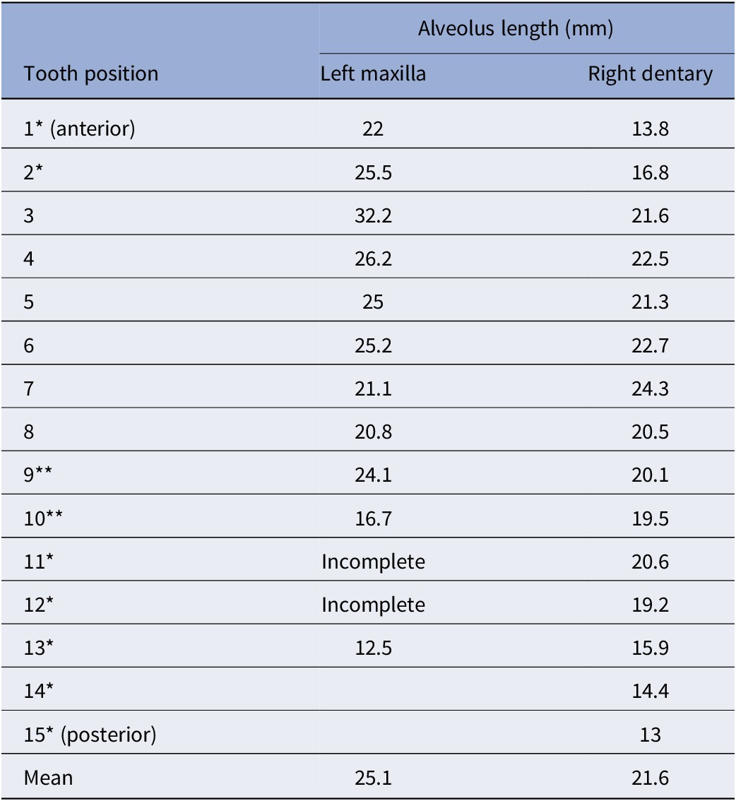


 Open access

