Down syndrome is the most frequent autosomal aneuploidy that is compatible with postnatal life. It is characterised by a complex phenotype in which over 80 features occur with various degrees of expression and frequency.Reference Banjar 1 It is not uncommon to be associated with congenital heart disease and it can provide an important model to link certain genes to the pathways controlling heart development. Congenital heart disease occurs in 40–50% of cases with Down syndrome and about 30% of patients have several cardiac defects.Reference Barlow, Chen and Shi 2 , Reference Frid, Drott, Lundell, Rasmussen and Anneren 3 The severity of the underlying congenital heart disease adversely affects survival. People with Down syndrome without congenital heart disease survive relatively longer than those with congenital heart disease especially with complexity of the defects.Reference Marino 4 Despite that, the newer surgical techniques have significantly improved the 1-year survival for those who have early surgical correction. Recent studies have mainly focused on congenital heart disease in patients with Down syndrome. However, data on cardiac functions in Down syndrome children with structurally normal hearts are rare. Owing to the fact that life expectancy of individuals with Down syndrome is increasing and most of them now reach adulthood in the developed countries, and they are actively engaged in various exercises and sports, information about cardiac dysfunction in this patient group is clinically important. This is especially important to find an explanation for exercise intolerance, decreased work capacity, and increased morbidity and mortality rate among the Down syndrome population. The purpose of this study was to evaluate the cardiac functions by two-dimensional and tissue Doppler echocardiography in Down syndrome children without structurally heart disease.
Materials and methods
The study was conducted as a case–control study. A group of 85 children with Down syndrome between 7 and 13 years of age with clinically and anatomically normal heart were included in the study from March, 2006 to April, 2009 in the Cardiology Unit of the Pediatric Department. An age- and sex-matched cohort of 50 healthy children was studied as the control group. The control group inclusion criteria were the absence of a history of any cardiac or significant systemic disease, as well as the absence of any family history of chromosomal disorders.
All children were evaluated by a paediatric cardiologist. Any anatomic cardiac lesions – congenital and acquired – were excluded by clinical and echocardiographic examination. A thorough clinical examination, as well as assessment of thyroid functions and complete blood pictures, were performed. Exclusion of upper airway obstruction was performed by an otolaryngologist and lateral neck radiograph was performed to exclude adenoid hypertrophy. Chest X-ray post-anterior and lateral views were done in some cases to exclude chronic lung diseases. Chromosomal studies were conducted to confirm diagnosis and to know the karyotype of the Down syndrome children: non-disjunction or translocation types. Exclusion criteria were the presence of any anatomical cardiac defect – congenital or acquired – hypothyroidism, severe anaemia, upper airway obstruction, obstructive sleep apnoea, recurrent or chronic lung disease, or any significant systemic disease.
Echocardiography
Echocardiographic examination was done using Hewlett-Packard/Phillips SONOS 5500 systems – Palo Alto, Calfornia, USA – equipped with 2.5–5 megahertz transducers. M-mode and two-dimensional echocardiography – to assess left ventricular internal dimensions, ejection fraction, and fractional shortening – were done according to the recommendation of the American Society of Echocardiography.Reference Picard, Adams and Bierig 5 The ejection fraction was measured by the Teichholz formula based on short-axis measurements of the left ventricle inner diameter by M-mode.Reference Cevik, Değertekin, Başaran, Turan and Pektaş 6 To avoid intra-observer variability, two examinations were performed by the same operator for each patient within 1 week. All children were examined in a semi-supine, left lateral position. Pulmonary arterial flow was recorded with pulsed-wave Doppler, placing the sample volume centrally between the leaflets of the pulmonary valve in a short-axis view at the base of the heart. The mitral and tricuspid Doppler signals were recorded in the apical four-chamber view, with the Doppler sample volume placed at the tip of the mitral or tricuspid valve. Colour Doppler study for the cardiac valves was conducted to assess the severity of regurgitation. The pulmonary artery systolic pressure was estimated in some cases from the tricuspid regurgitation jet using the Bernoulli equation. Pulmonary artery systolic pressure was considered abnormal when it was greater than 30 millimetres of mercury.Reference Giardini 7
Tissue Doppler image
Using pulsed-wave angle-corrected colour-coded Doppler tissue imaging filters, the baseline was adjusted to low velocity range (−20 to 20 centimetres per second) with minimal gain setting to minimise background noise and to obtain the highest quality images. The sample volume was placed within the myocardium equidistant from the endocardial and epicardial borders. From the apical four-chamber planes, using pulsed-wave Doppler tissue imaging, the myocardial velocity curves of septal mitral valve annulus, lateral mitral valve annulus, and lateral tricuspid valve annulus were recorded. The electrocardiogram was connected and traced simultaneously to define the timing of cardiac cycle events. The beginning of QRS complex was used as a reference point.
Velocities and interval measurements
The systolic wave reflects the systolic function of either right or left ventricle. The early′/atrial′ ratio of tricuspid and mitral valve annulus reflects the diastolic function of the right and left ventricle, respectively. Isometric contraction time was defined as the time duration between the beginning of QRS complex in the electrocardiogram to the beginning of Doppler tissue imaging systolic wave. The isometric relaxation time was defined as the interval between the end of systolic wave and the beginning of the early′ wave. At least 10 cardiac cycles were recorded at a speed of 100 millimetres per second and the images were stored electronically. The Doppler tissue imaging measurements were indexed for children's heart size.Reference Ng, Thomas and Leung 8
All parents signed a written informed consent before enrolment into the study. The local Institutional Research Review and Ethics Committee reviewed and approved the study protocol.
Statistical analysis
The power level of the number of cases in the study was more than 90%. Data are presented as mean (±standard deviation) values. The two-way analysis of variance with repeated measures was used to identify statistically significant differences in the different parameters among the groups. For all analyses, a statistical significance of p-value less than 0.05 was used. Wilcoxon's signed-rank test was used to assess the normality of distributions of the data. The Bonferroni correction/adjustment procedure was performed to avoid “significance” due to chance only, in multiple comparisons with echocardiographic parameters. The analysis was performed using TexaSoft, WINKS SDA Software, Sixth Edition, Cedar Hill, Texas, 2007.
Results
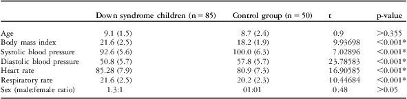
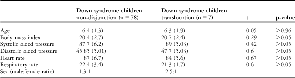
The demographic and clinical characteristics of children with Down syndrome and the control group are shown in Tables 1 and 2. The presence of Down syndrome was confirmed in all cases by chromosomal examination, which revealed 78 children with non-disjunction type of Down syndrome and seven children with Translocation type. There were no significant differences between Down syndrome children and the controls with regard to age and sex. The body mass index and pulse and respiratory rates were significantly higher in Down syndrome children than in the controls (p < 0.001), whereas systolic and diastolic blood pressure were significantly lower in Down syndrome children than in controls (p < 0.001). There were no significant differences in demographic and clinical data between children with Down syndrome due to non-disjunction and those with Down syndrome due to translocation.
Comparison of demographic data in Down syndrome children and control group.

*Means that p-value is significant
Demographic data of children with both types of Down syndrome.

Echocardiography
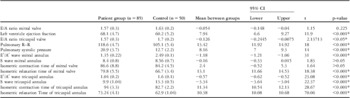
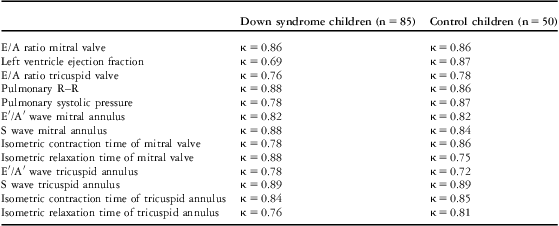
Echocardiographic data of children with Down syndrome and the controls are shown in Table 3. The intra-observer agreement of echocardiographic measurements is shown in Table 4. The left ventricular ejection fraction, which reflects left ventricular systolic function, was significantly higher in Down syndrome children than the controls (p < 0.001). There were no significant differences in systolic wave and isometric contraction time measured at mitral valve annulus by tissue Doppler (p > 0.05 for both).
Comparison of echocardiographic features in Down syndrome children and control group.

E/A = early/atrial; E′/A′ = early′/atrial′; S = systolic
*Means that p-value is significant
The intra-observer agreement of echocardiographic measurements between children with Down syndrome and control group.

E/A = early/atrial; E′/A′ = early′/atrial′; S = systolic
κ = Cohen Kappa; agreement is poor if κ ≤ 0.20, fair if κ 0.20 ≤ 0.40, moderate if κ 0.41 ≤ 0.60, substantial if κ 0.61 ≤ 0.80, and good if κ > 0.80
The early/atrial of mitral valve showed no significant difference between the two groups, but the tissue Doppler derived early′/atrial′ ratio was significantly reduced and the isometric relaxation time was significantly prolonged in Down syndrome children than in controls (p < 0.001). For right ventricle, the systolic wave measured at the tricuspid valve annulus showed significant reduction (p < 0.001), whereas isometric contraction time showed significant lengthening (p <0.001) in children with Down syndrome than in the controls. Both early/atrial and early′/atrial′ ratios at the tricuspid valve annulus showed significant reduction (p < 0.05 and 0.001, respectively), whereas the isometric relaxation time showed significant lengthening (p < 0.001) in children with Down syndrome than in the controls.
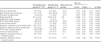
For pulmonary pressure, the pulmonary acceleration time (pulmonary R–R) was significantly prolonged in Down syndrome children than in the control group (p < 0.001). The pulmonary arterial systolic pressure was significantly higher in Down syndrome children than the controls. Table 5 showed no significant difference in any of the echocardiographic parameters in children with non-disjunction or translocation types of Down syndrome.
Comparison of echocardiographic features in Down syndrome children with non-disjunction type and translocation type.

E/A = early/atrial; E′/A′ = early′/atrial′; S = systolic
Discussion
Congenital heart diseases are a common problem in children with Down syndrome. However, despite being structurally normal, the heart may be affected in those children. They are more liable to the common cardiovascular risk factors – sedentary life, obesity, and hypertension – than those without Down syndrome.Reference Pastore, Marino, Calzolari, Digilio, Giannotti and Turchetta 9 Therefore, the aim of this study was to assess the presence of any significant echocardiographic changes in the cardiac function in a subset group of children with Down syndrome and structurally normal heart.
This study showed that children with Down syndrome had statistically but not clinically significant lower systolic and diastolic blood pressure than control children. This was in agreement with the results of Morrison et alReference Morrison, McGrath, Davidson, Brown, Murray and Lever 10 who found lower blood pressure in Down syndrome adults than in controls. They considered it as a feature of the disease rather than due to the protected surrounding environment. The low blood pressure noted in children with Down syndrome may be due to trisomy of type-1-angiotensin II receptor gene, resulting in its underexpression.Reference Sethupathy, Borel and Gagnebin 11 , Reference Wiseman, Alford, Tybulewicz and Fisher 12
Our study showed that children with Down syndrome had statistically but not clinically significant higher respiratory and heart rates than the control children, which was in agreement with the work of Pastore et al who found higher heart rates in Down syndrome children than controls.Reference Picard, Adams and Bierig 5 The differences in respiratory and pulse rate, as well as the blood pressure, may be related to autonomic cardiac dysregulation in Down syndrome, which may be due to the autonomic dysfunction occurring at a central, brain stem, site as a result of the genetic disorder.Reference Iellamo, Galante and Legramante 13
This study showed that the left ventricle ejection fraction by conventional echocardiography was significantly higher in Down syndrome children than the controls (p < 0.001), which is similar to the findings of Russo et al who studied 22 Down syndrome patients without congenital heart disease and found evident left ventricular hyperkinesia. They attributed these hyperkinesias to the reduced afterload and not to intrinsic abnormalities of the myocardium.Reference Russo, Pacileo and Marino 14
The absence of intrinsic myocardial abnormalities of left ventricle in the current study was supported by a lack of significant difference between systolic wave and isometric contraction time of the mitral valve annulus measured by tissue Doppler between Down syndrome children and the controls. The systolic wave, which reflects the longitudinal systolic function, is positively correlated with measurements of left ventricle ejection fraction. It is less load dependent, whereas the isometric contraction time is inversely correlated with myocardial contractility.
The current study showed no significant difference in the left ventricular diastolic function in Down syndrome children compared with the controls by the conventional pulsed Doppler (early/atrial ratio p > 0.05), but tissue Doppler showed a significant decrease in the early′/atrial′ ratio of mitral valve annulus and prolongation of left ventricle isometric relaxation time in the Down syndrome children than in the controls (p < 0.001). This may be due to the higher sensitivity of the tissue Doppler to detect diastolic dysfunction than the conventional Doppler. The prolonged isometric relaxation time, which is inversely correlated to myocardial relaxation, may indicate that the diastolic dysfunction, which observed in Down syndrome children, is due to impaired cardiac muscle relaxation. However, the difference in left ventricle Doppler echocardiographic findings between Down syndrome children and control group was of no clinical significance.
The right side of the heart showed a significant reduction of the early/atrial ratio at the tricuspid valve measured by pulsed Doppler and reduction of systolic wave and early′/atrial′ ratio measured by tissue Doppler, with significant prolongation of right ventricle isometric relaxation time in children with Down syndrome than observed in the controls. The reduction of the systolic and diastolic cardiac functions in children with Down syndrome observed in this study may be explained by autonomic cardiac control dysregulation,Reference Goulopoulou, Baynard and Collier 15 , Reference Fernhall and Otterstetter 16 abnormality in the cardiac muscle fibres with increased cardiac muscle fibre size and reduced cell number,Reference Recalde, Landing and Lipsey 17 or overexpression of calcineurin.Reference Molkentin, Lu and Antos 18 – Reference Arron, Winslow and Polleri 21 Overexpression of calcineurin may lead to cardiac muscle hypertrophy.
Children with Down syndrome are at increased risk of developing pulmonary arterial hypertension even without structural heart lesion. Pulmonary arterial hypertension may develop in Down syndrome children secondary to chronic upper airway obstruction, abnormal pulmonary vasculature growth, alveolar hypoventilation, and recurrent pulmonary infections.Reference Banjar 1 In the current study; the pulmonary R–R was significantly shorter and the pulmonary arterial systolic pressure was significantly higher in children with Down syndrome than in the controls. We excluded cases with congenital or acquired cardiac disease and cases with chronic upper airway obstruction, as well as chronic or recurrent lung problems that may affect pulmonary pressure. This may indicate that the cause of the higher pulmonary arterial systolic pressure may be intrinsic factors inside the lung rather than extrinsic factors. In an autopsy study of six patients with Down syndrome; Schloo et alReference Schloo, Vawter and Reid 22 found reduction in the alveolar count, persistence of the foetal double capillary network in the lung, and reduction in the cross-sectional area of the vascular bed. All these factors are important in the pathogenesis of pulmonary hypertension. It may also result from abnormal nitrous oxide production owing to impaired vascular endothelial function, which is possibly related to increased oxidative stress.Reference Cappelli-Bigazzi, Santoro and Battaglia 23
In this study, there was no significant difference in the echocardiographic findings in children with the dysjunction or translocation type of Down syndrome. However, the small number of children with translocation type of Down syndrome may give rise to inaccurate statistical results. Therefore, we advise including more cases in a further study.
Limitation of the study
Although there were statistical differences in many of the data examined between normal and Down syndrome children, the values in both groups in many of the data examined are within normal limits. This may make the findings of limited clinical significance. However, these findings may help to understand why children with Down syndrome have limited physical fitness and are more liable to many cardiovascular risks.
Conclusion
Despite having an apparently normal heart, children with Down syndrome may have silent disturbed cardiac functions, which may be discovered by two-dimensional or tissue Doppler echocardiography. This may have an important clinical implication, especially before involving Down syndrome children in surgery or strenuous exercise.
Acknowledgements
The authors greatly appreciate the generous support of Professor Osama A. Tolba, the Head of Pediatric Department, Tanta University, for his critical reading and drafting the manuscript. This study was performed in the Cardiology Unit, Pediatric Department, Faculty of Medicine, Tanta University, Egypt, a non-profit organisation, without any commercial support.