Introduction
Inclusion of live cells in additive manufacturing processes has seen tremendous progress in the last few years. Mammalian cells need to be kept in a soft, aqueous environment when embedded in a biomaterial matrix—this does not allow for the fabrication of structurally well-defined volumetric cell-laden constructs by means of three-dimensional (3D) bioprinting. Reference Malda, Visser, Melchels, Jungst, Hennink, Dhert, Groll and Hutmacher1 Thus, creating macroscopic objects was found to be more complicated than initially expected. Simple upscaling is not possible due to the mismatch between the mechanical properties needed for cell embedding and manufacturing of 3D objects with high shape fidelity, hence several strategies have been investigated to overcome this problem.
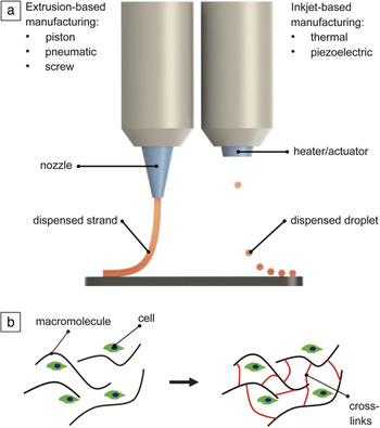
Two main technologies can be distinguished—inkjet- and extrusion-based. Figure 1a shows schematic representations of both technologies and the respective terminology. For bioprinting with inkjet-like printers, either single-cell suspensions (not suitable for manufacturing of volumetric structures), cell aggregates or cells encapsulated in hydrogel beads can be utilized as building blocks. This technology allows for achieving high cell densities, and hence it is advantageous for bioprinting of artificial organs in which close cell–cell contacts are crucial for proper function. The disadvantage of utilizing cell aggregates as building blocks is the need to produce large numbers of cells and assemble them into spherical aggregates; both are cost-intensive and time-consuming procedures. For this type of cell-printing, the term “bioassembly” has been suggested to distinguish it from extrusion-based bioprinting. Reference Groll, Boland, Blunk, Burdick, Cho, Dalton, Derby, Forgacs, Li, Mironov, Moroni, Nakamura, Shu, Takeuchi, Vozzi, Woodfield, Xu, Yoo and Malda2 Norotte and co-workers used the bioassembly technology, for example, to create hollow, blood vessel-like morphologies by arranging spherical cell aggregates in 2009. Reference Norotte, Marga, Niklason and Forgacs3
Principles of (a) (left) extrusion- and (right) inkjet-based 3D printing enabling manufacturing of volumetric constructs. (b) Hydrogel formation after ionic or covalent cross-linking of the bioink (cell suspension in less viscous (bio)polymer solution), essential for stabilization of volumetric bioprinted structures.

Utilizing extrusion-based bioprinting where cells (or small cell aggregates) are typically suspended in (bio)polymer hydrogels has made the manufacturing of volumetric structures easier and less expensive. Since the applicability of biomaterials in extrusion-based 3D printing is actually limited only by their viscosity, a wide range of materials can be utilized. In addition, 3D printers for this technology are already commercially available for less than USD$10,000, and construction kits are available for even less. Combinations of hydrogels and cells are called bioinks. As previously mentioned, after the extrusion process, bioinks must form soft hydrogels to support cell survival and maintenance over longer cultivation periods. In Figure 1b, hydrogel formation from a cell-laden bioink is shown schematically. Low-viscosity materials are not suitable for the manufacturing of large, and still structurally well defined, constructs and novel strategies had to be developed to enable both—successful bioprinting and fabrication of volumetric tissue-like objects. This article describes the most important and effective approaches to overcome this problem, and also the need to provide open macropores or perfusable channels in 3D tissue constructs to allow oxygen and nutrient supply to the embedded cells.
As 3D bioprinting moves from being a field of research toward real clinical applications, we define the term “volumetric” in this article to only include technologies that, in principle, can be used for fabrication of cell-laden constructs with a volume of at least 1 cm Reference Norotte, Marga, Niklason and Forgacs3 (10 × 10 × 10 mm3). Although the studies selected for review in this article do not all describe macroscopic objects of this defined size, the selected technology should, in estimation, be able to fabricate constructs with such dimensions. We have classified the selected studies based on the methodology and have distinguished between those utilizing special printing technologies, modified bioinks, or additional supporting materials. The last can be extruded with the bioinks either in an alternating fashion (separate bioink and support strands) or in a combined manner, leading to strands with core–shell morphology. These approaches are listed in Table I , Reference Tabriz, Hermida, Leslie and Shu4–Reference Mistry, Aied, Alexander, Shakesheff, Bennett and Yang7 with a schematic and an example taken from literature, respectively. Other articles in this issue describe in detail new research directions in bioink development (see the Rutz et al. article in this issue) Reference Rutz, Lewis and Shah8 as well as questions of perfusion/vascularization of bioprinted tissue constructs (see the Huang et al. article in this issue). Reference Huang, Zhang and Liu9
Principles of fabricating volumetric tissue constructs by extrusion bioprinting approaches and respective examples. The cell-laden bioink strands are shown in red in the schematics.

Note: PCL, poly(ε-caprolactone).
It is worth mentioning that the morphology of mammalian tissues and organs is quite diverse and different solutions have to be developed to mimic those as closely as possible, in terms not only of structural aspects, but also with regard to the composition of the respective extracellular matrices. Reference Pati, Gantelius and Svahn10 Different technologies must be used to print, for example, nonvascularized but mechanically stable articular cartilage constructs containing only one cell type (chondrocytes, [i.e., cartilage cells]) in contrast to highly vascularized, multiple cell types containing organs such as the liver or kidney.
Finally, we point out that the first studies on 3D bioprinting of nonmammalian cells have been published, which might open up new applications (e.g., in the field of biotechnology). Reference Lode, Krujatz, Brüggemeier, Quade, Schütz, Knaack, Weber, Bley and Gelinsky11 For these, diverging novel methods and materials will require further development.
Technical solutions
Specific printing environment
In order to overcome the limitations of common 3D bioprinting setups concerning structural stability, choice of material, dispensing systems, and concepts, modifying the procedural setup are essential. Different innovative approaches have been established to prevent the structure from deliquescing by providing a supportive inert solvent-based (e.g., dispensing into fluorocarbon) or additional particle-based printing environment (e.g., gelatin microparticle bath), or by in situ-compatible cross-linking mechanisms.
Although most currently used setups enable dispensing into air and onto a firm surface, a proposed solution is to use specific dispensing environments to enable immediate temporary stabilization of extruded strands, thus preventing deliquescence. The selected dispensing medium stabilizes the printed structure, maintaining its shape while being extruded. The surrounding medium should not mix with the dispensed material, affect or degrade the material in a destabilizing way, nor interfere with the cytocompatibility. For example, for highly hydrophilic hydrogels such as agarose, hydrophobic and completely inert fluorocarbons have been used, Reference Duarte Campos, Blaeser, Weber, Jäkel, Neuss, Jahnen-Dechent and Fischer12 enabling the manufacturing of a variety of differently shaped constructs and mediating scaffold stability over several months.
Another option is to utilize an additional hydrogel bath with gelatin microparticles acting as a sacrificial Bingham fluid, which shows viscous behavior at higher shear stress, and enables stabilization of the extruded structure. Reference Hinton, Jallerat, Palchesko, Park, Grodzicki, Shue, Ramadan, Hudson and Feinberg13 This idea is referred to as freeform reversible embedding of suspended hydrogels as the dispensed, cell-loaded alginate (alginic acid salts frequently used as biopolymer-hydrogels in biological application) hydrogel is simultaneously cross-linked ionically by CaCl2 added to the gelatin gel. After printing and stabilization, the gelatin is melted by increasing the temperature to 37°C and finally removed from the bioprinted structure.
For thermoresponsive materials such as gelatin and gelatin blends, their thermosensitivity is used to induce gelation and initial stability via temperature-controlled printer hardware. Zhang et al. reported alginate-gelatin gels that gelled at 3–10°C directly after deposition of the strands, the continuously extruded hydrogel filaments used to build the scaffold layers. Reference Zhang, Yan, Ouyang and Sun14 Another study described utilization of an alginate-gelatin blend, including hydroxyapatite supplementation Reference Wüst, Godla, Müller and Hofmann15 gelling on a 10°C cooling plate after being dispensed from a 40°C temperature-controlled cartridge system. Printers equipped with cooling plates or chambers can also be applied to induce in situ solidification of non-thermoresponsive materials such as alginate on a stage at –10°C. Reference Ahn, Lee, Lee and Kim16 In most cases, the proposed procedures require an additional cross-linking step for providing long-term stability of the constructs. New ways are opening up by combining techniques for specific materials, such as two-step gelation (thermic/ionic) after dispensing an alginate-gelatin hydrogel into a granular carbopol (polyacrylic acid particles) support bath. Reference Jin, Compaan, Bhattacharjee and Huang17
In situ cross-linking approaches
To fabricate volumetric tissue constructs, another aspect of stability optimization during the printing process that should be considered is the cross-linking procedure. Ideally, the bioink needs to be cross-linked directly following dispension from the nozzle, which remains a challenge, particularly for air-dispensing setups. To enable immediate contact between the printed ink and a cross-linking reagent that induces gelation, one approach applies a continuous spray of a CaCl2 aerosol Reference Ahn, Lee, Bonassar and Kim18 on open-porous alginate-based scaffolds.
Tabriz and co-workers described utilization of an alginate gel, precross-linked with a low concentration of calcium ions prior to extrusion and being further ionically cross-linked by lowering the building platform along the z-axis into a CaCl2 bath during the biofabrication process. Reference Tabriz, Hermida, Leslie and Shu4
Many concepts utilize UV-/photocross-linkable hydrogels for bioprinting as they allow for defined and reproducible control of spatiotemporal polymerization. Photoreactive chemical groups are incorporated in the materials to enable covalent cross-linking in response to UV illumination. Reference Pereira and Bártolo19 However, the most critical aspect of UV-cross-linked cell-laden scaffolds is the impact of the illumination on DNA structures, by both the UV light and the radicals (radicals generated by UV interaction with the respective-photo initiators) accounting for both UV-A and UV-B wavelength ranges. UV-A is generally considered less disruptive than the shorter wavelength UV-B. With Irgacure 2959 investigated as the photoinitiator, the expected adverse effects of photopolymerization on DNA integrity and cell cycle reentry (i.e., ability of cells to proliferate) of human mesenchymal stem cells (hMSCs) were confirmed as being stronger for monolayer cultures compared to hMSCs embedded in hydrogels. Reference Fedorovich, Oudshoorn, van Geemen, Hennink, Alblas and Dhert20
Early approaches to translate the photocross-linking of hydrogel blends that had been applied for years to a bioprinting-compatible setup were performed using the nonionic triblock copolymer (poloxamer) Lutrol-F127, enabling both thermogelation and covalent photocross-linking. Reference Fedorovich, Swennen, Girones, Moroni, van Blitterswijk, Schacht, Alblas and Dhert21 Later, those concepts were used for simultaneous cross-linking of synthetic hydrogels while being dispensed, such as for poly(ethylene glycol) dimethacrylate gels for cartilage tissue engineering. In this case, increased cell viability was obtained for in situ cross-linking outperforming previous post-printing approaches. Reference Cui, Breitenkamp, Finn, Lotz and D’Lima22 However, when considering translational approaches for clinical applications, the risk of DNA damage needs to be assessed.
One approach to at least reduce DNA interruption is employing ultrafast methods, where the cross-linking time could be reduced. Wang and co-workers recently investigated possible strategies in this direction but did not produce volumetric bioprinted structures. Reference Wang, Jin, Dai, Holzman and Kim23 Another current study suggested in situ cross-linking via photopermeable dispensing nozzles, generalizable for different bioinks such as gelatin methacryloyl (GelMA) and polyethylene glycol diacrylate (PEGDA), Reference Ouyang, Highley, Sun and Burdick24 and wavelengths in the UV and visible light range. This enables simultaneous cross-linking and extrusion without affecting viscosity (i.e., viscosity does not decrease by potential UV-induced molecular chain break and does not increase due to unspecific cross-linking), even along with core–shell structured extrusion.
Internal structure stabilization (modified bioinks)
The simplest technique to build up volumetric structures by additive manufacturing is to increase the viscosity of the hydrogel bioink. A printed construct consisting of a high-viscosity bioink will not collapse before further processing, for example, cross-linking. Generally, high-viscosity bioinks allow fabrication of scaffolds with high shape fidelity. However, tailoring the viscosity is a critical issue, as just a plain increase in the polymer concentration leads to stiff hydrogels unsuitable for cell encapsulation. Reference Malda, Visser, Melchels, Jungst, Hennink, Dhert, Groll and Hutmacher1 Overall, the criteria for development of bioinks with high viscosity are ambitious and diverse. Reference Jungst, Smolan, Schacht, Scheibel and Groll25
Two main strategies have been investigated to enhance the shape fidelity and stability of cell-laden constructs—modifying the bioink composition or modifying single components of the ink to increase their cross-linking density. The effects of adding a second material to the original bioink on the properties of the resulting blend can be numerous. Changes in the rheological properties are consequential, and shear thinning behavior or printing fidelity are affected strongly by blend composition.
Markstedt et al. infiltrated an alginate sol with nanofibrillated cellulose (NFC) in a 80:20 ratio (Table I, Category 2). Reference Markstedt, Mantas, Tournier, Martinez Avila, Hagg and Gatenholm5 They observed shear thinning behavior, necessary for 3D printing, and a viscosity, which enabled the scaffold to maintain its shape during the printing process until cross-linking of the alginate component occurs. The insoluble NFC remained inside the hydrogel matrix.
In contrast, further studies show that the viscosity can also be enhanced temporarily by incorporation of materials that vanish from the matrix after printing. These materials are not fixed but stay soluble and diffuse into the cell culture medium, leaving behind a cross-linked low polymer content hydrogel that is especially qualified for cell incorporation. Schütz et al. mixed 9 wt% methylcellulose in a cell-laden 3 wt% alginate bioink. Reference Schütz, Placht, Paul, Brüggemeier, Gelinsky and Lode26 After dispensing, carried out in air, the alginate was cross-linked ionically with calcium ions, while the soluble methylcellulose was shown to diffuse out of the scaffold. Printed scaffolds showed high shape fidelity, and the blend could be printed with a large (>50) number of layers. After methylcellulose vanished from the scaffold, the cross-linked alginate maintained its shape and presented a low concentration polymer matrix, consisting of only the alginate part, to the cells.
Even macroporous, cell-laden constructs could be fabricated with this bioink blend. It was demonstrated that hMSCs could be differentiated toward adipogenic lineage (fat tissue) after printing. A similar approach was investigated by blending 6% alginate with 13% of the triblock-copolymer poloxamer Pluronic F127. Reference Armstrong, Burke, Carter, Davis and Perriman27 The blend was printed at 37°C, which is the solidifying temperature of the Pluronic F127. The scaffold fabricated from this bioink remained stable at physiological temperature. After printing, the alginate component was cross-linked by calcium ions at room temperature, and the destabilized Pluronic vanished from the scaffold. Similar results were obtained for gelatin in alginate–gelatin blends, in which utilization for bioprinting is discussed in the “Technical solutions” section. Reference Zhang, Yan, Ouyang and Sun14,Reference Wüst, Godla, Müller and Hofmann15 Both methylcellulose and Pluronic left behind a distinct intrastrand micropore structure that could be advantageous for the oxygen and nutrient supply of the embedded cells during further cultivation or implantation.
The other strategy focuses on chemical modification of the bioink components to enable faster cross-linking or higher cross-linking density, which is important for accurate bioprinting. Reference Pereira and Bártolo19 We provide a brief overview of the three precross-linking techniques that enhance the printing behavior of cell-laden hydrogels.
Photopolymerization enhances the stability of printed structures. Reference Schuurman, Levett, Pot, van Weeren, Dhert, Hutmacher, Melchels, Klein and Malda28 For example, gelatin Type B can be reacted with methacrylic anhydride to obtain GelMA, which is the most commonly applied photosensitive hydrogel in bioprinting applications. Billiet et al. demonstrated that its dual cross-linking capabilities, physical cross-linking by cooling down immediately after printing and photoinitiated cross-linking shortly after printing, allowed perfect scaffold construction from 10–20% weight/volume GelMA pastes. Reference Billiet, Gevaert, de Schryver, Cornelissen and Dubruel29 Building large constructs with high printing fidelity and a connected internal pore network was possible. Cell encapsulation of the hepatocarcinoma (liver cancer) cell line and printing of the cell-laden hydrogel revealed a cell viability of more than 97%, obtained immediately after printing as well as after cultivation for 14 days. Reference Billiet, Gevaert, de Schryver, Cornelissen and Dubruel29
Rutz and co-workers synthesized gel-phase bioinks by slight cross-linking of gelatin and fibrinogen solutions prior to printing (precross-linking) with poly(ethylene glycol) (PEG) ending in two reactive groups, called PEGX (PEG with X as placeholder for the reactive groups). Reference Rutz, Hyland, Jakus, Burghardt and Shah30 The precross-linking enhanced the printing properties; for long-term cell culture, an additional post-printing cross-linking step was applied to ensure full cross-linking of the hydrogel. Kesti et al. created a blend by ionic precross-linking with a low concentration of divalent metal ions that was shown to be beneficial for extrusion of an alginate (2%)–gellan gum (3%) blend. Such prestabilized structures of this blend were robust during the printing process and further strengthened by a supply of more divalent cations, which cross-link alginate and gellan gum, from a co-extruded support structure. Reference Kesti, Eberhardt, Pagliccia, Kenkel, Grande, Boss and Zenobi-Wong31
The cell vitality of all approaches ranges between 50 and 90%, however, the number of living cells was demonstrated to be dependent on the cell position inside a 3D construct with a greater number of living cells near the periphery, likely due to limitations of oxygen supply and duration of keeping the cells under nonphysiological conditions. Reference Kesti, Eberhardt, Pagliccia, Kenkel, Grande, Boss and Zenobi-Wong31
External stabilization (bioink plus support)
Hydrogels with high polymer content cannot be used for cell encapsulation due to their limited water content, which is most important for the cell environment. Multichannel (multimaterial) printing is a method to combine both a tough and robust, grid-like structure as a mechanical support with a soft, cell-containing hydrogel. For this, at least two materials are loaded in separate cartridges. First, cell-free, viscous materials are printed as a grid. After finishing the grid of the first layer, the less viscous, cell-laden hydrogel is printed inside the voids of the grid. Schuurman et al. first demonstrated this concept in 2011 Reference Schuurman, Khristov, Pot, van Weeren, Dhert and Malda32 by printing thermoplastic poly(ε-caprolactone) (PCL) as the rigid grid next to a cell-containing alginate hydrogel; this was repeated by other groups for several bioprinting applications. Reference Pati, Jang, Ha, Won Kim, Rhie, Shim, Kim and Cho33,Reference Kang, Lee, Ko, Kengla, Yoo and Atala34 With additional usage of PEG as the sacrificial material, PCL as the grid structure, and an alginate bioink, this technique allowed for the construction of real 3D printed tissues, such as cartilage in the shape of a human ear (Table I, Category 3). Reference Lee, Hong, Jung, Shim, Oh and Cho6
As soft bioinks fill up the pores of the grid, large volumetric constructs generated by these methods might lead to problems, because the oxygen supply is limited for cells inside the scaffold. Moreover, in the case of an intended implantation, an open-porous structure would be needed for vascularization (i.e., ingrowth of blood capillaries from the surrounding tissue).
Kolesky et al. Reference Kolesky, Homan, Skylar-Scott and Lewis35 demonstrated an advanced concept to directly achieve volumetric tissue structures with perfusable, vessel-like pore channels by combining 3D printing, bioprinting, and conventional cell-seeding techniques. The 3D pore channel system was fabricated from Pluronic F-127 as the sacrificial material and subsequently internally endothelialized (formation of the blood vessel lining by endothelial cells) with human umbilical vein endothelial cells, which constitutes the overall concept of inducing vascularization in thick constructs. Perfusion of this microcapillary network allowed survival of embedded cells in macroscopic constructs of ca. 25 × 25 × 10 mm3 volume during a six-week time period.
Kim and co-workers developed scaffolds with open pores by extruding a cell-containing and precross-linked 3% alginate-based bioink directly on top of printed PCL strands. Reference Kim, Lee, Yang, Choi, Lee, Hwang, Jung, Yoon and Kim36 The bioink surrounded the PCL strands and the hydrogel did not clog the macropore structure of the PCL grid, which remained open even after swelling of the hydrogel. Lee et al. printed PCL strands next to an internally stabilized cell-laden alginate, which was precross-linked by calcium ions. Reference Lee, Ahn, Bonassar and Kim37 The open macropore structure of this approach can be adjusted by changing the layer orientations or the strand-to-strand distance. Cells seemed to tolerate the direct contact to the hot PCL melt, because they were protected by the surrounding hydrogel.
Melchels et al. took an alternative approach and engineered a synthetic poloxamer (Pluronic F127) hydrogel with high viscosity and good printing fidelity that was printed at room temperature together with a cell containing GelMA bioink, kept at 37°C, and achieved cell viabilities greater than 80% after printing and greater than 90% after 14 days in cell culture. Reference Melchels, Blokzijl, Levato, Peiffer, de Ruijter, Hennink, Vermonden and Malda38
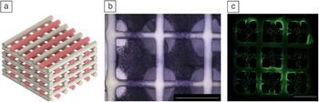
For bone applications, calcium phosphate mineral phases are suitable materials, due to their high biocompatibility, bone-like composition, and mechanical strength. Pasty, extrudable calcium phosphate cements (CPCs) that can set to biodegradable, nanocrystalline hydroxyapatite are well suited for 3D printing. Reference Lode, Meissner, Luo, Sonntag, Glorius, Nies, Vater, Despang, Hanke and Gelinsky39 Lode et al. printed this CPC together with an internally stabilized, hMSC-laden alginate-methylcellulose blend bioink, mentioned previously, thereby achieving open pores in vertical and lateral directions and volumetric constructs that could be printed with high accuracy ( Figure 2 ). Over time, hMSCs migrated from the bioink to the CPC at the interface region of the two materials, and attached to the CPC surface and started to proliferate there. Reference Lode, Gelinsky, Eichmann, Kynast and Witt40
Example of external stabilization. A volumetric bone tissue construct, fabricated with a self-setting, pasty calcium phosphate cement (CPC). (a) Schematic, showing the scaffold design (white = CPC strands, red = cell-laden hydrogel strands); (b) micrograph after MTT (3-(4,5-dimethylthiazol-2-yl)-2,5-diphenyltetrazolium bromide) staining for metabolic activity of encapsulated cells, one day after printing dark violet dots represent vital cells, embedded in the hydrogel strands, CPC strands are white. (c) Fluorescence micrograph, taken 21 days after printing; live/dead staining: live cells (>90%) show green, dead cells show red fluorescence; CPC strands exhibit green autofluorescence. Reference Lode, Gelinsky, Eichmann, Kynast and Witt40 Scale bars in (b–c) = 3 mm; the whole scaffold is 10 × 10 × 5 mm3.

Modification of strand morphology (core–shell bioprinting)
For retaining the viability of cells during bioprinting, low concentrated bioinks are generally preferred. However, for printing volumetric tissue constructs, low concentrated (less viscous) bioinks need to be extensively modified, or alternative strategies (previously mentioned) need to be employed to stabilize the construct, at least intermediately, until the printing process is completed and the bioink gels or is cross-linked. A relatively simple yet efficient strategy would be rendering intrastrand stability during the extrusion/printing process.
Encapsulating a low concentrated bioink with a highly concentrated (highly viscous) biomaterial within a single strand (i.e., forming a core–shell strand) would support printing of subsequent layers without extensively altering the shape and size of the whole construct (see Table I, Category 4). As two materials (spatially separated) are extruded as a single strand using coaxial nozzles, additive effects of their intrinsic properties are observed. Reference Perez and Kim41 Also, independent functions of the two materials can be simultaneously utilized, for example, as a dual drug delivery system by selectively loading the core and the shell with different drugs or growth factors. Reference Akkineni, Ahlfeld, Lode and Gelinsky42
The properties of the constructs, such as mechanical strength and release kinetics, can be easily tuned for specific biomedical applications by altering the composition of core or shell materials. Onoe et al. demonstrated this by using a microfluidic system for inclusion of various cells in the core (consisting of natural extracellular matrix proteins) encapsulated by Ca–alginate shell that resulted in high order organization of the cells and formation of respective meter-long functional tissue fibers. Reference Onoe, Okitsu, Itou, Kato-Negishi, Gojo, Kiriya, Sato, Miura, Iwanaga, Kuribayashi-Shigetomi, Matsunaga, Shimoyama and Takeuchi43 However, such long cell fibers would still need to be organized in three dimensions to form a volumetric tissue construct.
Alternatively, extrusion-based bioprinting of cell-laden core–shell structures can be used for fabrication of volumetric tissues. Cells can be loaded in either the core Reference Akkineni, Ahlfeld, Lode and Gelinsky42,Reference Yeo, Lee, Chun and Kim44 or the shell material Reference Ahn, Lee and Kim45,Reference Raja and Yun46 to fabricate cell-laden volumetric tissue constructs. If cells are located in the shell region, the stabilizing core can consist of either a stiff biopolymeric material Reference Ahn, Lee and Kim45 or a self-setting calcium phosphate suspension. Reference Raja and Yun46 Other materials for this purpose will most likely be established in future.
Different cell types encapsulated in the core successfully reorganized to form functional tissue strands (e.g., formation of tubular structures from endothelial cells, functional cardiomyocytes [cardiac muscle cells], and conducting cortical neural cells Reference Onoe, Okitsu, Itou, Kato-Negishi, Gojo, Kiriya, Sato, Miura, Iwanaga, Kuribayashi-Shigetomi, Matsunaga, Shimoyama and Takeuchi43 ) or were differentiated into a specific lineage (e.g., differentiation of human adipose [body fat]-derived stem cells to cells showing high levels of liver-specific gene expression Reference Yeo, Lee, Chun and Kim44 ). Constructs having two different cell types spatially separated in a single strand can potentially be fabricated using core–shell extrusion printing. The two cell types can independently reorganize to form a functional tissue or aid in interacting with each other, leading to the formation of complex tissues.
Combination of several categories
Advancements in bioinks and 3D printing technologies have led to easier and more efficient bioprinting of volumetric tissue constructs by employing more than one strategy. hMSC-laden 3D scaffolds with high resolution (strand diameter of ∼150 µm) were printed for cartilage tissue-engineering applications. Reference Costantini, Idaszek, Szöke, Jaroszewicz, Dentini, Barbetta, Brinchmann and Swieszkowski47 Low concentrated bioink solutions (consisting of photocurable bioinks + alginate) with a high concentration of hMSC suspension were used for printing. Instantaneous gelation of alginate (by simultaneous extrusion of CaCl2 using a coaxial needle) led to printing of stable 50-layered constructs. Further stabilization of the construct was achieved by UV cross-linking of the respective components in the bioinks. Jia et al. fabricated perfusable vascular constructs Reference Jia, Gungor-Ozkerim, Zhang, Yue, Zhu, Liu, Pi, Byambaa, Dokmeci, Shin and Khademhosseini48 by coaxial printing of photocurable bioinks and alginate blends with CaCl2 solutions. Mistry and co-workers have simultaneously employed strategies mentioned earlier in the “technical solutions” and “internal structure stabilization” sections for printing cell-laden volumetric constructs. Reference Mistry, Aied, Alexander, Shakesheff, Bennett and Yang7
Conclusions
Various approaches concerning bioprinting of volumetric tissue constructs have been proposed, and the field is still developing quickly. By carefully considering all aspects—the extrusion process, bioink composition, and cytocompatible gelation protocols—suitable strategies for different types of tissues and organs can be employed. We still have a long way to go until macroscopic, mechanically robust tissue equivalents can be fabricated by 3D bioprinting technologies to be used in human therapies.
One limitation is that most of the studies published so far have utilized immortalized, cancer-derived human or mammalian cell lines that are known to be more resistant to stress, which may occur during bioprinting or gelation, as compared to primary cells. As immortalized cell lines (cells in a culture that are genetically modified for indefinite proliferation) cannot be applied in clinical settings, more research needs to be performed on bioprinting using patient-derived, primary cells. Certainly, new biomaterials and material combinations, as well as novel technological solutions, will be introduced to the field, and we will surely see continued strong progress. An open question today remains whether mankind will be able to fabricate fully functional, complex tissue equivalents by means of 3D bioprinting. The question of how to fabricate volumetric 3D constructs will be one of the key challenges.

David Kilian has been a doctoral candidate at the Center for Translational Bone, Joint and Soft Tissue Research at the Medical Faculty of Dresden University of Technology (TU Dresden), Germany, since 2016. He obtained his MSc degree in regenerative biology and medicine from the Center for Regenerative Therapies at TU Dresden. His current research interests include 3D bioprinting for osteochondral tissue engineering, and investigating the cellular behavior within 3D bioprinted volumetric hydrogel constructs. Kilian can be reached by email at david.kilian@tu-dresden.de.

Tilman Ahlfeld has been a doctoral candidate at the Center for Translational Bone, Joint and Soft Tissue Research, Dresden University of Technology (TU Dresden), Germany, since 2013. His current research interests focus on developing printing strategies for bone-tissue engineering, developing bioinks, 3D printing of calcium phosphates, and multimaterial extrusion. Ahlfeld can be reached by email at tilman.ahlfeld@tu-dresden.de.

Ashwini Rahul Akkineni has been a doctoral candidate in the field of 3D printing for tissue-engineering applications at the Center for Translational Bone, Joint and Soft Tissue Research, Dresden University of Technology (TU Dresden), Germany, since 2012. He earned his bachelor’s degree in biotechnology at Jawaharlal Nehru Technological University, Hyderabad, India, and his master’s degree in nanobiophysics at TU Dresden. His research focuses on 3D printing of calcium phosphate cements, naturally derived hydrogels, developing and characterizing new bioinks, and developing co-extrusion 3D printing for biomedical applications. Akkineni can be reached by email at Ashwini_Rahul.Akkineni@tu-dresden.de.

Anja Lode has been a member of the Center for Translational Bone, Joint and Soft Tissue Research, since 2010, and heads the cell culture and bioanalytics laboratory at Dresden University of Technology (TU Dresden), Germany. She studied biology at the University of Potsdam, Germany, and earned her PhD degree in genetics at TU Dresden. From 2002 to 2010, she worked in the Tissue Engineering and Biomineralization Group at the Max Bergmann Center of Biomaterials (Institute for Materials Science, TU Dresden). Her current research focuses on cell–biomaterial interactions, extrusion-based additive manufacturing, 3D bioprinting, tissue engineering, and drug delivery. Lode can be reached by email at anja.lode@tu-dresden.de.

Michael Gelinsky has been a full professor at the Medical Faculty of Dresden University of Technology (TU Dresden) and head of the newly founded Center for Translational Bone, Joint and Soft Tissue Research, Germany, since 2010. He earned his PhD degree in bioinorganic chemistry at Freiburg University, Germany. He then worked at the Institute for Materials Science and headed the Tissue Engineering and Biomineralization Group at the Max Bergmann Center of Biomaterials of TU Dresden. His current research focuses on biomimetic materials for musculoskeletal regeneration, tissue engineering, and extrusion-based additive manufacturing, including 3D bioprinting. Gelinsky can be reached by phone at +49 351 458-6695 or by email at michael.gelinsky@tu-dresden.de.