Introduction
Ménière's disease is one of the most common vestibular disorders, with an estimated prevalence of 46–200 per thousand.Reference Minor, Schessel and Carey1 It is classically characterised by episodic vertigo, tinnitus and low frequency sensorineural hearing loss. Disease is restricted to one ear in the majority of patients,Reference Lustig, Yeagle, Niparko and Miner2 although estimates of bilaterality range from 2.3 to 73 per cent.Reference Castellano3, Reference Oosterveld and Greven4 Nonetheless, the incidence of profound, bilateral hearing loss in patients with Ménière's disease remains low. Although hearing loss is fluctuant and progressive, even patients with end-stage Ménière's disease rarely develop profound sensorineural hearing loss. According to Watanabe et al., pure tone average hearing thresholds drop below 61 dB in only 10 per cent of cases, and below 81 dB in less than 5 per cent of cases.Reference Watanabe, Mizukoshi, Shojaku, Watanabe, Hinoki and Kitahara5 Similarly, Stahl examined patients with severely disabling Ménière's disease and found that, even in this population, only 1 per cent of patients were classified as deaf.Reference Stahl6
Severe Ménière's disease has several treatment options, both pharmaceutical and surgical. For patients suffering incapacitating vertigo combined with unaidable hearing in the affected ear, labyrinthectomy remains the treatment of choice.Reference Zwolan, Shepard and Niparko7 In cases of bilateral Ménière's disease, however, a second contralateral labyrinthectomy is considered a drastic measure, as any residual hearing will be lost and permanent oscillopsia will develop. Furthermore, the effectiveness of cochlear implantation following surgical labyrinthectomy, especially after a significant delay, has been questioned.Reference Zwolan, Shepard and Niparko7
Cochlear implantation has been used to rehabilitate profoundly deafened adults for more than 25 years.Reference Wareing and O'Connor8 Since its early development, its role has continued to expand.
We present a rare case of successful cochlear implantation following bilateral surgical labyrinthectomies, which illustrates the potential role of cochlear implantation in rehabilitating even such challenging patients.
Case report
A 59-year-old woman was initially referred to our clinic with a right tympanic membrane perforation. However, during this appointment it was noted that she had a long history of bilateral Ménière's disease which had led to bilateral, sequential surgical labyrinthectomies. The second labyrinthectomy had been performed in 1989. Although this procedure had significantly improved the patient's vertigo, it had also resulted in profound, bilateral sensorineural hearing loss. As expected, she had experienced post-operative oscillopsia, but despite this was able to ambulate well. Unfortunately, the second procedure had also resulted in a complete iatrogenic facial nerve injury, for which she had undergone multiple aesthetic procedures.
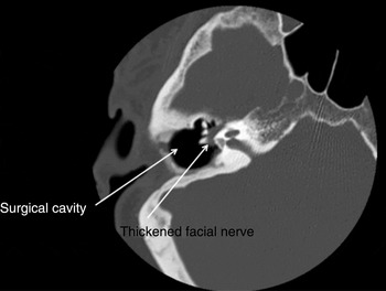
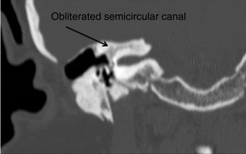
Computed tomography of the temporal bones showed a well aerated right middle ear with evidence of expansion of the fallopian canal in the tympanic segment of the right ear, presumably at the site of the original facial nerve injury (Figure 1). Sclerosis and obliteration of the right semicircular canal were observed, but the cochlea was intact and appeared patent (Figure 2). The left side showed similar sclerosis but was otherwise normal.
Axial computed tomography scan of the right mastoid cavity, showing thickening of the facial nerve.

Axial computed tomography scan of the left mastoid cavity, showing sclerosis and obliteration of the semicircular canals.

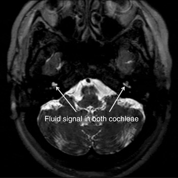
Following a tympanoplasty, the patient was seen by the cochlear implantation assessment team. Her audiometric results were found to meet the criteria for cochlear implantation. Her pre-operative hearing in noise test (HINT) score was 0 per cent. A pre-operative magnetic resonance imaging scan was performed to confirm cochlear patency (Figure 3); both cochleae were patent. The decision was made to undertake implantation on the right side, due to the pre-existing facial nerve paralysis.
Axial, high resolution, T2-weighted magnetic resonance imaging scan of the posterior fossa, showing patency of the left and right cochlear canals, with a normal fluid signal in both cochleae.

Surgery was undertaken using a standard post-auricular approach. No intra-operative difficulties were encountered.
Following surgery, the patient was followed closely by the implant audiologists. Her six month HINT score was 60 per cent.
At the time of writing, the patient remained very satisfied with her implant.
Discussion
There are several traditionally cited potential contraindications to cochlear implantation in patients who have undergone surgical labyrinthectomy.
Firstly, there is the issue of neural function retention in the cochlear nerve and spiral ganglion.Reference Wareing and O'Connor8 Damage or degeneration of these structures would lead to a poor outcome following cochlear implantation. However, several studies have illustrated excitability of the post-labyrinthectomy ear. Electron microscopy has demonstrated intact spiral ganglion neurons (albeit with smaller axonal diameters and nuclei).Reference Nadol and Thornton9 Furthermore, a histological study of post-labyrinthectomy temporal bones found consistent survival of spiral ganglion cells, despite extensive hair cell loss.Reference Chen, Linthicum and Rizer10
• Labyrinthectomy is the treatment of choice for debilitating Ménière's disease with unaidable hearing in the affected ear
• The effectiveness of post-labyrinthectomy cochlear implantation has been questioned, especially if there is a significant delay between the procedures
• This case shows that delayed cochlear implantation, even decades after labyrinthectomy, can be a viable option with significant benefit for selected patients
The second obstacle to cochlear implantation involves post-labyrinthectomy changes in the vestibule and cochlea. Following surgery, the vestibule often becomes obliterated due to accumulation of fibrous and bony tissue.Reference Wareing and O'Connor8 Similarly, the cochlea can become ossified, leading to difficulties in implantation.Reference Wareing and O'Connor8 Some authors have suggested that these problems can be avoided by performing implantation immediately following labyrinthectomy. However, in the more common case of initial labyrinthectomy followed by later failure of the other ear, this is not a viable solution.Reference Wareing and O'Connor8 In order to overcome this problem, Kemink et al. have suggested that a plastic obturator be implanted in the scala tympani to maintain its patency should cochlear implantation be later required.Reference Kemink, Kileny, Niparko and Telian11 However, cases such as ours illustrate that, even 20 years post-labyrinthectomy, cochlear implantation may be successful without such measures.
The strongest evidence regarding the viability of cochlear implantation following labyrinthectomy may lie in the growing number of successful cases. Kveton et al. have described a patient who underwent cochlear implantation six weeks after labyrinthectomy due to a papillary adenocarcinoma which had invaded the bony labyrinth.Reference Kveton, Abbott, April, Drumheller, Cohen and Poe12 Similarly, Zwolan et al. have described a woman with prelingual, bilateral sensorineural hearing loss who underwent cochlear implantation combined with simultaneous labyrinthectomy, with a good outcome.Reference Zwolan, Shepard and Niparko7 Facer et al. have reported a case of cochlear implantation performed 18 months after labyrinthectomy for acoustic neuroma,Reference Facer, Facer, Facer Fowler, Brey and Peterson13 while Thedinger et al. have described successful implantation of a patient 15 years after labyrinthectomy for endolymphatic hydrops, in whom a contralateral acoustic neuroma had developed.Reference Thedinger, Cueva and Glasscock14 Both cases had good results. Finally, Lustig et al. have reviewed nine patients with Ménière's disease who underwent cochlear implantation, one following labyrinthectomy.Reference Lustig, Yeagle, Niparko and Miner2 On average, post-operative auditory function was substantially improved from pre-operative levels. However, at two-year follow up, the post-labyrinthectomy patient was not doing as well on audiometric testing as the other patients. Nonetheless, this patient demonstrated an objective improvement from her pre-operative status, including an 89 per cent score in Central Institute for the Deaf (CID) sentences, and remained satisfied with her implant.Reference Lustig, Yeagle, Niparko and Miner2
Conclusion
While several successful cases of post-labyrinthectomy cochlear implantation have been reported, very few of these patients had any significant delay between labyrinthectomy and implantation. Prior to our case, the longest reported delay was 15 years, and the majority of previous reports described patients who had received implants within weeks to months of labyrinthectomy.
Despite the theoretical risk of difficulties due to ossification of the cochlea and obliteration of the vestibule, our patient obtained an excellent outcome.
This case illustrates the fact that delayed cochlear implantation, even decades after labyrinthectomy, can remain a viable solution with significant benefits for selected patients.