Introduction
The new mineral gorbunovite, CsLi2(Ti,Fe+3)(Si4O10)F2, belonging to the trioctahedral-mica subgroup, was found in the alkaline rocks of the Darai-Pioz massif [the Rasht (formerly Garm) district, Central Tajikistan]. It is named after the Russian chemist, Academician Nikolai Petrovich Gorbunov (Николай Петрович Горбунов) (1892–1938), head of the Tajik-Pamir expedition, this first expedition that geologically surveyed many remote areas of Central Asia, including the Darai-Pioz massif. Gorbunovite (Russian Cyrillic: горбуновит) is a titanium analogue of sokolovaite CsLi2Al(Si4O10)F2, garmite CsLiMg2(Si4O10)F2 and kreiterite CsLi2Fe+3(Si4O10)F2 and a caesium analogue of orlovite KLi2Ti(Si4O10)(ОF)2. Gorbunovite was approved as a new mineral species by the Commission on New Minerals, Nomenclature and Classification of the International Mineralogical Association, IMA No. 2017-040, Agakhanov et al., Reference Agakhanov, Pautov, Pekov, Karpenko, Siidra, Sokolova, Hawthorne, Muftakhov and Kasatkin2017a) The holotype sample of gorbunovite is deposited in the systematic collection of the Fersman Mineralogical Museum of the Russian Academy of Sciences (Moscow) with catalogue no. 94187. The approved mineral symbol for gorbunovite is Gbv (Warr, Reference Warr2021).
Occurrence and associated minerals
Gorbunovite was found in rock samples of the Upper Darai-Pioz alkaline massif (39°27’N, 70°43’E), located on the southern slope of the Alai Range in the joint area of three sub-latitudinal ranges: the Turkestan, Alai and Zeravshan Ranges. The Darai-Pioz massif itself is difficult to access due to the mountainous terrain, and the majority of mineralogical investigations have been done on material collected from the deposits of glacial moraine associated with the massif.
The first data on the Darai-Pioz massif were obtained by the Tajik-Pamir Expedition headed by N.P. Gorbunov in 1932–1936 (Moskvin, Reference Moskvin1937). Under the leadership of Yu.A. Arapov the Alai and Turkestan ranges, including the Darai-Pioz massif, were prospected for rare metals during 1946–1947. The most complete work on the massif was done in the 1960s to 1990s by V.D. Dusmatov, as a result of which a 1:10000 scale geological map was compiled. Extensive data on petrography and rock geochemistry were obtained, and in collaboration with E.I. Semenov, A.F. Efimov and A.P. Khomyakov, a unique mineralisation of lithium, boron, zirconium and rare earth elements was revealed. Many publications are devoted to the mineralogy of the Darai-Pioz massif (Dusmatov, Reference Dusmatov1968; Reference Dusmatov1970; Reference Dusmatov1971; Semenov and Dusmatov, Reference Semenov and Dusmatov1975; Ganzeev et al., Reference Ganzeev, Dusmatov, Efimov and Akramov1976; Belakovskiy, Reference Belakovskiy1991; Dusmatov and Morozov, Reference Dusmatov and Morozov1993; Grew et al., Reference Grew, Belakovskiy, Fleet, Yates, Mc.Gee and Marquez1993; Vladykin et al., Reference Vladykin, Dusmatov and Kovalenko1995; Vladykin and Dusmatov, Reference Vladykin and Dusmatov1996; Pautov et al., Reference Pautov, Agakhanov, Sokolova and Ignatenko1996; Pautov and Agakhanov, Reference Pautov and Agakhanov1997; Reguir et al., Reference Reguir, Chakhmouradian and Evdokimov1999; Agakhanov et al., Reference Agakhanov, Pautov, Karpenko, Bekenova and Uvarova2011, etc.). One of the most interesting features of the Darai-Pioz massif is the variety of caesium minerals (Agakhanov, Reference Agakhanov2010). To date, 13 distinct caesium minerals have been discovered in the rocks of the massif, caesium kupletskite (Efimov et al., Reference Efimov, Dusmatov, Hanseev and Kataeva1971) later renamed kupletskite-(Cs) Cs2NaMn2+7Ti2(Si4O12)2O2(OH)F (Bayliss, Reference Bayliss2007), telyushenkoite CsNa6[Be2(Si,Al,Zn)18O39]F2 (Agakhanov et al., Reference Agakhanov, Pautov, Belakovskiy, Sokolova and Hawthorne2003), zeravshanite Na2Cs4Zr3[Si18O45]·2H2O (Pautov et al., Reference Pautov, Agakhanov, Uvarova, Sokolova and Hawthorne2004), senkevichite CsKNaCa2TiO[Si7O18](OH) (Agakhanov et al., Reference Agakhanov, Pautov, Uvarova, Sokolova, Hawthorne and Karpenko2005), sokolovaite CsLi2Al(Si4O10)F2 (Pautov et al., Reference Pautov, Agakhanov and Bekenova2006), kirchhoffite, CsBSi2O6, (Agakhanov et al., Reference Agakhanov, Pautov, Karpenko, Sokolova and Hawthorne2012), mendeleevite-(Ce) Cs6[Ce22Ca6](Si70O175)(OH,F)14(H2O)21 (Pautov et al., Reference Pautov, Agakhanov, Karpenko, Sokolova and Hawthorne2013), odigitriaite CsNa5Ca5[Si14B2O38]F2 (Agakhanov et al., Reference Agakhanov, Pautov, Sokolova, Hawthorne, Karpenko, Siidra and Muftakhov2016), mendeleevite-(Nd) Cs6[Nd23Ca7](Si70O175)(OH,F)19(H2O)16 (Agakhanov et al., Reference Agakhanov, Pautov, Sokolova, Hawthorne, Karpenko, Siidra and Garanin2017b), fluorapophyllite-(Cs) CsCa4(Si8O20)F·8H2O (Agakhanov et al., Reference Agakhanov, Pautov, Kasatkin, Karpenko, Sokolova, Day and Britvin2019), kreiterite CsLi2Fe3+Si4O10F2 (Agakhanov et al., Reference Agakhanov, Pautov, Pekov, Karpenko, Siidra, Sokolova, Hawthorne, Faiziev, Kasatkin, Kulikova and Muftakhov2025), garmite CsLiMg2(Si4O10)F2 (Pautov et al., Reference Pautov, Agakhanov, Pekov, Karpenko, Siidra, Sokolova, Hawthorne and Faiziev2022a) and gorbunovite CsLi2(Ti,Fe)Si4O10(F,OH,O)2 described here.
Gorbunovite was found in non-rounded or slightly rounded boulders (up to 2 m in size) of a rock composed mainly of granular quartz, which were described in detail by Pautov et al. (Reference Pautov, Agakhanov, Uvarova, Sokolova and Hawthorne2004; Reference Pautov, Agakhanov, Pekov, Karpenko and Siidra2022b and references therein). Unfortunately, neither we nor previous geologists have found it in situ in bedrock outcrops. Moreover, no contacts of these quartz boulders with any other rock has been ever observed, which makes it even more difficult to establish their origin. For this reason, we will call them simply ‘quartz lumps’, as has already been done in many publications on Darai-Pioz mineralogy.
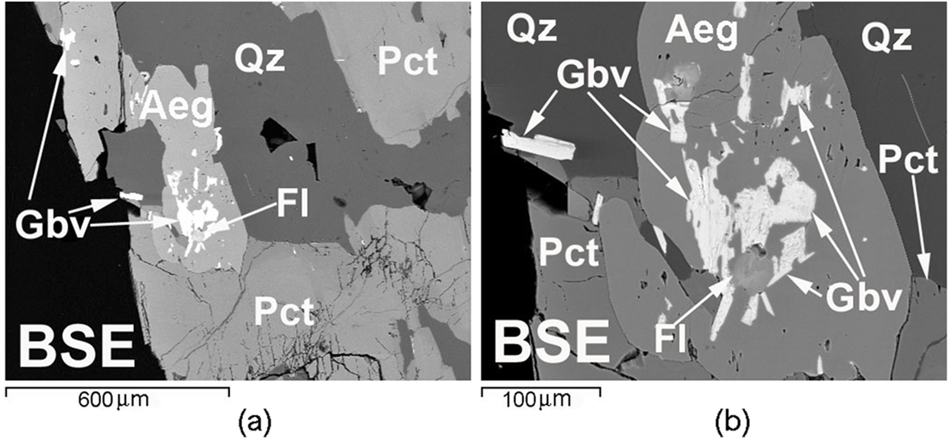
Gorbunovite occurs in polymineralic quartz–pectolite aggregates from 1 to 15 cm in the largest dimension, sometimes embedded in ‘quartz lumps’. They are composed of brown or greyish-brown fine- to medium-grained polymineralic aggregates, consisting of Mn-bearing pectolite (up to 3.5 wt.% MnO), quartz, Sr-rich fluorite (up to 21 wt.% SrO), aegirine (with chemical composition close to the end-member), polylithionite, datolite and a range of rare minerals. Within these pectolite aggregates, the distribution of gorbunovite is extremely uneven. Most commonly, gorbunovite is confined to the boundaries of aegirine with quartz or, sporadically, pectolite. Small gorbunovite individuals form ingrowths in aegirine crystals (Fig. 1). In general, this mica is very rare: it is found only in a few samples.
Gorbunovite forms lamellar grains from 2 μm to 0.1 mm, rarely up to 0.2 mm in the largest dimension. The thickness of its grains varies from fractions of a micron to 20 μm. Fan-shaped intergrowths of lamellar crystals are common. For the most part, the grains of gorbunovite are deformed.
SEM (BSE) image. (a) General view of lamellar aggregations of gorbunovite (Gbv) in aegirine (Aeg) with fluorite (Fl) from the quartz (Qz) – pectolite (Pct) aggregate; (b) enlarged area.

Physical properties
Grains of gorbunovite are colourless, in aggregates the mineral is white, and visually is indistinguishable from other micas of the association. Lustre is vitreous, the streak is white. Cleavage is perfect on {001}, lamellae are flexible. Mohs hardness is 2½; the average micro-indentation hardness VHN is 87 kgf/mm2 (with a range from 82 to 111 kgf/mm2 from 10 measurements), measured using a PMT-3 instrument at a loading of 20 g, calibrated on NaCl. The density of the mineral was determined by flotation of the grains in Clerici solution. The measured density, D meas is 3.28 (2) g/cm3, and the calculated density D calc is 3.302 g/cm3. Gorbunovite fluoresces pale yellow in shortwave ultraviolet light. The 2V angle measured on a Fedorov stage is = –30(5)°, 2Vcalc. = –44°. The small size of the mineral individuals, low birefringence and deformation of the grains did not allow better measurement. The optical axes plane is almost perpendicular to the cleavage plane {001}. The mineral is colourless in transmitted light. Pleochroism is not observed. Gorbunovite is biaxial (–) with α = 1.609(2), β = 1.621(2) and γ = 1.623(2). Measurements were done at λ = 589 nm using a rotating needle. Dispersion is weak, r > v.
Chemical composition
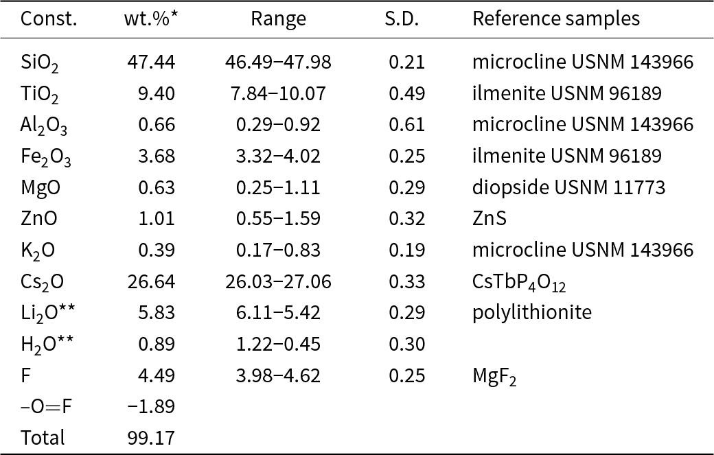
The chemical composition of gorbunovite was analysed using a JCXA-733 JEOL electron-microprobe analyser (EMPA) in the laboratory of the Fersman Mineralogical Museum and a Cameca IMS-4F ion-mass spectrometer (SIMS) at the Institute of Microelectronics and Information Mathematics of the Russian Academy of Sciences (Yaroslavl’, Russia). All elements except fluorine, lithium and hydrogen were determined using an Si(Li) energy-dispersive detector and the INCA Energy 350 analysis system of Oxford Instruments. The analyses were done at 20 kV accelerating voltage, 2 nA probe current, 5 μm probe diameter and 100 s of spectrum dwell time (excluding dead time). Fluorine concentration was measured using a wave-dispersion spectro- meter with an LDE1 crystal analyser at 10 kV accelerating voltage, 30 nA probe current and a 10 μm defocused electron beam.
Hydrogen and lithium in gorbunovite were determined using SIMS according to the methods reported by Smirnov et al. (Reference Smirnov, Sobolev, Batanova, Portnyagin, Simakin and Potapov1995). A primary 16O2– ion beam focused to a 10–15 μm diameter spot was used. The current intensity of the bombardment ions was 3–8 nA. Element concentrations were calculated from the intensity ratio of element/30Si+ ion currencies, using calibration constants obtained on reference samples.
For a more correct estimation of the lithium content in gorbunovite, an internal laboratory reference sample of polylithionite from the Darai-Pioz alkaline massif with a known Li content, was used. Both the standard sample and gorbunovite were placed in a same polished section and were analysed under the same conditions and this made it possible to correct the obtained lithium data.
The valence of iron in gorbunovite was not determined, however it was determined in kreiterite CsLi2Fe3+Si4O10·(F,OH)2 (which is the Fe analogue of gorbunovite) by measuring oxygen directly using EMPA with wavelength dispersive spectroscopy. Although the relative error in oxygen determination was too large, it was shown unambiguously that iron is present as Fe3+ in kreiterite. It was not possible to find out the valence of iron in gorbunovite by the same method of oxygen measuring, as there is four times less iron than in kreiterite. It seems probable that there is an isomorphic series between kreiterite and gorbunovite, and that the valence of Fe (in full or partly) in gorbunovite is the same as in kreiterite.
The chemical composition of gorbunovite is given in Table 1.
Chemical composition for gorbunovite (wt.%)

Notes: *Average of 10 analyses; ** SIMS analysis data; S.D. – standard deviation
The empirical formula of gorbunovite, calculated on the basis of 12 (O + F + OH), is:
(Cs0.95K0.04)Σ0.99Li1.96(Ti0.59Fe+30.23Mg0.08Al0.06Zn0.06)Σ1.02Si3.96O10(F1.19OH0.50O0.31)Σ2 and the simplified formula is CsLi(Ti,Fe+3)(Si4O10)(F,OH,O)2. The Gladstone-Dale compatibility index value from Mandarino (Reference Mandarino1981) for D meas. = 3.28 g/cm3 is (1 – K p/K c) = 0.073 (fair). The value of the compatibility index is probably strongly influenced by the variable degree of distortion of Ti–O polyhedra in different minerals. For example, such a problem exists when calculating the compatibility index for layered titanosilicates of the lamprophyllite group, as well as in orlovite, the titanium member of mica group, in which (1 – K p/K c) is even larger (Agakhanov et al., Reference Agakhanov, Pautov, Karpenko, Bekenova and Uvarova2011).
X-ray diffraction data
Single-crystal X-ray diffraction studies were done using a Bruker APEX II DUO diffractometer equipped with a CCD detector (MoKα radiation) at 50 kV and 0.6 mA. The following parameters of the monoclinic unit cell (space group C2/m, C2 or Cm) corresponding to the 1M polytype were obtained: a = 5.25 (15), b = 9.1(1), c = 10.7(7) Å, β = 99(3)°, V = 505(3) Å3 and Z = 2. Single-crystal X-ray diffraction showed gorbunovite to be a 1M polytype. The low quality of all tested crystals is due to deformation and splitting which prevented single-crystal structure refinement and the paucity of material which prevented the Rietveld refinement.
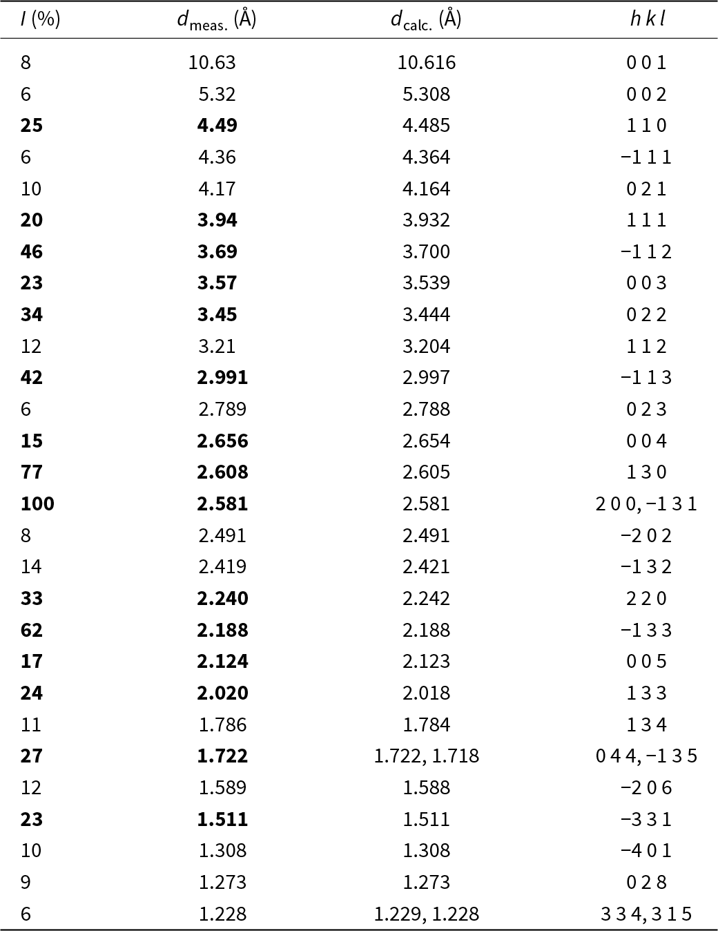
The powder X-ray diffraction pattern of gorbunovite was obtained using a Rigaku R-AXIS Rapid II single-crystal diffractometer equipped with a cylindrical detector using Debye-Scherrer geometry (d = 127.4 mm) at CoKα radiation. The results of calculation of the powder X-ray diffraction pattern are shown in Table 2. The unit-cell parameters, refined on the basis of powder data are: a = 5.236 (2), b = 9.054(4), c = 10.767(4) Å, β = 99.61(4)° and V = 503.3(6) Å3.
Powder X-ray diffraction data, for gorbunovite

Note: Bold indicates the strongest lines.
Raman spectroscopy
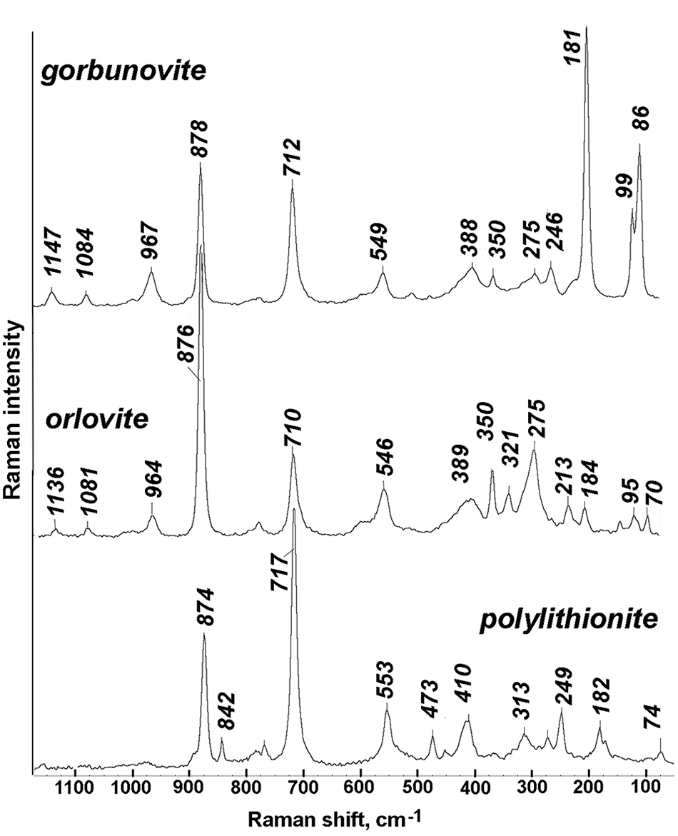
Raman spectra were obtained from a polished section of a randomly oriented crystal of gorbunovite, and from cleavage plates of chemically characterised orlovite and polylithionite (Fig. 2). Spectra were obtained at room temperature using a Thermo DXR2xi Raman imaging confocal microscope in the range 3400 cm–1 to 50 cm–1 with 2 cm–1 resolution, a 25 µm aperture, and a grating of 400 lines/mm. The excitation source was a laser with a wavelength of 785 nm, and the output power of the laser beam was 30 mW (excitation with a 532 nm laser beam caused intensive sample fluorescence). The back-scattered Raman signal was collected with a 100x objective; the signal acquisition time for a single scan was from 0.25 to 2 s and the signal was averaged from 5 to 30 scans. The spectra were processed using OMNIC software.
Raman spectra of gorbunovite, orlovite and polylithionite.

The Raman spectrum of gorbunovite, polylithionite and orlovite is shown in Fig. 2. Bands from 1150 to 800 cm–1, similar to other layered silicates, generally are stretching modes of the SiO4 tetrahedra (McKeown et al., Reference McKeown, Bell and Etz1999; Wang et al., Reference Wang, Hu, Zhang, Huang and Ni2004; Wang et al., Reference Wang, Hu, Zhang, Fontan, de Parseval Ph and Jiang2007). The intense band at 878 cm–1 is possibly diagnostic for this group of trioctahedral mica. Another intense line at 712 cm–1 is common in sheet silicates and corresponds to both Si–O stretching and O–Si–O bending deformations (Wang et al., Reference Wang, Freeman and Jolliff2015).
Raman lines below 600 cm–1 correspond to deformations of octahedra and interlayer complexes (Tlili et al., Reference Tlili, Smith, Beny and Boyer1989; Wang et al., Reference Wang, Freeman and Jolliff2015). Note that the 388 cm–1 band is also present in orlovite, and is attributed to possible associated deformations of the TiO6 octahedra because this band is absent in polylithionite and other Ti-free trioctahedral Li-micas. Differences in the intensities of the Raman bands in the range 80–200 cm–1 for gorbunovite, compared to those of orlovite and polylithionite, may be the result of different orientation: the spectra for polylithionite and orlovite were collected on cleavage planes whereas the spectrum of gorbunovite was collected on a randomly orientated grain.
Discussion
The formation of such unusual and rare trioctahedral micas as sokolovaite, orlovite, garmite, kreiterite and gorbunovite is connected directly to the genesis of both the quartz–pectolite aggregate itself and significantly the quartz rocks or ‘quartz lumps’ in which it was found. It can be assumed that the ‘quartz lumps’ of Darai-Pioz are fragments of quartz cores of specific peralkaline and supersaturated with silica highly agpaitic pegmatites. Unfortunately, we have not yet found any pegmatites with quartz cores at the Darai-Pioz, that are completely similar to ‘quartz lumps’, and therefore it is unclear whether such pegmatites exist at all. We hypothesise that the formation of ‘quartz lumps’ occurred from a water–silicate liquid (WSL) that arose during the interaction of an alkaline fluid, derived from carbonatites, which caused extensive fenitisation of the Darai-Pioz massif rocks, with quartz-containing rocks of the massif, which are granites and their pegmatites.
The mechanism of the formation of ‘quartz lumps’, as well as the formation of quartz–pectolite aggregates in them, is discussed in detail in the work of L.A. Pautov et al. (Reference Pautov, Agakhanov, Pekov, Karpenko and Siidra2022b). Several processes for the formation of pectolite aggregates with caesium mineralisation can be assumed: (1) the separation of an aqueous silicate liquid into two liquids, one of which, significantly enriched in Na, Ca and F, just gave rise to pectolite aggregates; (2) formation of pectolite aggregates from extruding residues from crystallisation of the quartz aggregate; and (3) contamination of carbonate melt clumps with WSL.
The origin of caesium minerals, including gorbunovite, is associated with the ability of WSL to ‘extract’ Cs and Li from a coexisting fluid (Smirnov, Reference Smirnov2015; Smirnov et al., Reference Smirnov, Thomas, Kamenetsky and Kozmenko2017). A very high Si/Al ratio and the presence of Li promotes the crystallisation of trioctahedral mica which contributes to the crystallisation of both K- and Cs-dominant tetra-silicate micas), the species diversity of which depends on the amount of cations in the surrounding fluid.
The following heterovalent isomorphism schemes maintain electroneutrality where the octahedral sheets are occupied by tri- and tetra-valent ions we assume:
Ti4+ + O2– → Fe,Al)3+ + (F,OH)–;
Ti4+ + Li+ + O2– → 2(Mg,Zn)2+ + (F,OH)–.
Gorbunovite is a caesium analogue of orlovite KLi2TiSi4O11F (Agakhanov et al., Reference Agakhanov, Pautov, Karpenko, Bekenova and Uvarova2011; Sokolova et al., Reference Sokolova, Hawthorne, Agakhanov, Pautov and Karpenko2018) and a titanium analogue of as many as three Cs–Li-micas, including sokolovaite CsLi2AlSi4O10F2 (Pautov et al., Reference Pautov, Agakhanov and Bekenova2006), garmite CsLiMg2Si4O10F2 (Pautov et al., Reference Pautov, Agakhanov, Pekov, Karpenko, Siidra, Sokolova, Hawthorne and Faiziev2022a) and kreiterite CsLi2Fe3+Si4O10(F,ОН)2 (IMA2019-041).
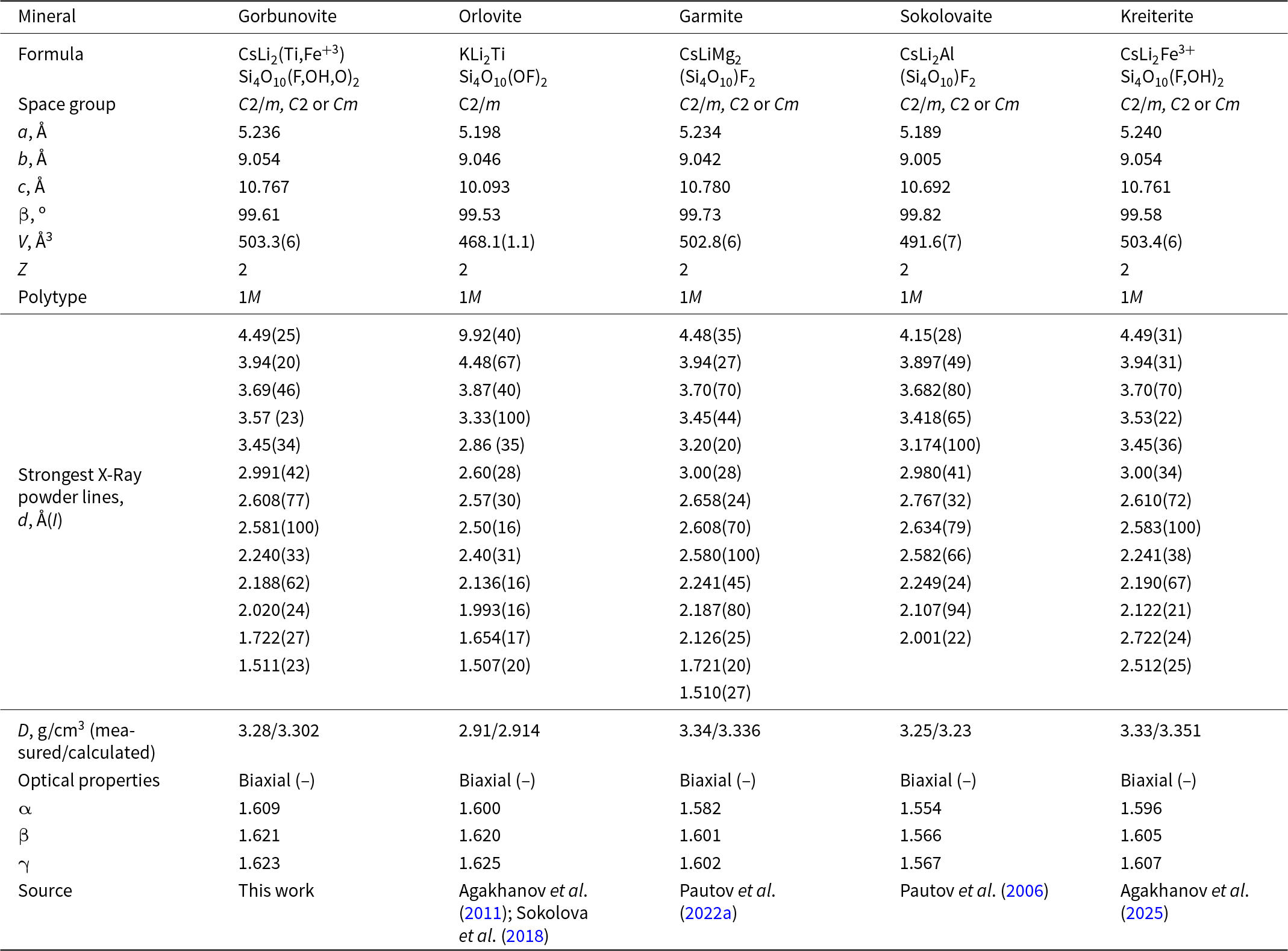
A comparison of gorbunovite with related minerals is given in Table 3. As expected, all caesium micas are characterised by significantly larger unit-cell dimensions compared to that of potassium analogues. This is due to the significantly larger ionic radius of the [12]Cs = 2.011 Å interlayer cation compared to [12]K = 1.729 Å (Hawthorne and Gagné, 2024). For the same reason, there are no significant isomorphic series between caesium and potassium mica in Nature. The unit-cell dimensions of gorbunovite are close to those of kreiterite as their ionic radii are reasonably similar ([6]Ti4+ = 0.605 Å and [6]Fe3+ = 0.649 Å). This is probably due to the fact gorbunovite includes Fe3+ as a secondary component (3.3–4.0 wt.% Fe2O3), and, on the other hand, Ti is a minor component in kreiterite (0.6–1.6 wt.% TiO2). Whether there is a continuous series between gorbunovite and kreiterite can only be shown by new findings and further study of these, so far rare, minerals.
Comparative data for gorbunovite, orlovite and trioctahedral caesium micas

Acknowledgements
Technical support by the X-Ray Diffraction Resource Center of Saint-Petersburg State University is gratefully acknowledged. The authors thank S.G. Simakin for the SIMS analysis of the new mineral, R.U. Sobirova for the organization of expedition transportation to Darai-Pioz, and all the participants of expeditions works, first of all P.V. Khvorov, T.K. Berkeliev, M.A. Shodibekov and N. Radjabov.
Funding statement
The work was done with the support of the Russian Science Foundation, grant 24-27-00225.
Competing interests
The authors declare none.