Introduction
Successive advances in lateral skull base surgery since the 1950s have enabled aggressive tumour excision and improved local control of both benign and malignant pathologies. However, patients undergoing temporal bone resections still have poor prognoses, with high recurrence rates.Reference Ward, Loch and Lawrence1–Reference Dean, White, Carter, Desmond, Carroll and McGrew4
Because of the involvement of critical bony and neurovascular structures, cancers of the temporal bone require specialised treatment, with surgery forming the mainstay, potentially supplemented by immunotherapy, chemotherapy and/or radiotherapy. Surgical approaches can broadly be classified into lateral temporal bone resection, subtotal temporal bone resection and total temporal bone resection.
Lateral temporal bone resection involves removing the ear canal and tympanic membrane, expanding to the inner ear and otic capsule in subtotal temporal bone resection and/or the petrous apex in total temporal bone resection. Lateral temporal bone resection is viewed as the standard minimum surgery for T1 and T2 disease.Reference Homer, Lesser, Moffat, Slevin, Price and Blackburn5 Subtotal temporal bone resection is necessary for tumours affecting the mastoid, middle ear, inner ear or lateral skull base.Reference Gidley and DeMonte6 In total temporal bone resection, the dissection extends beyond the subtotal temporal bone resection to include the petrous apex.Reference Gidley and DeMonte6 Depending on the grade and location of the tumour, resection may involve structures such as part or all of the pinna, periauricular skin, parotid and mandible, with or without neck dissection.Reference Homer, Lesser, Moffat, Slevin, Price and Blackburn5
Temporal bone resection is challenging not only because of the difficulty in preserving anatomical structures, such as the facial nerve in lateral temporal bone resection, while achieving clear margins, but also because of the complex reconstruction required. Adequate amounts of well-supplied tissue are crucial in repairing defects that result from loss of skin and bone, which can expose critical structures, including cranial nerves, major blood vessels, the brain and the dura. A sound reconstruction is necessary to prevent recognised complications, including cerebrospinal fluid leak, meningitis and potentially death.Reference Howard, Nagel, Barrs, Donald and Hayden7
There is presently a scarcity of literature concerning the long-term survival outcomes and rates of complications in patients who undergo temporal bone resection and subsequent reconstruction for a variety of indications. The varied outcomes of survival are mainly due to the heterogeneity of histological diagnoses. The aim of this study was to evaluate the clinical characteristics and survival rates of patients who underwent temporal bone resection at our tertiary skull base surgery centre in the UK.
Materials and methods
A retrospective review of patient charts was conducted for individuals who underwent temporal bone resection for malignancy at a UK tertiary centre from February 2004 to March 2024. This study involving human participants adhered to the ethical standards set by the institutional and national research committee, as well as the 1964 Helsinki Declaration, including its subsequent amendments or equivalent ethical guidelines. Approval for this study was granted by the University Hospitals Birmingham National Health Service (NHS) Foundation Trust Quality Improvement Department (registration number 21664). The study did not require formal ethics committee review after completing the National Decision Tool developed by the Medical Research Council Regulatory Support Centre in partnership with the Health Research Authority.8
The classification of temporal bone resection and the surgical techniques are described by Jackler.Reference Jackler9 Patient demographic details, including age, sex, histological diagnosis, cancer stage and surgical indications, were documented. Tumours were staged according to cancer type following the guidelines of the American Joint Committee on Cancer.Reference Amin, Edge, Greene, Schilsky, Brookland and Washington10 Evaluation encompassed the extent of lateral temporal bone resection, neck dissection status, reconstruction type and adjuvant therapies (chemotherapy, chemoradiation, radiotherapy). Post-operative complications and follow-up duration were assessed, while outcomes focused on surgical complications, disease-free survival and overall survival.
Statistical methods
Statistical analysis was conducted using R (R Project for Statistical Computing, Vienna, Austria) to explore and summarise the characteristics of patients undergoing temporal bone resection. Descriptive statistics were calculated to summarise the demographic and clinical characteristics of the patient cohort. This included measures of central tendency (mean, median) and dispersion (range) for continuous variables such as age. Categorical variables, including sex, cancer stage, facial nerve grade and diagnosis, were summarised using frequencies and percentages.
Survival analysis was performed using the Kaplan–Meier method to estimate overall survival and disease-free survival. The ‘survival’ package in R was used to create Kaplan–Meier curves with survival probabilities plotted against time. These plots were stratified by relevant clinical factors to visualise differences in survival outcomes between groups. Confidence intervals (95 per cent) were calculated and displayed on the Kaplan–Meier plots. Log-rank tests were conducted to compare survival distributions between different groups, such as cancer stages or treatment modalities. Cox proportional hazards regression models were employed to assess the impact of clinical and pathological factors on survival outcomes.
To evaluate the statistical significance of differences between groups for categorical variables, chi-square tests or Fisher's exact tests were used as appropriate. For continuous variables, student’s t-tests or Mann–Whitney U tests were applied, depending on the normality of data distribution. All statistical tests were two-sided, and a p value of less than 0.05 was considered statistically significant.
Results
Demographics and diagnosis
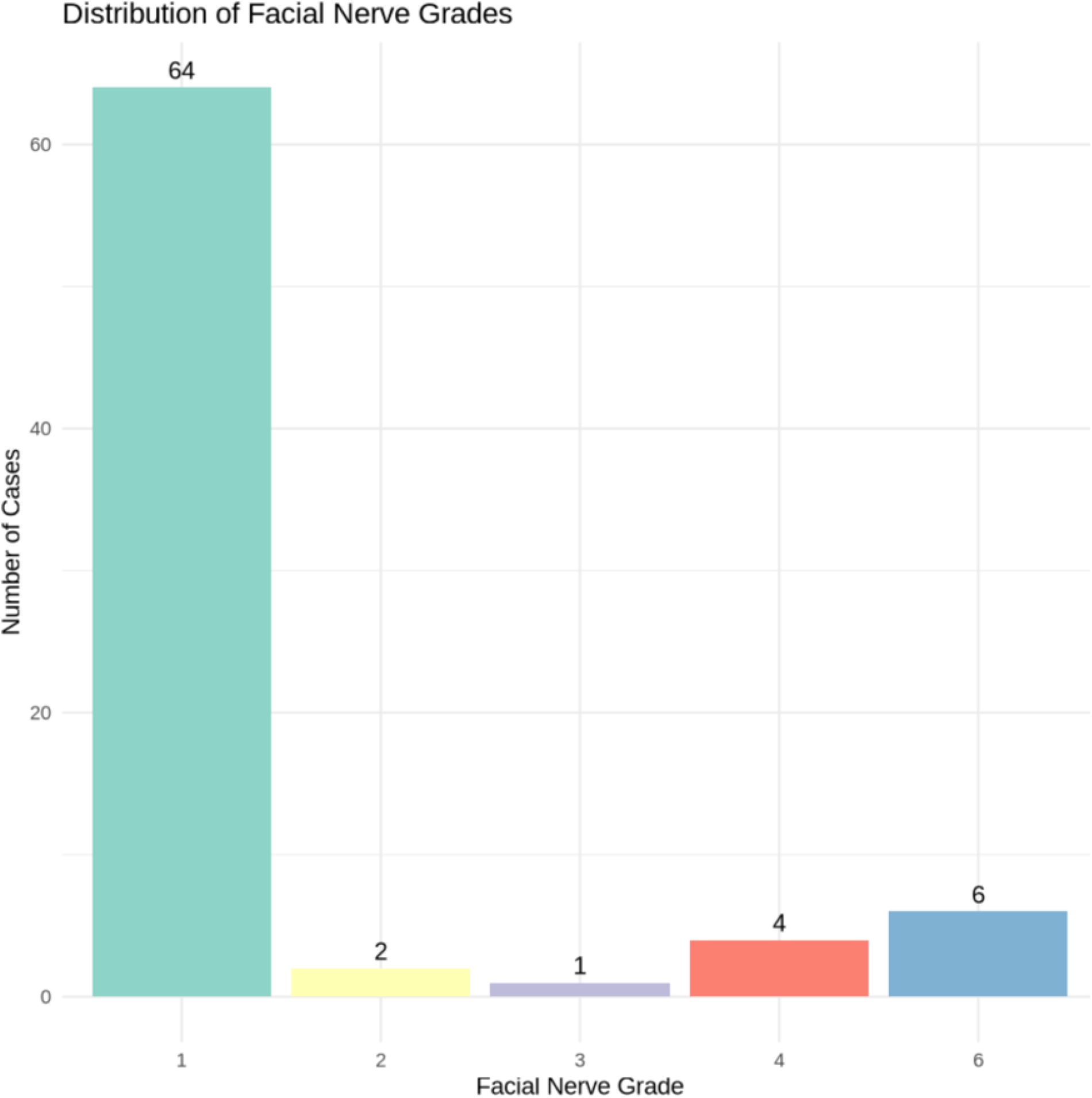
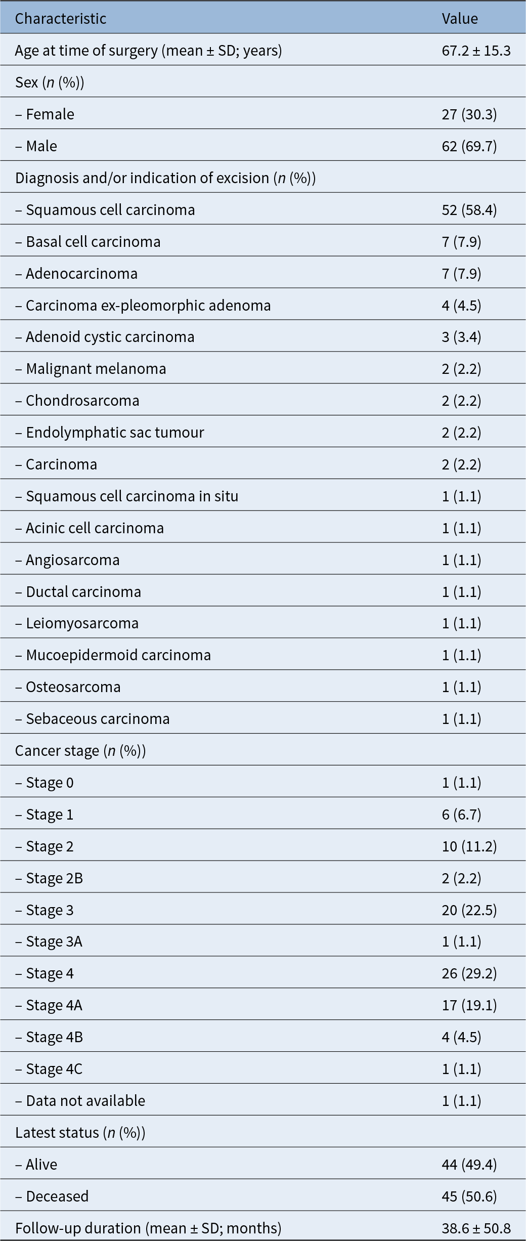
The patient population comprised 89 individuals, of which 62 were male (69.7 per cent) and 27 were female (30.3 per cent), with a mean age of 67.2 years (median age, 70 years; range, 24–91 years) (Table 1). Most patients (n = 64, 71.91 per cent) did not have any facial nerve palsy on their initial presentation to the ENT department (Figure 1). Cancer staging data were unavailable for one patient (1.12 per cent) because the medical records, dating back 20 years, were inaccessible. Among those with available data, 26 patients (29.2 per cent) were Stage 4, 20 patients (22.5 per cent) were Stage 3, 17 patients (19.1 per cent) were Stage 4A and 10 patients (11.2 per cent) were Stage 2. Other stages (1, 2B, 3A, 4B, 4C) each represented less than 7 per cent of the cohort.
Distribution of patients by facial nerve palsy classification on the House–Brackmann scale at initial presentation.

Demographic and clinical characteristics of patients undergoing lateral temporal bone resection

SD = standard deviation
Squamous cell carcinoma (SCC) was the most common diagnosis, affecting 52 patients (58.4 per cent), followed by basal cell carcinoma and adenocarcinoma, each found in 7 patients (7.9 per cent). Seventy patients presented with primary cases (78.7 per cent), 16 with recurrent disease (18.0 per cent) and 3 with metastatic disease (3.4 per cent). The most common primary site of malignancy was the external auditory canal, found in 38 patients (42.7 per cent), followed by the pinna in 22 patients (24.7 per cent) and the post-auricular skin in 3 patients (3.37 per cent). Out of the 89 patients, 70 (78.7 per cent) were primary cases, with a mean age of 66.8 years. Sixteen patients (18.0 per cent) had recurrent disease with a mean age of 68.6 years and 3 patients (3.4 per cent) had metastatic disease with a mean age of 69.0 years.
Surgical and adjuvant treatment
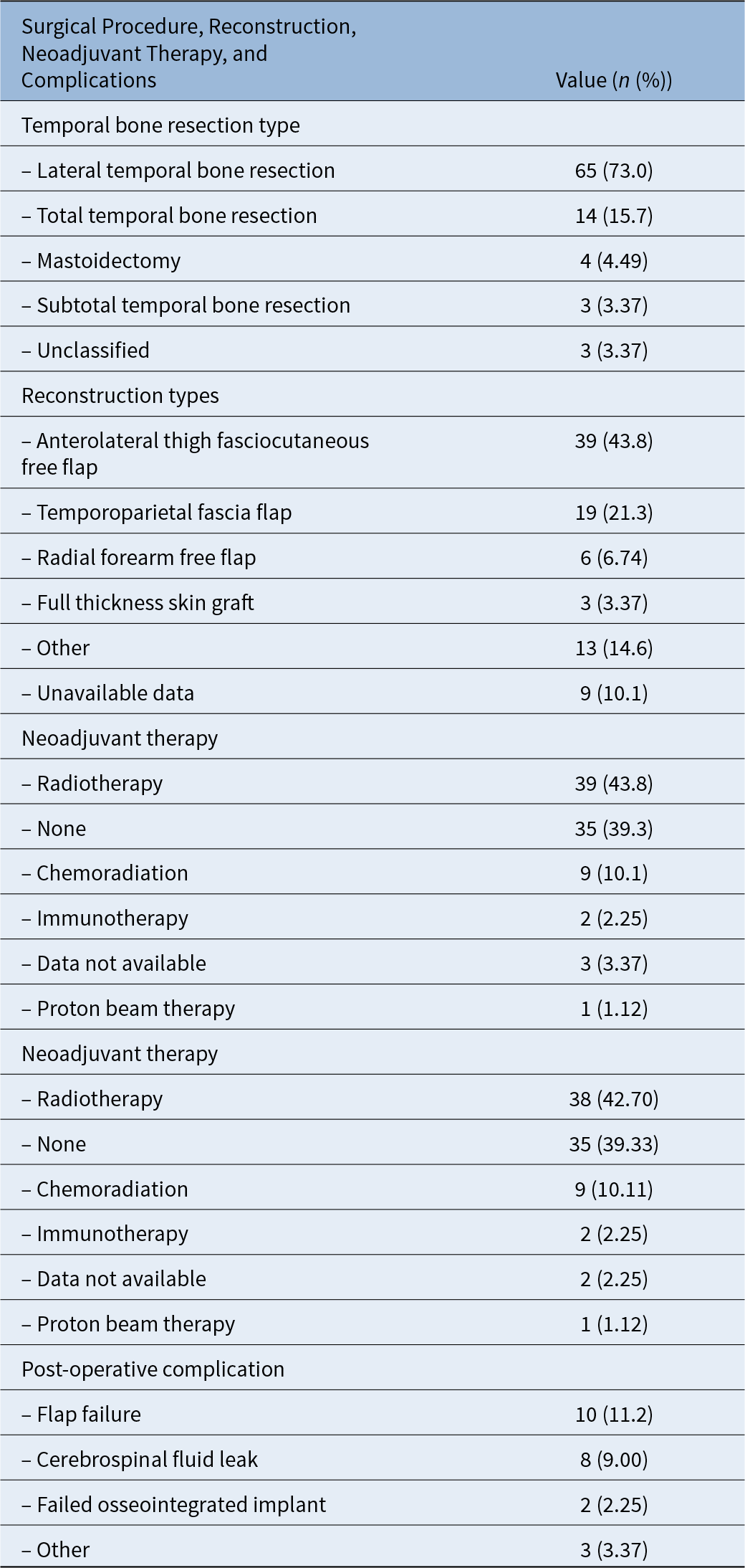
Lateral temporal bone resection was the most frequent surgical approach (n = 65, 73.0 per cent), followed by total temporal bone resection in 14 patients (15.7 per cent) (Table 2). Other types of resections, such as mastoidectomy and subtotal temporal bone resection, each accounted for less than 5 per cent of the cases. Reconstruction details were unavailable for 9 patients (10.1 per cent). Among those with available data, 39 patients (43.8 per cent) underwent an anterolateral thigh free flap, 19 patients (21.3 per cent) underwent reconstruction using a temporoparietal fascia flap and 6 patients (6.74 per cent) underwent a radial forearm free flap. Other reconstruction methods, such as local random pattern rotation flap and abdominal fat graft, each represented less than 4 per cent of cases (Table 2).
Summary of surgical procedures, reconstruction methods, neoadjuvant therapy and complications in temporal bone resection

Forty-nine patients (55.1 per cent) received adjuvant therapy and 39 patients (43.8 per cent) underwent radiotherapy, which was the most common adjuvant therapy. Also, 35 patients (39.3 per cent) received no adjuvant therapy, 9 patients (10.1 per cent) underwent chemoradiotherapy and 2 patients (2.25 per cent) received immunotherapy alone. Data on adjuvant therapy were unavailable for 3 patients (3.37 per cent) (Table 2).
Complication
Twenty-three patients (25.8 per cent) experienced some form of surgical complication, ranging from low harm (n = 1, 1.12 per cent of total cohort) to moderate harm (n = 14, 60.9 per cent) to severe harm (n = 8, 9.0 per cent) as defined by the NHS England National Patient Safety Grades of Harm.11 The most common complications were flap failure, which occurred in 10 cases, and cerebrospinal fluid leak, which occurred in 8 cases (Table 2). Most complications occurred either immediately (within 0 months) or very soon after surgery. Notably, the failed osseointegrated implants had the longest mean time to complication, averaging eight months post surgery. Furthermore, no cases of osteoradionecrosis were observed in patients who underwent radiotherapy. Nineteen patients (21.3 per cent) developed new post-operative facial paralysis that persisted long term, requiring facial reanimation treatment by the plastics team and physiotherapy.
Post-operative survival
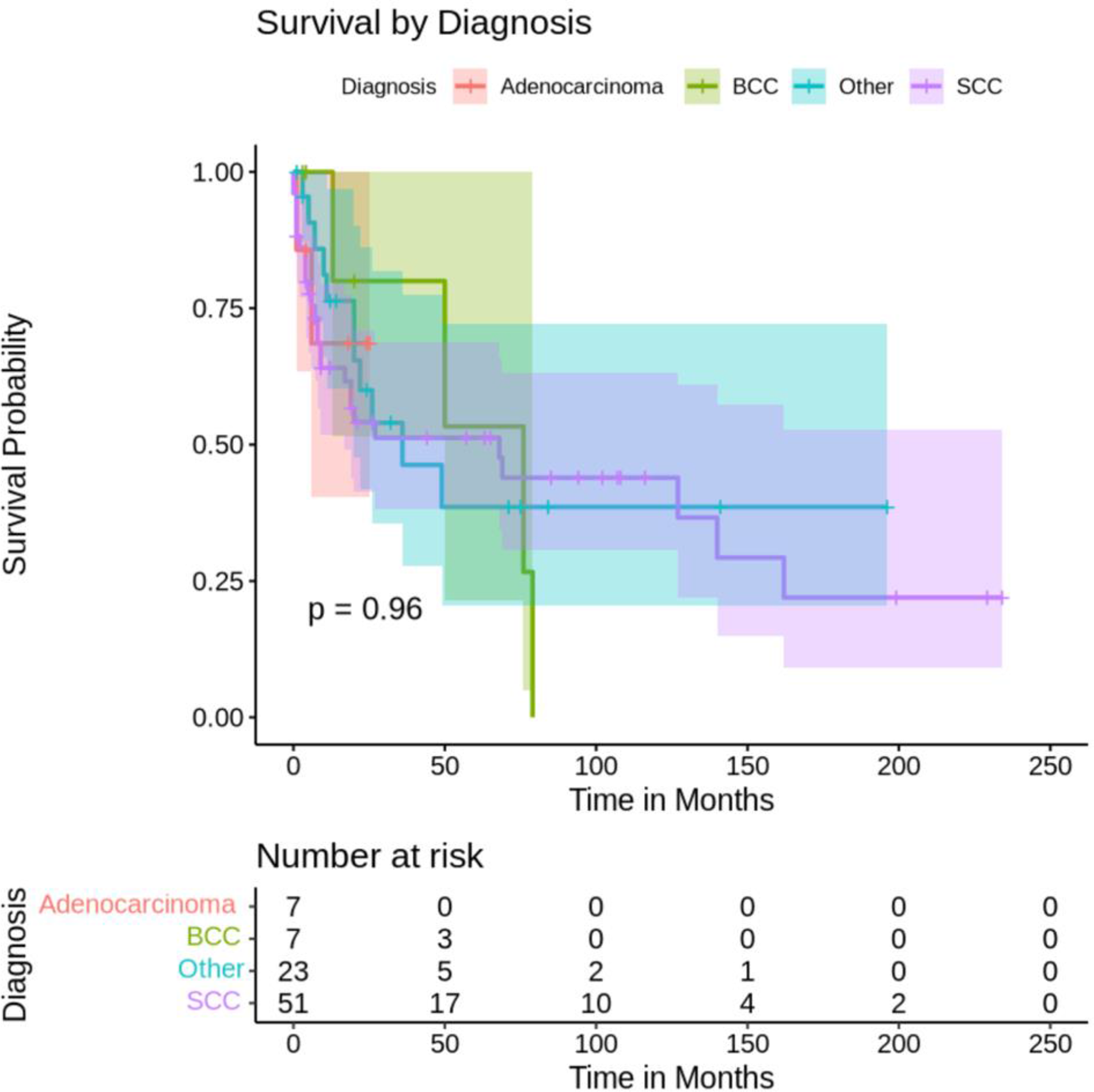
The follow-up duration for the patients had a mean of 38.6 months and a median of 19 months, with a range from 0 to 234 months. The median survival times varied among the different diagnoses (Figure 2). Squamous cell carcinoma had the longest median survival time of 78 months, while adenoid cystic carcinoma had a median survival time of 14 months. There was no statistically significant difference in survival between the diagnosis groups, partially because of the small number of patients with rare tumours (p = 0.9).
Kaplan–Meier survival curve by diagnosis: post-surgery survival probability over time (months). BCC = basal cell carcinoma; SCC = squamous cell carcinoma.

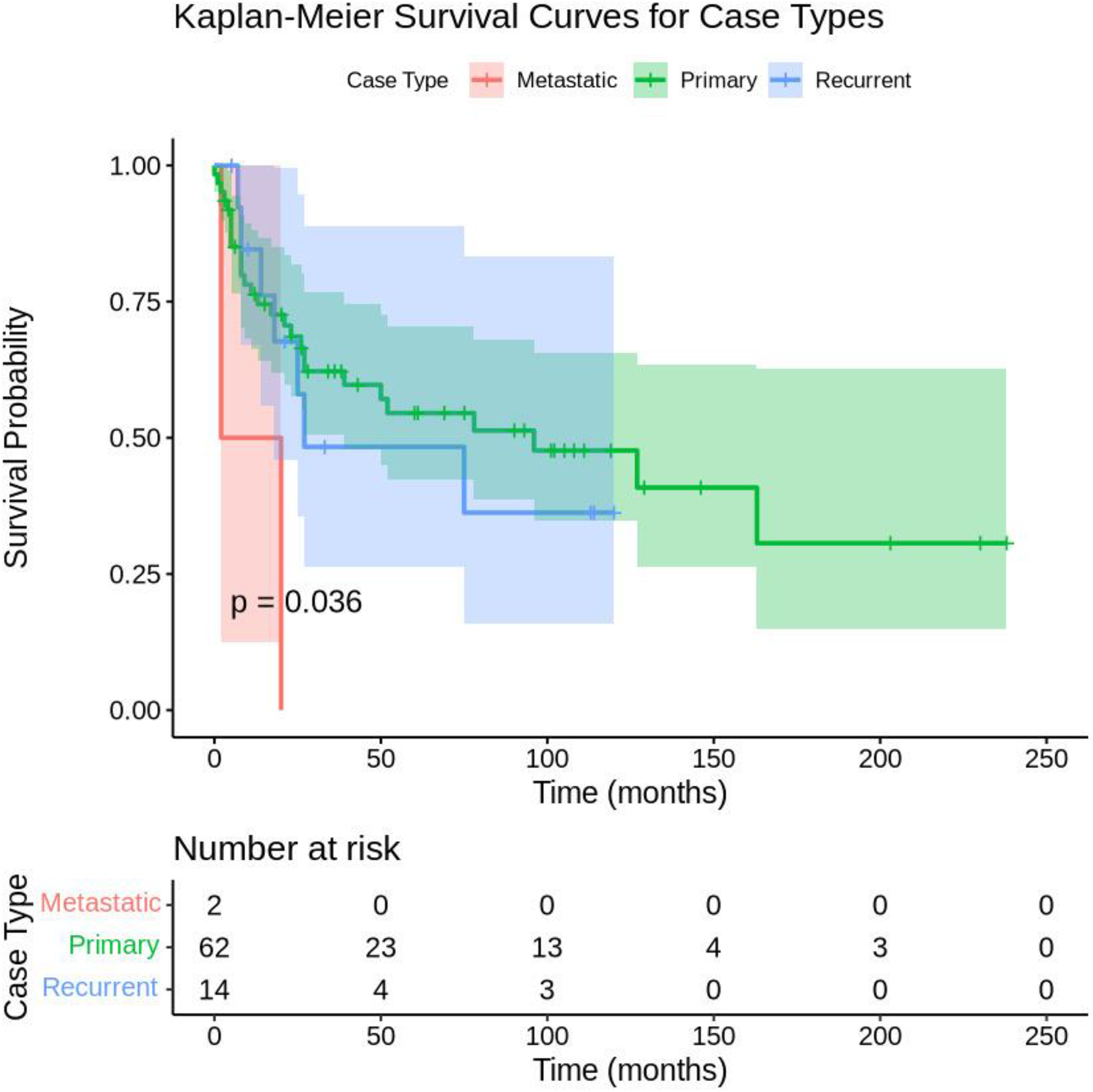
In the study, there were 3 metastatic cases with a median survival of 9 months, 70 primary cases with a median survival of 69 months and 16 recurrence cases with a median survival of 50 months (Figure 3). An observed trend indicates a difference in survival between the case types; however, it does not meet the threshold for statistical significance (p = 0.0684). For metastatic cases, more deaths were observed (n = 3) than expected (n = 0.91), which aligns with the generally poorer prognosis for metastatic disease. In primary cases, the number of observed deaths (n = 33) was slightly fewer than expected (n = 35.94). For recurrence cases, the number of observed deaths (n = 8) was close to the expected number (n = 7.15).
Kaplan–Meier survival curve by case type (primary, recurrence, metastatic): post-surgery survival probability over time (in months).

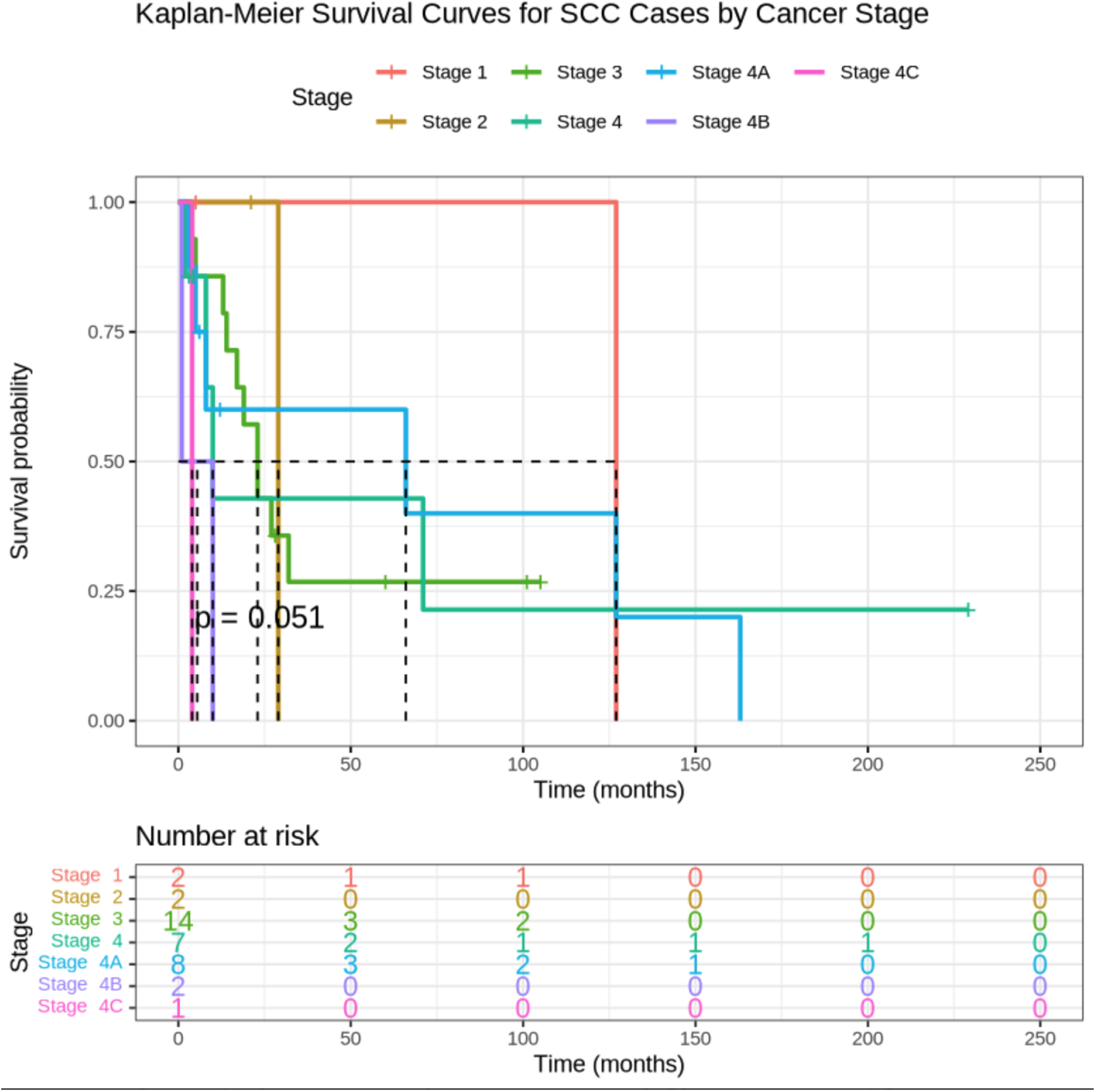
The association between cancer stage and survival outcomes was analysed separately for each histological subtype, reflecting differences in disease biology. For SCC, the most common subtype in our cohort, survival probability declines with increasing stage, with stage 3 demonstrating moderate survival and stage 4, including its subgroups, exhibiting substantially poorer outcomes (Figure 4). Estimates for very small groups (stages 1, 2, 4B, 4C) are limited by minimal sample sizes, resulting in reduced reliability. Lymph node status did not have a statistically significant impact on survival (Supplementary Figure S1).
Kaplan–Meier survival curve by stage of squamous cell cancer: post-surgery survival probability over time (in months).

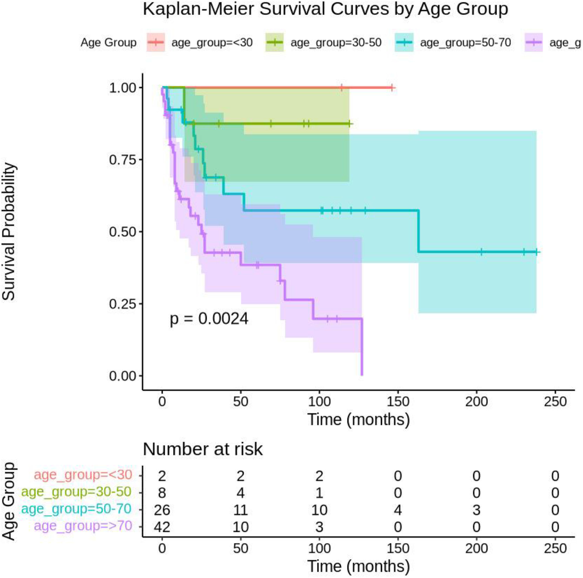
Age at time of surgery is significantly associated with survival outcomes (p < 0.001) (Figure 5). Younger age groups, particularly those under 30 years and between 30 and 50 years, have better survival probabilities compared with older age groups, specifically those aged 50–70 years and over 70 years. The positive coefficient for age (0.05149) indicates that as age increases, the hazard (risk of death) also increases. Specifically, for each year increase in age, the hazard increases by approximately 5.3 per cent.
Kaplan–Meier survival curve by age group: post-surgery survival probability over time (in months).

Discussion
The lateral skull base is a complex and anatomically challenging region, characterised by significant anatomical variability and diverse cancer growth patterns that can develop in this area.Reference Zanoletti, Marioni, Nicolai and Mazzoni12 Our understanding of the treatment results and survival outcomes following temporal bone resection is still limited and continues to evolve because of the infrequency and diversity of malignancies involving this region, whether they are primary, recurrent or metastatic.Reference Morris, Mehra, Shah, Bilsky, Selesnick and Kraus13
In this case series of 89 patients, the majority were male (69.7 per cent), with a mean age of 67.2 years. The mean follow-up duration was 38.6 months. Squamous cell carcinoma was the most common diagnosis, accounting for 58.4 per cent of cases, while primary cases made up 78.7 per cent of the cohort. Lateral temporal bone resection was the most frequent surgical approach, performed in 73.0 per cent of patients, and radiotherapy was the most common adjuvant therapy, administered to 43.8 per cent of patients. No cases of osteoradionecrosis were observed in patients who underwent radiotherapy in our case series. However, it should be noted that our case series only included patients with follow-up periods of less than 234 months and there are reports indicating that osteoradionecrosis can occur more than 10 years post radiotherapy.Reference Lovin, Hernandez, Elms, Choi, Lindquist and Moreno14
Furthermore, surgical complications were observed in 25.8 per cent of cases, with flap failure following reconstruction being the most frequent issue. This finding is consistent with previously published literature. For example, Bertelsen et al. reported that 25 per cent of patients who underwent lateral temporal bone resection experienced complications, most commonly cerebrospinal fluid (CSF) leaks, pneumonia and meningitis.Reference Bertelsen, Simsolo, Maceri, Sinha and Kokot15, Reference Ordoyne, Alvarez, Borne, Fabian, Adilbay and Kandula16 Furthermore, Gidley et al. reported that in their cohort of 157 patients who underwent temporal bone surgery for cancer, 31.2 per cent experienced at least one complication within the first 30 days post surgery, with wound infection and haematoma being the most common.Reference Gidley, Thompson, Roberts, DeMonte and Hanna17 Similarly, Hanasono et al. found that among 39 local or regional pedicled flap reconstructions and 211 free flap reconstructions, complications occurred in 29.6 per cent of patients, with seroma, wound infection and haematoma being the most prevalent.Reference Hanasono, Silva, Skoracki, Gidley, DeMonte and Hanna18
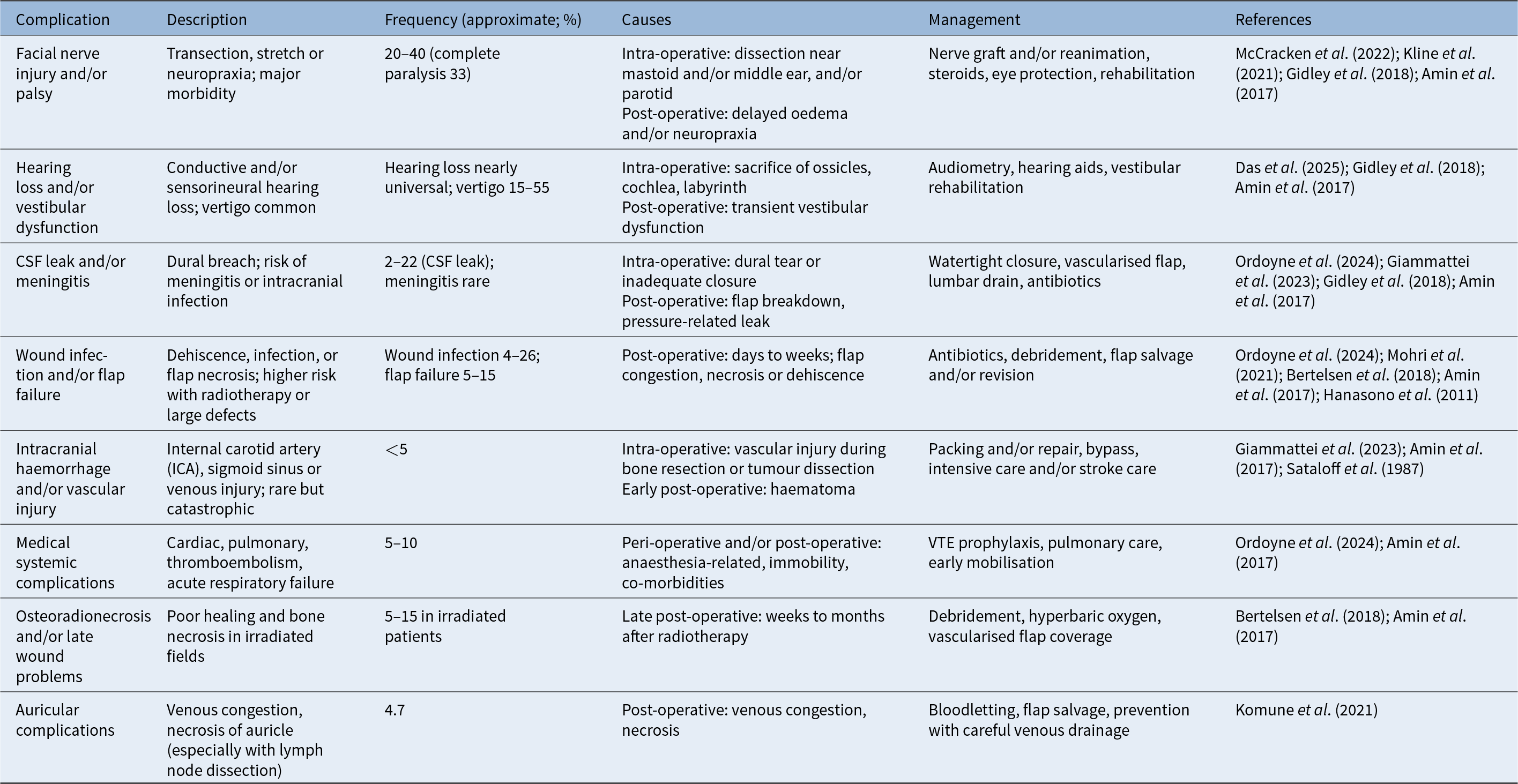
Other research reported acute post-operative complications such as pulmonary oedema, thrombosis, embolism and myocardial infarction, while longer-term issues within the first month post surgery included pulmonary embolism and multiorgan failure.Reference Ordoyne, Alvarez, Borne, Fabian, Adilbay and Kandula16, Reference Aslıer, Doğan, İkiz, Sarıoğlu, Akman and Güneri19 A notable 9 per cent of our patient cohort experienced complications classified as severe, all of which were CSF leaks. According to the NHS complication severity table,11 these were deemed severe as a result of the requirement for immediate life-saving clinical intervention. The most common surgical complications of temporal bone resection are summarised in Table 3.Reference Gidley and DeMonte6, Reference Bertelsen, Simsolo, Maceri, Sinha and Kokot15, Reference Ordoyne, Alvarez, Borne, Fabian, Adilbay and Kandula16, Reference Hanasono, Silva, Skoracki, Gidley, DeMonte and Hanna18, Reference Amin, Zayed, El-Dessouky, Said, El-Din and El-Anwar20–Reference Kline, Bhatnagar, Eisenman and Taylor27
Summary of common complications following temporal bone resection

CSF = cerebrospinal fluid
A recent nationwide analysis of 277 patients undergoing temporal bone resection with neck dissection in the USA included a mixed cohort with various pathologies and surgical types, including lateral, subtotal and total resections.Reference Ordoyne, Alvarez, Borne, Fabian, Adilbay and Kandula16 This study demonstrated lower overall complication rates but identified key risk factors for adverse outcomes. Anaemia was strongly associated with surgical complications, while dementia, congestive heart failure, prior stroke and liver disease significantly increased the risk of medical complications, prolonged hospitalisation, discharge to non-home facilities and higher costs. Together, these findings highlight that while temporal bone resection is generally safe, patients with pre-existing co-morbidities represent a vulnerable subgroup requiring tailored peri-operative management.
The overall 5-year survival rate of our cohort was 50 per cent and we analysed the survival times further based on different determinants. Firstly, the 5-year survival of the SCC cohort was 51.3 per cent, which is similar to previously published figures.Reference Brenet, Atallah, Guerlain, Moya-Plana, Verillaud and Kania28–Reference Masterson, Rouhani, Donnelly, Tysome, Patel and Jefferies30 Whilst the survival times of other histological diagnosis groups were different, these differences were not statistically significant. This may be attributed to the small sample sizes in certain groups, especially for adenoid cystic carcinoma. However, a cohort study by Kline et al., involving 26 patients with primary or secondary malignancies of the temporal bone, also found no significant differences in outcomes when comparing cutaneous versus non-cutaneous tumour types or their site of origin.Reference Kline, Bhatnagar, Eisenman and Taylor27 They suggest that tumours in the temporal region may exhibit comparable behaviour at advanced stages, and the malignancy type may not be a significant determinant of a patient's prognosis.Reference Kline, Bhatnagar, Eisenman and Taylor27 Further studies involving larger cohorts, extended follow-up periods and control for confounding variables will be valuable.
Moreover, we observed statistically significant differences in survival among primary, recurrent and metastatic cases, with primary cases showing the best prognosis, followed by recurrent and metastatic cases (Figure 3). This outcome is not surprising because recurrent and metastatic cases typically involve more aggressive cancer cells as a result of the principles of natural selection. Our findings are consistent with a meta-analysis of 39 case series and 12 case reports, which demonstrated that, on multivariate analysis, recurrent disease was independently associated with worse overall survival (p < 0.001).Reference McCracken, Pai, Cabrera, Johnson, Tamaki and Gidley21 The small sample size for metastatic cases and wide confidence intervals for both metastatic and recurrent groups indicate considerable uncertainty in these two survival estimates. It would be valuable for future studies to compare the survival outcomes of patients with recurrent primary or secondary temporal bone tumours that have metastasised from a distant site with the same histological cell.
Similarly, cancer stage for SCC was significantly associated with survival outcomes, showing a clear trend of worse survival at higher stages, particularly Stage 4. This aligns with findings from Sioufi et al., who reported that patients with early-stage temporal bone SCC (T1 and/or T2) had markedly higher 5-year overall survival rates (81.9 per cent) compared with those with advanced-stage disease (T3 and/or T4), whose 5-year survival dropped to 47.5 per cent (p < 0.0001).Reference Sioufi, Haynes, Gidley, Maniakas, Roberts and Nader29 Grouping into main stages improved our ability to identify trends, although small sample sizes in the lower stages still presented some limitations to interpretation due to the wide confidence intervals.
Furthermore, lymph node status demonstrated a statistically insignificant impact on survival trends in our cohort. Although T classification is generally recognised as an important prognostic indicator, the predictive value of lymph node involvement and surgical margin status remains debated.Reference Sioufi, Haynes, Gidley, Maniakas, Roberts and Nader29 Evidence from studies regarding the effect of nodal disease on outcomes is inconsistent. Investigations by Masterson et al.,Reference Masterson, Rouhani, Donnelly, Tysome, Patel and Jefferies30 Zanoletti et al.Reference Zanoletti, Marioni, Stritoni, Lionello, Giacomelli and Martini31 and Lobo et al.Reference Lobo, Llorente and Suárez32 have reported poorer survival among patients with nodal involvement, suggesting that it may serve as a negative prognostic factor. On the other hand, other literature shows that nodal disease is not consistently associated with survival outcomes.Reference Sioufi, Haynes, Gidley, Maniakas, Roberts and Nader29, Reference Gidley, Roberts and Sturgis33–Reference Xie, Zhang and Dai36 Further research is therefore needed to clarify the role of lymph node status in prognostication across different cancers of the lateral temporal bone.
Lastly, age was found to be a statistically significant predictor of survival outcomes, a finding consistent with a multicentre, prospective cohort study using the REFCOR national database in France that included 103 adult patients diagnosed with carcinomas of the external auditory canal between 2000 and 2016.Reference Brenet, Atallah, Guerlain, Moya-Plana, Verillaud and Kania28 In that study, patients over 65 years had more than twice the risk of death compared with younger patients, independent of tumour stage or treatment modality. Notably, age did not significantly affect event-free survival, indicating that older patients were not necessarily more prone to recurrence, but once complications or disease progression occurred, their overall survival was disproportionately reduced. This association can be explained by the higher prevalence of co-morbidities and reduced physiological reserves in older patients, which can limit recovery from highly invasive surgical procedures, chemotherapy or radiotherapy. Together, these findings underscore the importance of considering age not only when assessing prognosis in patients undergoing temporal bone resection but also when planning treatment strategies and post-operative rehabilitation. The significant impact of age on survival further highlights its role as a critical covariate in future analyses, particularly when evaluating the influence of other prognostic factors.
This study was principally constrained by the relatively modest sample size, the limited follow-up period and its single-centre setting. Further studies involving larger patient cohorts, ideally controlled for confounding variables, would be highly beneficial. Additional determinants of survival that could be examined include time to diagnosis, surgical techniques, the impact of ethnicity and the varying guidelines for adjuvant chemoradiotherapy, given the absence of a standardised international protocol for temporal bone cancers.
Conclusion
This case series contributes to the growing body of evidence on the outcomes of temporal bone resection for malignancy, highlighting the complexity and high morbidity associated with this rare group of cancers. Despite the limitations of a single-centre design and a modest sample size, our findings reinforce the prognostic significance of tumour origin, stage and patient age. These insights underscore the need for larger, multicentre studies with extended follow up to better inform clinical decision-making, improve patient selection and ultimately guide the development of more standardised treatment protocols for lateral skull base malignancies.
Supplementary material
The supplementary material for this article can be found at https://doi.org/10.1017/S0022215125103940.
Competing interests
None declared.