INTRODUCTION
Since Corliss (Reference Corliss1968) emphasized the significance of ontogenetic data in reconstructing protozoan phylogeny, many studies have increased our knowledge about ciliates evolution and phylogeny (Borror, Reference Borror1979; Wicklow, Reference Wicklow1981). Within the Euplotida Small & Lynn, 1985, studies of cortical development have provided critical evidence in determining homologous structures and morphogenetic patterns (Hill, Reference Hill1979, Reference Hill1981; Song, Reference Song1996; Hu, Reference Hu2008). Among euplotids, the Certesiidae Borror & Hill, Reference Borror and Hill1995 is the only monospecific family that comprises one genus and the sole species, i.e. Certesia and C. quadrinucleata Fabre-Domergue, Reference Fabre-Domergue1885 respectively (Fabre-Domergue, Reference Fabre-Domergue1885; Corliss, Reference Corliss1979; Borror & Hill, Reference Borror and Hill1995; Lynn, Reference Lynn2008). Since its original report, Certesia quadrinucleata has been studied with more or less details of live morphology (Bütschli, Reference Bütschli and Bronn1889; Sauerbrey, Reference Sauerbrey1928; Kahl, Reference Kahl1932). Vacelet (Reference Vacelet1960) reported one other species, C. ovata, which was subsequently considered as a junior synonym of C. quadrinucleata (Curds & Wu, Reference Curds and Wu1983; Wicklow, Reference Wicklow1983). In terms of infraciliature, data are only available for two populations (Wicklow, Reference Wicklow1983; Lin & Song, Reference Lin and Song2004). The morphogenesis of this species has been studied once based on one USA population (Wicklow, Reference Wicklow1983). Therefore, this species remained insufficiently investigated with respect to intraspecific morphological and developmental variance.
Prior to the present study, this species had never been found in Japan, thus a new record is herein presented, alongside a morphological and morphogenetic characterization of the Japanese population of C. quadrinucleata.
MATERIALS AND METHODS
Certesia quadrinucleata was collected from seawater in the Mie Port of Nagasaki during the period from October 2003 to February 2004 using the polyurethane foam unit method. It was found with low abundance. Water temperature was about 15–24°C, pH 8.1–8.2, salinity 33–35‰ and dissolved oxygen ~5.8–8.1 mg/l. After isolation, observations on living cells were carried out with bright field and differential interference contrast microscopy at 100–1250 ×. The protargol impregnation method according to Wilbert (Reference Wilbert1975) was used to reveal the infraciliature and nuclei. Measurements were carried out with an ocular micrometer. Drawings of impregnated specimens were conducted with the help of a camera lucida under 1250× magnification. In drawings of the progress of morphogenesis, parental cirri are shown as outlines and newly formed cirri are shown as filled outlines. Terminology and systematics are mainly according to Lynn (Reference Lynn2008). Two voucher slides (Nos. MP2003122201 and MP2004021201) have been deposited in the Laboratory of Protozoology, Ocean University of China.
RESULTS
Morphological characterization of Certesia quadrinucleata from Japan (Table 1; Figures 1–3)
MORPHOLOGY DURING INTERPHASE
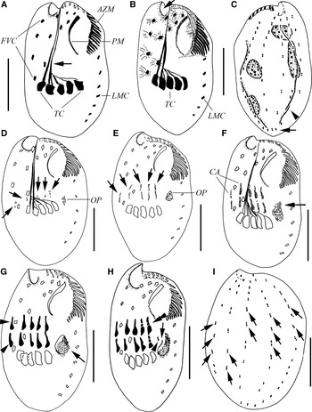
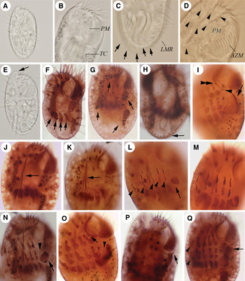
Size 45–75 × 20–40 µm in vivo, body irregular oval-shaped (Figure 3A); anterior end obliquely truncated from right to left with an angular protrusion on the anterior right (Figure 3E, arrow), posterior end rounded. Left side straight or slightly concave near mid-body while right one conspicuously convex. Dorsoventrally ~2:1 flattened; dorsal side convex, sometimes two ribs are visible, one on each side (Figure 1C, arrowhead); ventral side concave in median where cirri arise with one rib near each side. Pellicle rigid; no cortical granules observed. Cytoplasm colourless or brownish (owing to the presence of ingested diatoms). Constantly four macronuclear nodules, two along the right side and two along the left side of the cell; the posterior right nodule usually longer than the other three (Figures 1C; 3G, arrows); micronuclei not observed.
Morphology (A–C) and morphogenesis (D–I) of Certesia quadrinucleata after protargol impregnation (A, B, D–H, ventral view; C, I, dorsal view): (A) a representative individual with 11 frontoventral cirri, arrow denotes two well-developed connecting fibres of the transverse cirri; (B & C) the same specimen with 6 left marginal cirri, arrow in B shows anterior right protrusion, while in C marks the posterior end of leftmost dorsal kinety curving to right, arrowheads in B show fibres radiating from the base of cirri, arrowhead in C indicates dorsal rib; (D) very early stage, showing the formation of opisthe's oral primordium and cirral anlagen (arrows); (E) early stage, 5 streaks of cirral anlagen are formed and begin to divide into two parts (arrows), and oral primordium becomes slightly enlarged; (F) early stage, cirral anlagen continue to become wider and longer, oral primordium develops further (arrow); (G) early-middle stage, each streak becomes thicker (arrowheads), kinetosomes in oral primordium begin to reorganize in the subsurface pouch (arrow); (H) early-middle stage, showing fragmentation of cirral streaks starting from left to right (double-arrowhead), independent occurrence of the anlage for the leftmost frontoventral cirrus (arrowhead), separation of undulating membrane anlage from oral primordium (arrow); (I) middle stage, showing kinetosome proliferation within the parental dorsal kineties (arrows). AZM, adoral zone of membranelles; CA, cirral anlagen; FVC, frontoventral cirri; OP, oral primordium; PM, paroral membrane; LMC, left marginal cirri; TC, transverse cirri. Scale bar: 20 µm.

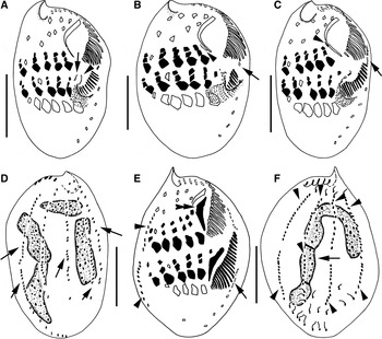
Morphogenesis of Certesia quadrinucleata after protargol impregnation (A–C, E, ventral view; D, F, dorsal view): (A) middle stage, showing further development of anlagen for the leftmost frontoventral cirrus (arrow) and paroral membrane (arrowhead) in the opisthe; (B) middle-late stage, denoting the independent occurrence of anlage for the leftmost cirrus (arrowhead) and dedifferentiation of the anterior-most left marginal cirrus (arrow) in the proter; (C, D) middle-late stage, the same specimen, showing the formation of the leftmost frontoventral cirrus (arrowhead) and the formation of left marginal cirral anlagen in the proter (arrow in C) and dorsal kineties anlagen within old structures (arrows in D); (E, F) late stage, the same cell, showing the independent formation of paroral membrane anlage in the proter (double-arrowhead) and occurrence of left marginal cirral anlagen in the opisthe (arrow in E), development of dorsal kineties anlagen (arrowheads) and the fused macronucleus (arrow in F). Scale bar: 20 µm.

Photomicrographs of Certesia quadrinucleata from life (A–E) and after protargol impregnation (F–Q); ventral (A–G & I–Q) and dorsal (H) views: (A) a representative cell, showing outline and cirral arrangement; (B, D) anterior part, showing adoral zone of membranelles, paroral membrane, frontoventral (arrowheads) and strong transverse cirri; (C) posterior portion, showing transverse cirri (arrows) and left marginal cirral row; (E) a cell denoting a conspicuous anterior right protrusion (arrow); (F) a specimen with 10 frontoventral and 4 transverse (arrows) cirri; (G) macronucleus comprising 4 nodules (arrows); (H) five dorsal kineties, arrow marks the posterior end of leftmost dorsal kinety curving to right; (I) middle divider, showing anlage for the leftmost frontoventral cirrus (double-arrowhead), dedifferentiation of paroral membrane at its posterior end (arrowhead), new adoral zone starting to curve to right (arrow); (J, K) specimens with 11 and 10 frontoventral cirri respectively, arrow marks well-developed connecting fibres extending to the anterior end of the body; (L) early divider, showing cirral anlagen originated epiapokinetally (arrowheads) and oral primordium occurred hypoapokinetally in a subsurface pouch (arrow); (M) middle divider, showing two sets of cirral anlagen; (N) middle divider, showing de novo formation of the anlage of the leftmost frontoventral cirrus (arrowhead) and new membranelles beginning to organize in the opisthe's oral primordium (arrow); (O) middle divider, showing de novo formation of the anlage of the leftmost frontoventral cirrus in the proter (arrow) and the anlage of paroral membrane in the opisthe (arrowhead); (P) late divider, showing left marginal cirral anlage in the proter (arrow); (Q) late divider, showing each streak generating 3 new cirri (arrowheads) and newly formed membranelles emerging from the subsurface pouch (arrow).

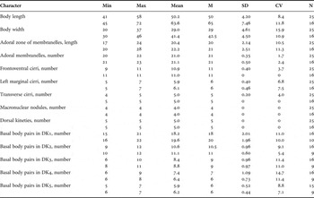
Morphometric data of Certesia qudrinucleata (upper rows, December population; lower rows, February population). All data based on protargol impregnated specimens.

CV, coefficient of variation in %; DK, dorsal kinety; Max, maximum; Mean, arithmetic mean; M, median; Min, minimum; N, number of specimens examined; SD, standard deviation; SE, standard error of the mean. Measurements in μm.
Buccal field wide and about 40% to 50% of body length (Figure 3A, E). Adoral zone of membranelles (AZM) consisting of 20–23 membranelles, cilia of which are 13 µm long in anterior portion. Prominent paroral membrane (PM) arched, base composed of many oblique kinetosomal arrays (Figures 1A, B; 3B, D & F).
Nine to eleven frontoventral cirri, about 16 µm long, arranged in two curved rows (Figures 1A; 3A, F, J, K; 3D, arrowheads); short fibres radiating from the base of cirri (Figure 1B). Usually five (Figures 1A, B; 3B, J, K), rarely four (Figure 3F, arrows) strong transverse cirri, positioned at posterior 1/3 of cell, with cilia about 35 µm long in vivo and projecting beyond the posterior end of the cell (Figures 1 & 7), the rightmost two with well-developed connecting fibres extending to the anterior end of the body, while the other three with fine fibres joining with the thick fibre (Figures 1A, 3J, K, arrow). Five to seven left marginal cirri in a row (Figures 1A, B, 3C), cilia 14 to 15 µm long. Constantly five dorsal kineties, of which the leftmost one extends over entire length of body with the posterior end curved to the right side along posterior margin, and others are posteriorly shortened (Figures 1C; 3H, arrow).
CORTICAL MORPHOGENESIS
Cortical morphogenesis commences by de novo development of the oral primordium (OP) of the opisthe as a dense cluster of kinetosomes anterior left of left-most transverse cirrus and to the right of the second anterior left marginal cirrus (Figures 1D, E; 3L, arrow). After initial OP formation, there is rapid kinetosomal proliferation (Figure 1F, arrow) until it grows into a subsurface pouch (Figures 1G, arrow; 3M; 3N, arrow). As the pouch enlarges, kinetosomes begin to align into the new membranelles of the opisthe (Figures 1H, 2A); membranelles alignment starts along the right side at the anterior end and proceeds towards left and posteriorly. As this continues, the membranelles zone twists into a descending spiral (Figure 3I, arrow). Kinetosomal region remains at the base of the spiral while anterior membranelles emerge from the pouch onto the cell surface (Figures 1H, 2A; 3O; 3Q, arrow). At this time, undulating membrane anlage (anlage I) arises from the right side of the OP as a long series of kinetosomes (Figures 1H, arrow; 2A, 3O, arrowhead). These developmental processes are not completed until just before the cell completes division (Figure 2B, C, E).
In the proter, the posterior portion of the parental paroral membrane becomes dedifferentiated and kinetosomes are incorporated in new anlage (Figures 1H, 3O; 3I, arrowhead), which finally develops into new paroral membrane to replace the old one (Figure 2E, double-arrowhead). The parental adoral zone of membranelles is inherited by the proter.
At the same time as the formation of the OP, the cirral anlagen of frontoventral and transverse cirri begin to form to the right of the buccal cavity as five short longitudinal series of kinetosomes. Anlagen II, III and IV appear anterior to the three leftmost transverse cirri, anlage V below and beside the cirrus V/2, and anlage VI close to and to the right of parental cirrus VI/2 (Figure 1D, arrows). With more kinetosomes joining, the cirral anlagen begin to elongate as five streaks and soon divide into two sets thus forming the cirral anlagen of the proter and the opisthe (Figures 1E, arrows; 3L, arrowheads). Parental cirri remain intact during the development of these cirral anlagen. As the cirral anlagen lengthen kinetosomes proliferate laterally, first near the middle and posterior end of each streak, then progressively toward the anterior end (Figures 1F; G, arrowheads; 3M, N). Subsequently streaks start to segment in a posterior to anterior direction and from left to right and finally form three new cirri each (Figures 1H, double-arrowhead; 2A–C, E; 3O; 3Q, arrowheads).
At the beginning of segmentation of the cirral streaks in the opisthe, an anlage appears de novo between the cirral anlagen and the undulating membrane anlage on the cell surface (Figures 1H, arrowhead; 2A, arrow; 3N, arrowhead), and will form the leftmost frontoventral cirrus (Figures 2B, C, E; 3I, O, Q).
When the cirral streaks finish the segmentation, a small anlage is formed de novo beside the posterior portion of the parental paroral membrane in the proter, which will contribute the leftmost frontoventral cirrus of the proter (Figures 2B, C, arrowhead; 3I, double-arrowhead; 3O, arrow); meanwhile, the anterior-most left marginal cirrus becomes dedifferentiated (Figure 2B, arrow). As kinetosomes proliferate, it lengthens and forms the anlage of left marginal cirri of the proter (Figure 2C, arrow). Subsequently, the anlage of left marginal cirri forms beside the next posterior marginal cirrus in the opisthe (Figure 2E, arrow). From each anlage differentiate five to seven marginal cirri.
At the time of cirral formation, kinetosome proliferation appears within the parental dorsal kineties at mid-body of the cell and then extends to both directions (Figures 1I, arrows; 2D, arrows; 2E, F, arrowheads).
In later divider, the fusion of macronuclear nodules is visible (Figure 2F, arrow).
DISCUSSION
Based on data available, C. quadrinucleata is a cosmopolitan marine species. The Japanese populations correspond very well with the original and subsequent populations in possessing an irregularly ovoid body shape, a prominent paroral membrane and usually five transverse cirri, eleven scattered frontoventral cirri, one row of left marginal cirri and four macronuclear nodules. In two populations, the February population is a little larger than the December one and has more stable morphometric data (Table 1). Intraspecific morphological variance, nevertheless, lies only in cell size and the number of adoral membranelles (Fabre-Domergue, Reference Fabre-Domergue1885; Kahl, Reference Kahl1932; Curds & Wu, Reference Curds and Wu1983; Wicklow, Reference Wicklow1983; Lin & Song, Reference Lin and Song2004). The current populations are smaller (41–72 µm long versus 66–92 µm long in the China population, 75–80 µm in the France population, 70–100 µm in the Germany population) and have fewer adoral membranelles (~21 versus ~24 in the China population and 25–27 in the USA population). In terms of nuclear apparatus, the number of macronuclear nodules is constant among the Japanese populations and the USA population, while variable in the China population.
Morphogenesis in the genus Certesia has previously been reported in a population of C. quadrinucleata from the USA (Wicklow, Reference Wicklow1983). The morphogenetic process in the Japanese population reported here is consistent with that of the USA population in the sense that: (1) the opisthe's oral primordium develops de novo in a subsurface pouch; (2) the parental AZM is completely inherited by the proter; (3) the old frontoventral cirri do not donate kinetosomes to any developing cirral anlagen; (4) the cirral anlagen of the frontoventral and transverse cirri are formed from the division of a primary primordium originated de novo; (5) the anlagen of left marginal cirri and dorsal kineties develop within parental structures; (6) the parental paroral membrane is replaced by the new structure derived from a newly formed anlage in the proter; and (7) the cirral anlagen II–VI differentiate into cirri in the same pattern of 3:3:3:3:3 (counted from left to right). These common characters support the conspecifity of these two forms. With reference to the origin of the leftmost frontoventral cirrus in both dividers, however, there is a disagreement. Based on the current observations on the Japanese populations, the anlage is formed de novo and completely independent from the oral primordium. This phenomenon was also recorded in species of Diophrys and Aspidisca (Hill, Reference Hill1979, Reference Hill1981; Hu, Reference Hu2008). But it is different from the observations on the USA population by Wicklow (Reference Wicklow1983). He stated that the anlage is derived from the anlage of paroral membrane, which, however, is not clearly illustrated (see Wicklow, Reference Wicklow1983, p. 264). Actually, it is too difficult to ascertain the origin of the leftmost frontoventral cirrus in the case of lacking critical morphogenetic stages, so one possibility may exist that Wicklow made a wrong interpretation.
Compared with several other euplotid genera, whose ontogeny are known (Hill, Reference Hill1979, Reference Hill1981; Song & Packroff, Reference Song and Packroff1993; Song, Reference Song1996; Hu & Song, Reference Hu and Song2003; Song et al., Reference Song, Wilbert, Chen and Shi2004; Hu, Reference Hu2008; Shao et al., Reference Shao, Ma, Gao, Al-Rasheid and Song2010), Certersia differs in the following respects: (1) the segmentation pattern of anlagen II–VI is 3:3:3:3:3 in the former (versus 3:3:3:3(2):2(1) in Euplotes, 3:3:2:2:1(2) in Aspidisca, 3:2:2:3:1 in Diophrys, 3:3:2:2:1(2) in Uronychia); (2) no caudal cirri are formed at the posterior end of dorsal kineties anlagen (caudal cirri formed in Euplotes, Diophrys and Uronychia); (3) marginal anlage is formed within the parental structure (versus de novo in Euplotes, Diophrys and Uronychia; no anlage formed in Aspidisca and Gastrocirrhus); (4) five cirral anlagen are present (versus more than five anlagen in Gastrocirrhus); and (5) no cirrus is formed at the anterior end of anlage of paroral membrane (versus one cirrus formed in Gastrocirrhus). These differences plus morphological characters are enough to support the separation of Certesiidae (Certesia as type genus) from Euplotidae (Euplotes as type genus), Aspidiscidae (Aspidisca as type genus), Gastrocirrhidae (Gastrocirrhus as type genus) or Uronychiidae (Uronychia as type genus) within the Euplotida.
ACKNOWLEDGEMENTS
This work was supported by the Natural Science Foundation of China (Project number 41176119) and a Marie Curie International Incoming Fellowship within the 7th European Community Framework Programme. I thank Dr Toshikazu Suzuki, Nagasaki University, Japan for his help in sampling.