INTRODUCTION
Acanthocheilonema reconditum (Grassi 1889) (Spirurida, Onchocercidae) is a vector-borne helminth of dogs with a global distribution (Otranto et al. Reference Otranto, Dantas-Torres, Brianti, Traversa, Petrić, Genchi and Capelli2013). Adult worms live in the subcutaneous tissues of infected dogs and gravid females lay thousands of microfilariae, which enter the bloodstream and remain there until ingestion by a competent vector (Grassi and Calandruccio, Reference Grassi and Calandruccio1890; Pennington and Phelps, Reference Pennington and Phelps1969; Farnell and Faulkner, Reference Farnell and Faulkner1978). Despite A. reconditum being either the sole (Mazzotti and Chabaud, Reference Mazzotti and Chabaud1962; Korkejian and Edeson, Reference Korkejian and Edeson1978; Minnaar and Krecek, Reference Minnaar and Krecek2001) or the most prevalent (Pennington and Phelps, Reference Pennington and Phelps1969; Pampiglione et al. Reference Pampiglione, Poglayen and Capelli1986; Saleh et al. Reference Saleh, Kirkpatrick, De Haseth and Lok1988; Ortega-Mora et al. Reference Ortega-Mora, Gomez-Bautista, Rojo-Vazquez, Rodenas and Guerrero1991; Aranda et al. Reference Aranda, Panyella, Eritja and Castella1998; Alves et al. Reference Alves, de Almeida Silva, Faustino, McCall, Supakonderj, Labarthe, Sanchez and Caires1999; Cringoli et al. Reference Cringoli, Rinaldi, Veneziano and Capelli2001; Giannetto et al. Reference Giannetto, Poglayen, Gaglio and Brianti2007) filarioid infesting dogs in some geographical areas (e.g. the Mediterranean Basin, Middle East, South Africa, South America and Oceania), information on its biology and life history is scarce. Different arthropod species have been suggested as competent vectors for this filarioid. Developing or infective forms of A. reconditum have indeed been found in the mallophagan lice Heterodoxus spiniger (Enderlein 1909) and Linognathus setosus (Olfers 1816) (Nelson, Reference Nelson1962; Pennington and Phelps, Reference Pennington and Phelps1969) and in the fleas Ctenocephalides canis (Dugès 1832), Ctenocephalides felis (Bouché 1835), Echidnophaga gallinacea (Westwood 1875), Pulex irritans (Linneo 1758) and Pulex simulans (Baker 1895) (Grassi and Calandruccio, Reference Grassi and Calandruccio1890; Calandruccio, Reference Calandruccio1892; Newton and Wright, Reference Newton and Wright1959; Pennington, Reference Pennington1971; Korkejian and Endeson, Reference Korkejian and Edeson1978; Brianti et al. Reference Brianti, Gaglio, Napoli, Giannetto, Dantas-Torres, Bain and Otranto2012). The cat flea, C. felis, is regarded as the main vector of this filarioid (Grassi and Calandruccio, Reference Grassi and Calandruccio1890; Newton and Wright, Reference Newton and Wright1959; Nelson, Reference Nelson1962; Brianti et al. Reference Brianti, Gaglio, Napoli, Giannetto, Dantas-Torres, Bain and Otranto2012). In this species Grassi and Calandruccio (Reference Grassi and Calandruccio1890) suggested that the larvae develop to the infective stage in fat body cells. However, scant information is available on the developmental time of A. reconditum larvae inside the flea, as well as on the route of infection. In fact, it is not yet clear if infective larvae are transmitted through the flea-bite or whether the dog acquires the infection by eating the infected arthropods (Lindemann and McCall, Reference Lindemann and McCall1984).
The flea C. felis, a widely distributed ectoparasite of cats and dogs, is a known biological vector of several pathogens (Hornok et al. Reference Hornok, Meli, Perreten, Farkas, Willi, Beugnet, Lutz and Hofmann-Lehmann2010; Eisen and Gage, Reference Eisen and Gage2012), including bacteria (e.g. Bartonella henselae, Rickettsia felis, Yersinia pestis) (Macaluso et al. Reference Macaluso, Pornwiroon, Popov and Foil2008; Hornok et al. Reference Hornok, Meli, Perreten, Farkas, Willi, Beugnet, Lutz and Hofmann-Lehmann2010; Reif et al. Reference Reif, Kearney, Foil and Macaluso2011; Lappin et al. Reference Lappin, Davis, Hawley, Brewer, Morris and Stanneck2013; Rebeil et al. Reference Rebeil, Jarrett, Driver, Ernst, Oyston and Hinnebusch2013), viruses (e.g. feline leukaemia virus) (Vobis et al. Reference Vobis, D'Haese, Mehlhorn and Mencke2003) and helminths (e.g. Dipylidium caninum) (Boreham and Boreham, Reference Boreham and Boreham1990; Fourie et al. Reference Fourie, Crafford, Horak and Stanneck2012). Artificial feeding systems have been developed for haematophagous arthropods and a variety of protocols exist for different species (Rutledge et al. Reference Rutledge, Ward and Gould1964; Hastriter et al. Reference Hastriter, Robinson and Cavanaugh1980; Hastriter and Cavanaugh, Reference Hastriter and Cavanaugh1981; Fahrner and Barthelmess, Reference Fahrner and Barthelmess1988; Hunt and McKinnon, Reference Hunt and McKinnon1990; Kogan, Reference Kogan1990; Waladde et al. Reference Waladde, Ochieng and Gichuhi1991; Richman et al. Reference Richman, Koehler and Brenner1999). For example, the so-called ‘artificial dog’ is a feeding system commonly used to breed cat fleas, although only the Ctenocephalides felis georgi strain seems to feed successfully via this system (Wade and Georgi, Reference Wade and Georgi1988). Nonetheless, artificial feeding systems are of importance as they reduce the use of live animals in research and minimize variables associated with natural infestation procedures. The artificial feeding systems above have been successfully used to infect fleas with bacteria (Eisen et al. Reference Eisen, Wilder, Bearden, Montenieri and Gage2007; Reif et al. Reference Reif, Kearney, Foil and Macaluso2011; Bouhsira et al. Reference Bouhsira, Ferrandez, Liu, Franc, Boulouis and Biville2013; Kernif et al. Reference Kernif, Leulmi, Socolovschi, Berenger, Lepidi, Bitam, Rolain, Raoult and Parola2014), but never with larvae of nematodes.
Our research aimed at studying the development of A. reconditum inside fleas experimentally infected through the ‘artificial dog’ feeding system, instrumental to the elucidation of: (i) the correlation between the concentration of microfilariae in the blood and the rate of flea infection; (ii) the developmental time from microfilariae to infective third-stage larvae; (iii) the anatomical localization of the developing larvae inside the fleas’ body.
MATERIALS AND METHODS
Infection of fleas
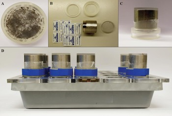
Fleas were infected by a modified version of the original ‘artificial dog’ (Wade and Georgi, Reference Wade and Georgi1988) in which, the perspex cage was removed and cylinders were enclosed by discs of the same material (Fig. 1). This device was placed inside a temperature-controlled room at 28 °C and 60% relative humidity. Blood used in the study was collected from a dog, naturally infected with A. reconditum, living in Messina (Sicily, Italy), an area endemic for this filarioid (Giannetto et al. Reference Giannetto, Poglayen, Gaglio and Brianti2007; Brianti et al. Reference Brianti, Gaglio, Napoli, Giannetto, Dantas-Torres, Bain and Otranto2012), with an estimated parasite load of 1800 microfilariae per mL. No other circulating microfilariae species were detected in the blood of the donor either by microscopy or molecular analysis (see below). After storage in tubes containing anticoagulant (lithium heparin), blood samples were maintained at 4 °C and shipped to the laboratory within 24 h to be diluted with dog blood negative for A. reconditum. Three different concentrations of microfilariae per mL were used, namely, 250±10 (low concentration, L), 500±10 (medium concentration, M) and 1500±10 (high concentration, H). Viability of microfilariae was assessed microscopically for all doses prior to flea infection.
Modified ‘artificial dog’ used in the study. (A) The plastic box containing the fleas; (B) Single components of the feeding system; (C) The flea box and, on its top, the iron cylinder sealing the plastic membrane used to separate fleas from the blood; (D) Units of the ‘artificial dog’ used in the study.

Fleas used in the study were from a laboratory colony of C. felis georgi, bred at the Bayer arthropod breeding department (Monheim am Rhein, Germany). A total of 2105 fleas were divided into three groups of approximately 700 fleas each and named as groups L, M, and H according to the concentration of microfilariae in the blood used for experimental infection.
On Day 0 (D0) fleas of each group were fed with infected blood doses (30 mL for each group) for 24 h. After the initial infected meal, fleas were fed daily throughout the study period (4 weeks) with cattle blood. A control group (C) of 200 fleas was fed at D0 with uninfected dog blood for 24 h and thereafter maintained throughout the study as above. Fleas were sampled every 24 h after infection in the first 2 days (D1 and D2) and thereafter at intermittent time-points (i.e. D4, D6, D8, D11, D13, D15, D18, D21, D25 and D28). About 33 fleas were sampled at each time-point from infected groups (i.e. L, M and H) and divided into three aliquots, of which 12 were stored in saline solution for dissection (see below), six were kept in ethanol 70% for PCR (see below), and 15 were preserved in 10% buffered formalin for histological analysis (see below). In all groups, dead fleas were regularly removed and counted at each time-point, and consequently divided in two aliquots for dissection and PCR according to group and time-point of sampling.
Flea dissection
Fleas were dissected in a few drops of saline solution under a stereomicroscope, and a drop of methylene blue (1%) was added in order to better visualize larval nematodes. The dissected material was observed under a microscope (400× magnification) for the presence of larvae (Brianti et al. Reference Brianti, Gaglio, Napoli, Giannetto, Dantas-Torres, Bain and Otranto2012), and, when retrieved, larvae were photographed with a digital camera (Zeiss Axiocam MRc, Carl Zeiss AG, Germany), mounted on the microscope (Zeiss Axioscop 2 plus, Carl Zeiss AG, Germany) and measurements made using AxioVision rel. 4.8 software (Carl Zeiss AG, Germany). The morphological diagnostic characters of developing larval stages and infective larvae were compared with those of A. reconditum (Calandruccio, Reference Calandruccio1892; Pennington and Phelps, Reference Pennington and Phelps1969; Bain and Beaucournu, Reference Bain and Beaucournu1974).
PCR amplification and DNA sequencing
Fleas were pooled according to the L, M and H groups, sex and time of sampling. Genomic DNA was extracted from fleas using the DNeasy Blood & Tissue Kit (Qiagen, GmbH, Hilden, Germany). PCRs were performed using the species-specific forward primer Arcox1F (5′-ATC TTT GTT TAT GGT GTA TC-3′), amplifying a region of the cytochrome oxidase subunit 1 (cox1) gene (about 589 bp) (Latrofa et al. Reference Latrofa, Weigl, Dantas-Torres, Annoscia, Traversa, Brianti and Otranto2012). The above forward primer was coupled with the reverse primer NTR (5′-ATA AGT ACG AGT ATC AAT ATC-3′) (Casiraghi et al. Reference Casiraghi, Bain, Guerrero, Martin, Pocacqua, Gardner, Franceschi and Bandi2004). PCR amplifications were performed in a final volume of 50 μL, using approximately 100 ng of genomic DNA (with the exception of the no-template control), 10 mm Tris HCl, pH 8·3 and 50 mm KCl, 2·5 mm MgCl2, 250 μ m of each dNTP, primer pairs at 100 μ m and 1·25 U of AmpliTaq Gold (Applied Biosystems, Foster City, CA, USA). The amplification protocol was performed in a thermal cycler (2720, Applied Biosystems, Foster City, CA, USA) as follows: 95 °C for 10 min (for polymerase activation), followed by 40 cycles of 95 °C for 1 min (denaturation); 58 °C for 1 min (annealing); 72 °C for 1 min (extension), followed by 7 min at 72 °C (final extension). Samples without DNA or with DNA from fleas of the control group were included in all PCR reactions. Amplicons were resolved in ethidium bromide-stained agarose (Gellyphor, EuroClone, Milan, Italy) gels (2%) and sized by comparison with Gene RulerTM 100-bp DNA Ladder (MBI Fermentas, Vilnius, Lithuania). Gels were photographed using a Gel Doc 2000 (Bio-Rad, Hercules, CA, USA). To confirm the identity of PCR amplicons, products were purified using QIAquick® Gel Extraction Kit (Qiagen, GmbH, Hilden, Germany) and then sequenced in both directions, employing the same primers as for PCR amplification, using the Taq Dye Deoxy Terminator Cycle Sequencing Kit (v.2, Applied Biosystems Inc.) in an automated sequencer (ABIPRISM 377; Applied Biosystems Inc.). The sequences were then identified by comparisons to sequences in the GenBank™ database (www.ncbi.nlm.gov), using the Basic Local Alignment Search Tool (BLASTn) (Altschul et al. Reference Altschul, Madden, Schäffer, Zhang, Zhang, Miller and Lipman1997).
Histology
Infected fleas, stored in 10% buffered formalin, were pooled by group of infection and time of sampling. Before embedding, each flea was completely covered for 20 min with thioglycolic acid salt in order to soften the outer chitin layer. Fleas were then routinely processed for paraffin embedding. Five-micrometre thick sections were cut and stained with haematoxylin and eosin (H&E).
Statistical analysis
Flea infection rates were assessed according to the dissection or PCR results, as the number of positive fleas/pools over the number of processed fleas/pools. Infection rates among groups, as resulting from dissection or PCR analysis, were investigated for statistically significant differences by Yates corrected chi-square test. Differences in the mortality rate among groups at different time-points were compared using one-way ANOVA. Statistical analyses were performed using Statistica 8.0 (StatSoft Inc., USA) and the level of significance was set at P<0·05.
RESULTS
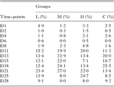
A total of 2305 (i.e. 2105 from infected and 200 from the control groups, respectively) fleas were fed via the ‘artificial dog’ system (Table 1). Overall 1214 live fleas from the infected groups were examined for the presence of A. reconditum larvae using different diagnostic tools. In the three groups infected with A. reconditum about a third of the fleas (n = 891) died before being sampled, resulting in an overall mortality rate of 42·3% (Table 1). A proportion of dead fleas from infected groups were dissected (n = 614; 68·9%), whilst the remaining (277; 31·1%) were pooled by sex and time of death, and molecularly processed. In the control group (C), 128 fleas died during the study, with an estimated mortality rate of 64% (Table 1). No statistically significant differences in mortality rates were detected amongst infected and control groups at any time-point (Table 2).
Number (n) and gender of live and dead fleas in each group (L = low concentration, M = medium concentration, H = high concentration and C = control group) along with the mean mortality rate

Flea mortality rates recorded at different time points (D1–D28) for each group (L = low concentration, M = medium concentration, H = high concentration and C = control group)

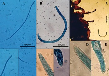
Upon dissection the three larval stages of A. reconditum were retrieved from infected fleas (Table 3), with first-stage (L1) found in a single male flea sampled from group L at D2 (Fig. 2A). L1 was identified based on body size and morphology (i.e. 275·2 μm in length and 5·6 μm in width), resembling those of blood-circulating microfilariae. The second larval stage (L2) was retrieved from a single male flea of group M sampled at D13 (Fig. 2B). This larval stage is considerably larger (i.e. 950×26 μm), showing typical features such as the cephalic knob on the anterior end and a caudal end rounded with the cuticle detached from the sheath (Fig. 2B). The infective third-stage larvae (L3) were retrieved from two males of group H, at D15 examination. The L3 again showed a considerable increase in body size (i.e. 1150×28 μm), its morphometric features (e.g. tail lapplets) were consistent with those described for the A. reconditum infective stage by Calandruccio (Reference Calandruccio1892), Nelson (Reference Nelson1962) and Bain and Beaucournu (Reference Bain and Beaucournu1974) (Fig. 2C). On dissection, no larvae were found in the 614 dead fleas. No statistically significant difference in the rate of infection was found amongst the three infected groups.
Developing stages of Acanthocheilonema reconditum inside the cat flea. (A) Developing first-stage larva retrieved from a flea 2 days post-infection (D2); note in the pictures below, magnifications of the anterior (left) and caudal (right) ends of the same larva; (B) Second-stage larva found in a flea 13 days post-infection (D13); note, in the pictures below, magnifications of the anterior (left) and caudal (right) ends of the same larva; (C) A second-stage larva localized to the abdomen of a dissected flea; (D) Anterior and (E) caudal ends of an infective third-stage larva retrieved from a flea 15 days post infection (D15).

Number of dissected fleas in the three infected groups (L = low concentration, M = medium concentration and H = high concentration) along with the number and stage of Acanthocheilonema reconditum larvae found. Legend: L1 = first-stage larval; L2 = second-stage larval; L3 = infective third-stage larval

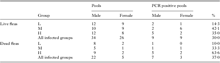
Eighteen (30%) of the 60 pools, derived from 264 live fleas, tested positive at the PCR analysis (Table 4). Although not statistically significant, positive pools were higher in group M (8/19; 42·1%) and H (7/20; 35·0%) than in group L (3/21; 14·3%). Ten (37%) of the 27 pools, derived from dead fleas (277), tested positive for A. reconditum (Table 4). A statistically significant difference was recorded for the frequency of dead flea positive pools, with the highest in group H (P = 0·0377), whereas no difference was recorded in the frequency of positive pools between sexes.
Pools of live and dead fleas tested by PCR for Acanthocheilonema reconditum

L = low concentration, M = medium concentration, H = high concentration.
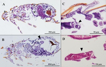
A total of 12 pools, collected from infected groups at D2 (n = 3), D13 (n = 3), D15 (n = 3) and D25 (n = 3), were histologically examined (Fig. 3A). Two round structures (26 μm in diameter), referring to cross-sections of the larval body, were detected in the sub-cuticle region of one female flea (group H) at D13. These structures were characterized by an outer layer of cuboidal to flattened cells and a central rudimental alimentary canal and were consistent, by measurements and morphology, with a late second-stage larva of A. reconditum. The larva was embedded in the back muscle outside of the alimentary canal (Fig. 3B–D).
(A) Flea histology haematoxylin and eosin stained, note the location of the following anatomical sites: brain (B), thoracic ganglia (G), midgut (M), rectal ampulla with rectal pads (R) and fat body cells (F); (B) Histology (50×) of a flea examined 13 days post-infection (D13);the arrow indicates the location in which the cross-section of a developing larva of Acanthocheilonema reconditum was evident; (C) Magnification of B; note cross-sections of the larval body (arrow); (D) Deeper sections of C; cross-sections (arrow) of the larval body morphometrically consistent with a second-stage larva of A. reconditum.

DISCUSSION
The ‘artificial dog’ feeding system was used here for the first time to infect C. felis flea with A. reconditum larvae, therefore providing evidence for their development in this arthropod species. The overall rate of A. reconditum infection in dissected fleas (0·9%) was lower than that reported in naturally infected fleas (i.e. 70·5 and 5·1% in Pennington and Phelps, Reference Pennington and Phelps1969; Brianti et al. Reference Brianti, Gaglio, Napoli, Giannetto, Dantas-Torres, Bain and Otranto2012, respectively). This could be accounted for by the short time of artificial feeding on infected blood (i.e. 24 h), which is usually much longer under natural conditions. However, employing the ‘artificial dog’ avoids the use of experimental animals as infection models for this parasite and to define larval developmental times via infections that all begin at the same time.
Not surprisingly, a higher infection rate was detected by PCR analyses than by dissection with 30 and 37% molecularly positive for A. reconditum in live and dead flea pools, respectively. In pools of live fleas a higher infection rate, not statistically significant, was recorded in groups fed with medium or high levels of microfilariae, whilst in pools of dead fleas a significantly higher frequency (P = 0·0377) was recorded in group H (i.e. fed with the highest level of microfilariae).
These findings suggest that a suitable concentration for flea infection with A. reconditum larvae could be set between 500 and 1500 microfilariae per mL.
Developing L1, found at D2, completed their moult into L2 at D13, therefore developing to the infective L3 stage at D15. This finding suggests that A. reconditum completes its life cycle in C. felis maintained under laboratory conditions in about 15 days, which is a shorter time span than that of other filarioids infesting dogs, such as Dirofilaria immitis, Acanthocheilonema dracunculoides and Cercopithifilaria bainae which develop in their appropriate vectors in 18 and 30 days, respectively (Olmenga-Garcìa et al. Reference Olmenga-Garcìa, Rodrìguez-Rodrìguez and Rojo-Vàzques1993; Viera et al. Reference Viera, Villar, Cecilian, Simon, Simon and Genchi2001; Brianti et al. Reference Brianti, Otranto, Dantas-Torres, Weigl, Latrofa, Gaglio, Napoli, Brucato, Cauquil, Giannetto and Bain2011). Conversely, the developmental period of A. reconditum in the cat flea overlaps that of Dirofilaria repens (i.e. 9–15 days), in mosquitoes (Viera et al. Reference Viera, Villar, Cecilian, Simon, Simon and Genchi2001). Nonetheless, pools positive at PCR were found consistently throughout the observation period (i.e. D1–D28) suggesting that A. reconditum larvae were alive in the fleas throughout the entire study period. Since the digestion process of a blood meal in flea is very rapid (i.e. cat fleas can ingest and eliminate blood equivalent to 15 times its body weight per day; Dryden and Gaafar, Reference Dryden and Gaafar1991) it is unlikely that A. reconditum DNA could be retained in the fleas’ intestine throughout the study period.
The survival rate of infected fleas observed in this study is similar to that recorded in other flea colonies (Osbrink and Rust, Reference Osbrink and Rust1984; Dryden, Reference Dryden1989, Reference Dryden1993), with no difference in the mortality rate between infected and control groups at each time-point (Table 2). Although arthropod-transmitted pathogens may cause alterations in the vector's fitness, thus playing an important role in the dynamics of transmission (Hurd, Reference Hurd2003; Morick et al. Reference Morick, Krasnov, Khokhlova, Gutiérrez, Fielden, Gottlieb and Harrus2013), findings herein presented suggest that the development of A. reconditum larvae does not impair the survival of C. felis. This is similar to that recorded for C. bainae in the dog tick Rhipicephalus sanguineus sensu lato (Brianti et al. Reference Brianti, Gaglio, Napoli, Giannetto, Dantas-Torres, Bain and Otranto2012; Ramos et al. Reference Ramos, Giannelli, Brianti, Annoscia, Cantacessi, Dantas-Torres and Otranto2013), and different from other filarioids (e.g. Brugia spp., D. immitis and Onchocerca spp.), which reduce the survival and fecundity of their vectors (Kershaw et al. Reference Kershaw, Lavoipierre and Chalmers1953; Duke, Reference Duke1962; Husain and Kershaw, Reference Husain and Kershaw1971). On histological examination, the presence of larvae, embedded in the back muscle in the sub-cuticular region of the abdomen, confirmed findings of a previous study in which infective larvae were found near flea body fat cells (Calandruccio, Reference Calandruccio1892). Larval localization inside the flea (i.e. outside the digestive tract) and the large size of the infective stage (1150×28 μm) should render it difficult for L3 to pass through the mouth apparatus. Therefore, it could be speculated that fleas most likely act as an intermediate host and that the transmission occurs via their ingestion, as has also been suggested by a previous study in which dogs were experimentally infected with A. reconditum L3 on the vestibular mucosa (Lindemann and McCall, Reference Lindemann and McCall1984).
Histological analysis of fleas was conducted with an easy and innovative method, in which, instead of the use of special storage solutions (e.g. Duboscq-Brasil) or vacuum-embedding procedures (Rothschild et al. Reference Rothschild, Schlein and Ito1986; Ismaeel et al. Reference Ismaeel, Garmson, Molyneux and Bates1998), thioglycolic acid salt was successfully employed to soften the hard chitin layer of the fleas.
This paper demonstrates that the ‘artificial dog’ may be successfully used to infect the cat flea with A. reconditum larvae, reducing the use of live animals in experiments. In artificially infected fleas, maintained under laboratory conditions, the development of A. reconditum to L3 infective stage takes 15 days, one of the shorter times among filarioids of dogs. Although further confirmatory studies are needed, the data presented here confirm that fleas act as an intermediate host of A. reconditum and suggest that the transmission likely occurs via the ingestion of infected fleas.
ACKNOWLEDGEMENTS
The authors acknowledge the Unit of Arthropod Breeding of Bayer Animal Health and its personnel for the support and resources provided for this study. Dr Bronwyn Evelyn Campbell (University of Bari, Italy) is acknowledged for her critical reading of the manuscript.