INTRODUCTION
Nobenedenia girellae has been reported as an ectoparasite on many species of marine fish, particularly in tropical areas like Mexico and California (Crane, 1972; Brooks and Mayes, 1975; Gaida and Frost, 1991; Goldberg et al. 1991). Infection of Japanese cultured marine fish was first noted in 1991. Ogawa et al. (1995) confirmed that N. girellae was introduced to Japan through imported amber jack (Seriola dumerili). N. girellae infects the body surface of many species of marine fish, such as tiger puffer Takifugu rubripes, Japanese flounder Paralichthys olivaceus, spotted halibut Verasper variegatus and amber jack Seriola dumerili (Ogawa and Yokoyama, 1998; Hirazawa et al. 2004). Active feeding, by large populations of capsalid monogeneans on the mucus and epithelial cells of host fish, can cause haemorrhage, inflammation and mucus hyperproduction (Paperna, 1991). Heavily infected fish may stop eating, the colour of their bodies darkens and they swim erratically and rub against the net, which may result in dermal ulceration and subsequent bacterial invasion (Leong and Colorni, 2002). This parasite is now recognized as a relevant pathogen in finfish aquaculture due to the high mortality of host fish, low host-specificity and wide distribution. However, information on N. girellae is as yet limited. The disinfection of skin parasites has been carried out by bath treatment in freshwater, formalin or copper sulphate (Thoney and Hargis, 1991; Okabe, 2000; Kim et al. 2001). Oral administration of medication such as praziquantel to host fish has also been examined (Hirazawa et al. 2000, 2004). However, many of these chemicals are highly toxic to fish (Schmahl and Taraschewski, 1987; Pickering, 1998). Recent studies reported decreases in hatching rate and abnormalities during development of marine fish parasites exposed to water of reduced salinity (Ogawa, 1998; Yoshinaga et al. 2000: Umeda and Hirazawa, 2004). Although a freshwater bath is a simple treatment method, long-term exposure causes severe damage also to the host fish (Kim and Choi, 1998; Roberts and Powell, 2003a,b). As for the oral administration of chemical treatments, the safety issues for human consumption are still questionable. Furthermore, use of chemical treatments in aquaculture facilities has become one of the major causes of environmental contamination (McHenery et al. 1991). Hence, a new helminth disinfection technology that is simpler and safer than the present technologies should be established. Because N. girellae is not able to survive in fresh water or in low salinity, we hypothesized that metallic ions abundant in seawater, such as sodium, potassium, calcium and magnesium, are likely to have major roles in the survival of N. girellae. Therefore, in this research, we focused on the possible role of the concentration of these ions in seawater and on the survival rate of N. girellae on and off the host.
MATERIALS AND METHODS
Source of N. girellae
N. girellae were maintained on host juvenile spotted halibut Verasper variegatus. Using forceps, adult parasites were carefully removed from the body surface of 1-year-old spotted halibut (73·2–99·6 g) reared at the Marine Biological Technology Center, Nippon Suisan Kaisha, Ltd.
Assay seawater
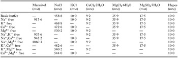
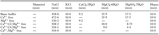
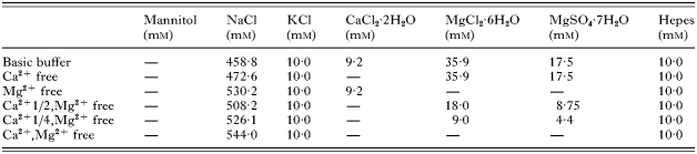
The basic buffer used for in vitro trials (parasites were removed during treatments) and in vivo trials (parasites were maintained on hosts during treatments) contained 458·8 mM NaCl, 10·0 mM KCl, 9·2 mM CaCl2·2H2O, 35·9 mM MgCl2·6H2O, 17·5 mM MgSO4·7H2O and 10·0 mM Hepes. The osmotic pressure of this buffer was 1027 mOsm. The pH of the buffer was adjusted to pH 8·0 by adding an appropriate amount of NaOH or KOH. We prepared buffers in which only a single ion (Na+, K+, Ca2+ and Mg2+) was omitted. We made buffers from which 2 metallic ions were removed (details of buffer composition are shown in Table 1). We prepared buffers containing only 50% (Mg2+ to 18·0 mM MgCl2·6H2O, 8·8 mM MgSO4·7H2O from 35·9 mM MgCl2·6H2O, 17·5 mM MgSO4·7H2O) or 25% (9·0 mM MgCl2·6H2O, 4·4 mM MgSO4·7H2O) of the magnesium concentration of seawater. Buffers containing 50% (4·6 mM CaCl2·2H2O from 9·2 mM CaCl2·2H2O) or 25% (2·3 mM CaCl2·2H2O) of the calcium concentration were also prepared. Buffers that changed the concentrations of Mg2+ and Ca2+ were combined and 4 different combinations of buffers were prepared as shown in Table 2. The osmotic potential of these buffers was adjusted to that of seawater by adding an appropriate amount of NaCl. The osmotic pressure of the Na+-free buffer was adjusted to 1027 mOsm by adding an appropriate amount of mannitol.
Table 1. Compositions of buffers showing removal of 1 or 2 ions simultaneously for the in vitro assays (The osmotic pressure of these buffers was 1027 mOsm. The osmotic pressure of these ions-free buffers was adjusted by adding an appropriate amount of NaCl. The osmotic pressure of the Na+-free buffer was adjusted by adding an appropriate amount of mannitol. The pH of buffers was adjusted to pH 8·0 by adding an appropriate amount of NaOH or KOH.)

Table 2. Composition of buffers that changed the concentration of calcium and magnesium ions for in vitro assays (1/2 or 1/4 indicate that a concentration was changed a half or a quarter of basic buffer. For example Ca2+ 1/2,Mg2+ free indicates that buffer included a half of basic buffer (4·6 mM) CaCl2·2H2O, 0 mM MgCl2·6H2O and 0 mM MgSO4·7H2O. The osmotic pressure of these buffers was 1027 mOsm. The osmotic pressure of these buffers was adjusted by adding an appropriate amount of NaCl. The pH of buffers was adjusted to pH 8·0 by adding an appropriate amount of NaOH.)

In vitro trials
Buffers that removed single metal ions or combinations of pairs of ions, or changed the concentration of Ca2+ and Mg2+ were tested in in vitro trials. Natural seawater that was sand-filtered and ultraviolet-irradiated (Flonlizer 4-L unit; approximately 50 000 μW s/cm2; Chiyoda Kohan, Tokyo, Japan), basic buffer and distilled water as a control were also tested in vitro. Approximately 20 adult parasites were put into each well of a 12-well plate (Becton Dickinson Labware, USA) containing 2 ml of natural seawater. Wells containing the parasites were washed twice with 10 ml of natural seawater. Any eggs that were deposited were removed at this time. We checked the integrity of N. girellae by stereomicroscopic observation after collection with forceps. We only used N. girellae that moved normally, had laid eggs and were not opaque. All parasites were incubated for up to 120 min at 25 °C. Percentage survival of N. girellae was examined after 5, 10, 15, 20, 25, 30, 60 and 120 min of incubation using a stereomicroscope. Each treatment was carried out in triplicate, and results from these 3 wells were averaged.
Transmission electron microscopic observations
For ultrastructural examination, parasites were fixed in 2% glutaraldehyde, 3% paraformaldehyde in 0·1 M sodium cacodylate buffer (pH 7·2) for 1 day at 4 °C, and post-fixed with 1% osmium tetroxide in 0·1 M sodium cacodylate buffer (pH 7·2) for 1 h at room temperature. Specimens were dehydrated through a graded acetone series and embedded in Quetol-812 epoxy resin (Nisshin-EM, Tokyo, Japan). Ultrathin sections were cut transversely, stained with uranyl acetate and lead citrate, and observed under a transmission electron microscope (H 7100 Hitachi) at 75 kV.
In vivo trials
Six halibut infected by N. girellae were placed in 6 separate 4 litre tanks. The infected fish were maintained at 25 °C for 1 h in natural seawater and in the buffers listed in Table 3, which were identified as effective in in vitro trials. The number of parasites was counted for each fish under a stereomicroscope, and the percentage survival was calculated. This test was carried out 3 times.
Table 3. Composition of buffers for in vivo assay (1/2 or 1/4 indicate that a concentration was changed a half or a quarter of basic buffer. For example Ca2+ 1/2,Mg2+ free indicates that buffer included a half of basic buffer (4·6 mM) CaCl2·2H2O, 0 mM MgCl2·6H2O and 0 mM MgSO4·7H2O. The osmotic pressure of these buffers was 1027 mOsm. The osmotic pressure of the ions-free buffers was adjusted by adding an appropriate amount of NaCl. The pH of the buffers was adjusted to pH 8·0 by adding an appropriate amount of NaOH.)
Statistical analysis
Results were analysed using Bonferroni/Dunn tests and one-way ANOVA. All calculations were performed using StatView statistical software (Abacus Concepts, USA).
RESULTS
In vitro trials
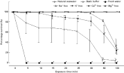
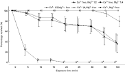
None of the N. girellae exposed to natural seawater and basic buffer died during the experimental period, whereas N. girellae exposed to distilled water died within 5 min (Fig. 1). Compared with the group treated with natural seawater, the percentage survival of N. girellae treated with Na+-free and Ca2+-free buffers started to decrease after 30 min of exposure, and then the percentage survival of both treatment groups significantly declined to about 30% after 120 min exposure (P<0·05).

Fig. 1. Changes in the percentage survival of Neobenedenia girellae in buffers, in which one of the main metal ions was removed. The symbols show the average percentage survival, the bars indicate standard errors. Significant differences in the percentage survival are indicated by * (P<0·05).
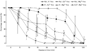
The most effective treatment to decrease the percentage survival of N. girellae was Ca2+/Mg2+-free buffer, with a decrease to 24·2% after only 10 min of exposure, compared with natural seawater (Fig. 2). All N. girellae died after 20 min exposure to Ca2+/Mg2+-free buffer. In the Na+/Mg2+-free buffer, it took more than 15 min for the percentage survival to show a significant decrease compared with natural seawater, and the percentage survival was 31·8%. The percentage survival was 32·7% after 25 min exposure to Na+/Ca2+-free buffer, and 28·5% after 20 min exposure to Na+/K+-free buffer. It required more than 30 min for the percentages of survival of N. girellae in K+/Ca2+-free buffer to decrease significantly. It took more than 60 min to see significant decrease of the percentage survival in K+/Mg2+-free buffer.

Fig. 2. Changes in the percentage survival of Neobenedenia girellae in buffers, in which combinations of 2 of the 4 main metal ions were removed. The symbols show the average percentage survival, the bars indicate standard errors. Significant differences in the percentage survival are indicated by * (P<0·05).
A change in concentration of Ca2+ or Mg2+ to 50% or 25% of that in basic buffer resulted in a decline in survival of N. girellae (Fig. 3). The percentage survival of the N. girellae exposed to Ca2+/Mg2+-free buffer significantly declined to under 24·2% after 5 min compared to natural seawater (P<0·05). There was no difference in the percentage survival of N. girellae among the groups exposed to buffer with reduced concentrations of Ca2+, whereas percentage survival of N. girellae significantly declined after 120 min of exposure to buffer with reduced Mg2+ concentrations.

Fig. 3. Changes in the percentage survival of Neobenedenia girellae in response to different concentrations of calcium and magnesium ions in the basic buffer. The symbols show the average percentage survival, the bars indicate standard errors. Significant differences in the percentage survival are indicated by * (P<0·05).
Transmission electron microscopic observations
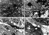
We studied the central region of N. girellae by transmission electron microscopy. This area consists of vitelline and hypodermis cells. In the control group exposed to natural seawater, both cell types showed the typical architecture of cell organelles, and no gaps or holes were found in cell-cell junctions or in the tegumental syncytium (Fig. 4A). In the freshwater-exposed N. girellae, many large gaps were observed in the cells. Cell nuclei contracted and the cell organelles were different from the control. Moreover the tegumental syncytium showed large holes (Fig. 4B). In N. girellae exposed to Ca2+-free, or to Ca2+/Mg2+-free buffer, intracellular gaps were observed also, with a large gap in the cell-to-cell junctional area. However, contraction of the cell was not observed and the cell organelles appeared similar to those in control animals. There were no holes or gaps in the tegumental syncytium (Fig. 4C and D).

Fig. 4. Cross-sections of the central part of different specimens of Neobenedenia girellae. (A) Worm exposed to natural seawater. (B) Worm exposed to fresh water. (C) Worm exposed to artificial seawater without calcium ions. (D) Worm exposed to artificial seawater without calcium and magnesium ions. N, nucleus; IG, intracellular gap; arrow indicates intercellular junction.
In vivo trials
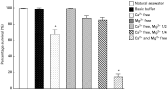
In the groups exposed to buffers from which either calcium ions, or magnesium and calcium ions, had been removed, percentage survival declined significantly (P<0·05) compared with the group exposed to natural seawater (Fig. 5). The percentage survival of the group exposed to the Ca2+/Mg2+-free buffer were 14·7% after 1 h. Most of the N. girellae were dead or appeared to be weakened. Following exposure to the deionized buffers used for the trials, abnormal swimming or bleeding of the gills was not observed in spotted halibut.

Fig. 5. Changes in the Neobenedenia girellae percentage survival after in vivo trials. The columns show the averages, the bars indicate the respective standard errors. Significant differences in the percentage survival are indicated by * (P>0·05).
DISCUSSION
In marine fish aquaculture, high stocking densities can quickly lead to heavy infestations by monogenean ectoparasites. Various chemicals such as formalin, copper sulphate, potassium permanganate, sodium peroxycarbonate and trichlorfon have been used to control monogeneans (Thoney and Hargis, 1991). The US Food and Drug Administration has approved formalin in aquaculture since 1986 for the treatment of finfish and their eggs against external parasites, bacteria and fungal infections (Howe et al. 1995). However, many of these chemicals are highly toxic to fish and pollute the environment around fish farms (Schmahl and Taraschewski, 1987). In order to prevent infestation by N. girellae, cultured fish in floating offshore net pens have been routinely treated by bath treatment with freshwater or diluted hydrogen peroxide approximately every 20 days in the high water temperature season. This treatment required additional labour and subjects the fish to additional stress (Kim and Choi, 1998; Roberts and Powell, 2003a,b). Therefore, a new method to control parasitic diseases in cultured fish without the use of chemicals and potential hazard for the fish is requested. This study focused on Na+, K+, Ca2+ and Mg2+ in seawater to establish a new method of disinfection N. girellae that is safe for the cultivated fish, for consumer, and for the environment.
In this study, the results of in vitro trials showed that all N. girellae exposed to freshwater died within 5 min. The effectiveness of killing N. girellae with freshwater bath treatments was proven. Na+, K+ and Ca2+ are indispensable for this organism to maintain homeostasis. Although buffers without Na+, K+ or Ca2+ had significant antiparasitic effects after 60 to 120 min of treatment, they were not as effective as freshwater treatment. Since buffers in which a single ion was removed were relatively ineffective, buffers in which 2 metallic ions were removed were used for in vitro trials.
The latter buffers were more effective. Specifically, removing Ca2+ and Mg2+ from the basic buffer significantly decreased the percentage survival of N. girellae after only 10 min of treatment. These results suggest that Ca2+/Mg2+-free buffer has enough antiparasitic effect against N. girellae to approximately equal the freshwater bath treatment.
We then tested buffers with different concentrations of Ca2+ and Mg2+. While Ca2+/Mg2+-free buffer strongly decreases the percentage survival of N. girellae after 5 min, reducing the concentration of Ca2+ and Mg2+ did not show the same antiparasitic effect as the Ca2+/Mg2+-free buffer. Hence, reducing the concentration of Ca2+ and Mg2+ is not crucial, whereas complete of Ca2+ and Mg2+ was necessary for an effective method for the control of N. girellae.
Freshwater bathing of cultured fish had been used previously to prevent infection by monogenean parasites (Kaneko et al. 1988). A decrease in hatching rates and abnormalities during development of marine fish parasites exposed to water of reduced salinity has also been reported (Ogawa, 1998; Yoshinaga et al. 2000: Umeda and Hirazawa, 2004). However, no studies have indicated why monogenean parasites are killed by freshwater treatments. Thus we examined morphologically how freshwater and ion-free buffer treatments affect N. girellae using transmission electron microscopy. These results demonstrate that N. girellae died as a result of changes in osmotic pressure that caused cells of N. girellae to disrupt when exposed to distilled water. Interestingly, cell nuclei of N. girellae were not contracted when exposed to Ca2+-free buffer and Ca2+/Mg2+-free buffer. Under these conditions, larger gaps were generated especially between the vitelline producing cells and surrounding cells, but the integumental syncytium showed no holes. These observations suggest that N. girellae treated with distilled water died due to change of osmotic pressure, while treatment with Ca2+-free or Ca2+/Mg2+-free buffer inflicted damage based on other mechanisms, possibly related to shortage of Ca2+ after Ca2+ removal. Takeichi and Okada (1972) described a very important role for Ca2+ in the functionality of cell junctions. It was reported that the function of cadherin, a building-block of intercellular junctions, depended on the availability of calcium ions (Lagunowich et al. 1990; Inuzuka et al. 1991). Cadherins exist in invertebrates, also reported by (Ghersi and Vittorelli, 1990; Oda et al. 1994; Wong et al. 1996; Hill et al. 2001). Therefore, cadherin may be invaluable in the maintenance of cell junctions also in N. girellae. By exposing N. girellae to Ca2+-free buffer, Ca2+ is depleted from the living body, and the activity of cadherin may be weakened, resulting in destabilized cell junctions. Ca2+ and Mg2+ are indispensable for survival, because our ultrastructural observations demonstrated that the most effective weakening of the intercellular junction appeared to be the main cause of death. Future studies using molecular biological and biochemical techniques should focus on the relation between cadherin and Ca2+ in N. girellae.
N. girellae is a parasite adhering to the body surface of host fish. Therefore, changes in the probability of survival of N. girellae by exposure to these buffers should be accompanied by investigating the response of the host to the treatment. The percentage survival of N. girellae exposed to Ca2+/Mg2+-free buffer during in vivo experiments on the host fish was 14·7%, which is similar to the results of in vitro trials. Therefore, a sufficient antiparasitic effect can be expected by using Ca2+/Mg2+-free buffers under in vivo circumstances. Importantly, the host fish, spotted halibut, showed no abnormalities after exposure to various buffers for the entire examination period.
Previous studies reported that osmoregulatory disturbances are associated with stress in fish (Wood et al. 1983; Ackerman et al. 2000; Roberts and Powell, 2003a). Roberts and Powell (2003b) reported that low concentrations of Ca2+ and Mg2+ in the water changed the structure of the mucous layer. In this study, the gills of treated host fish showed no alterations compared to non-treated fishes. Hence, from this result we can conclude that Ca2+/Mg2+-free buffer affected N. girellae, had minimal impact on the host fish.
In conclusion, bath treatment with the Ca2+/Mg2+-free buffer had antiparasitic effects similar to bath treatment with freshwater. Depleting Ca2+ and Mg2+ in N. girellae could cause weakening and necrosis of vitelline and hypodermis cells. Moreover, treatment effects with Ca2+/Mg2+-free buffer appeared to be minimal on the cultured fish compared with freshwater treatment. Hence, this disinfection method using Ca2+/Mg2+-free buffer for 60 min would be a safer method for cultured fish as a food source and environment than the previous methods.
We thank Dr R. W. Schulz (Universiteit Utrecht, The Netherlands), Dr M. Kusakabe, Dr I. Nakamura (University of Washington, USA) and Miss. Fritzie T. Celino (Ehime University, Japan) for critical reading of the manuscript.