INTRODUCTION
Acute nociceptive pain is instructive and warns against imminent or existing tissue damage, whereas chronic pain has no known defensive or beneficial function and remains one of the most vexing challenges in medicine. In the United States alone, the direct economic costs of acute and chronic pain conditions are estimated to cost $560–635 billion annually (Institute of Medicine Report, 2011). Although major advances have been made toward understanding the fundamental processes that underlie acute pain, little is known about the cellular and molecular processes initiated by acute pain that lead to the progression to chronic pain, which is characterized by pain persisting long after the tissue damage has healed. Paradoxically, the degree of injury is not necessarily predictive of the severity or chronicity of the pain, and the development of chronic pain following injury is highly variable between individuals. Considerable evidence suggests that the transition from acute to chronic pain is a consequence of alterations in the cellular, molecular and anatomical organization of nociceptive neural networks in the spinal dorsal horn and brain (Woolf and Salter, Reference Woolf and Salter2000; Scholz and Woolf, Reference Scholz and Woolf2002; Latremoliere and Woolf, Reference Latremoliere and Woolf2009; Voscopoulos and Lema, Reference Voscopoulos and Lema2010). Thus, an acute injury can have profound effects on the nociceptive neural circuitry, transforming the CNS from conveying normally protective acute nociceptive signals into a pathologically altered system in which pain can occur spontaneously and responses to innocuous and noxious stimuli are amplified.
Among the most severe and debilitating types of chronic pain conditions is neuropathic pain which can arise following lesions to the somatosensory nervous system caused by trauma, infection, or pathology (Zimmermann, Reference Zimmermann2001; Scholz and Woolf, Reference Scholz and Woolf2002; Gwak and Hulsebosch, Reference Gwak and Hulsebosch2009). Such damage to a nerve can instigate a series of cellular and molecular changes that directly affect neuronal plasticity, leading ultimately to altered synaptic connectivity and the reorganization of peripheral and central nociceptive circuitry (Woolf and Salter, Reference Woolf and Salter2000; Scholz and Woolf, Reference Scholz and Woolf2002; Latremoliere and Woolf, Reference Latremoliere and Woolf2009). These changes have profound effects on pain transmission by suppressing mechanisms that inhibit pain and by enhancing mechanisms that facilitate pain in the central nervous system (CNS). The consequent shift in inhibitory and excitatory control can cause a pathological amplification that alters the modality of sensory input and output from the spinal cord to elicit exaggerated pain responses that typifies chronic pain conditions (Costigan et al., Reference Costigan, Scholz and Woolf2009).
A principal locus of this pathologically altered activity arises from the nociceptive neurons in lamina I of the spinal dorsal horn. The action potential discharge of these neurons is normally evoked only in response to noxious peripheral stimulation (Keller et al., Reference Keller, Beggs, Salter and De Koninck2007). However, after peripheral nerve injury the output of lamina I neurons is transformed such that innocuous stimulus can evoke action potential discharges, and response to noxious stimulation is greatly exaggerated. In addition, after peripheral nerve injury lamina I neurons exhibit spontaneous bursting in the absence of overt stimulation, whereas in uninjured animals these neurons are normally silent. These changes in the output of lamina I neurons provide a neural basis for the three cardinal signs of neuropathic pain in humans (Woolf and Salter, Reference Woolf and Salter2000): mechanical allodynia (discharge in response to innocuous stimulation), hyperalgesia (exaggerated response to noxious stimulation) and spontaneous pain (spontaneous bursting in the absence of an overt stimulus). The symptoms of neuropathic pain are often resistant to the current available treatments, which have typically been directed against cellular targets in neurons. Failure of the current cadre of drugs in treating the sequelae of neuropathic pain has intensified the search for new molecular players and cellular substrates that can be targeted for development of novel pharmacological therapies. This search has generated a rapidly growing body of evidence that indicates the interactions between neurons and glia are critical in establishing and maintaining neuropathic pain (Inoue and Tsuda, Reference Inoue and Tsuda2006; Milligan and Watkins, Reference Milligan and Watkins2009; Gwak and Hulsebosch, Reference Gwak, Hulsebosch, Costa and Villalba2010; Beggs and Salter, Reference Beggs and Salter2010; Grace et al., Reference Grace, Rolan and Hutchinson2011). Microglia, in particular, have emerged as key players in the initiation and in the expression of neuropathic pain (Watkins et al., Reference Watkins, Milligan and Maier2001; Watkins and Maier, Reference Watkins and Maier2003; Tsuda et al., Reference Tsuda, Shigemoto-Mogami, Koizumi, Mizokoshi, Kohsaka and Salter2003, Reference Tsuda, Inoue and Salter2005; Inoue and Tsuda, Reference Inoue and Tsuda2006; Trang et al., Reference Trang, Beggs and Salter2011).
Spinal microglia respond to peripheral nerve injury
Microglia originates from embryonic macrophages derived from the yolk sac during late prenatal development (Ginhoux et al., Reference Ginhoux, Greter, Leboeuf, Nandi, See and Gokhan2010). In the adult CNS, microglia comprises 5–10% of the total glial population (Kreutzberg, Reference Kreutzberg1996; Lawson et al., Reference Lawson, Perry, Dri and Gordon1990; Nakajima and Kohsaka, Reference Nakajima and Kohsaka2001). Likened to the electricians of the CNS, microglia is able to modulate the electrical activity within neuronal circuits (Graeber, Reference Graeber2010). In the surveillance mode of activity, microglia possesses small soma bearing thin ramified processes that cover large non-overlapping territories throughout the brain and spinal cord (Kreutzberg, Reference Kreutzberg1996; Bushong et al., Reference Bushong, Martone, Jones and Ellisman2002). In response to stimuli that potentially disrupt homeostasis, microglia rapidly extends their processes toward the site of neural damage, forming a barrier between healthy and injured cells that limit damage in the CNS (Davalos et al., Reference Davalos, Grutzendler, Yang, Kim, Zuo and Jung2005; Nimmerjahn et al., Reference Nimmerjahn, Kirchhoff and Helmchen2005). The rapid microglia responsiveness precedes a more slowly developing series of changes in morphology, gene expression, function and proliferation (Kreutzberg, Reference Kreutzberg1996; Nakajima and Kohsaka, Reference Nakajima and Kohsaka2001; Hanisch and Kettenmann, Reference Hanisch and Kettenmann2007; Kettenmann et al., Reference Kettenmann, Hanisch, Noda and Verkhratsky2011). These changes are observed in spinal microglia in rodent models of peripheral nerve injury caused by compression, ligation, or transaction (Liu et al., Reference Liu, Tornqvist, Mattsson, Eriksson, Persson and Morgan1995; Tsuda et al., Reference Tsuda, Shigemoto-Mogami, Koizumi, Mizokoshi, Kohsaka and Salter2003; Zhang and De Koninck, Reference Zhang and De Koninck2006; Echeverry et al., Reference Echeverry, Shi and Zhang2008; Calvo and Bennett, Reference Calvo and Bennett2011; Calvo et al., Reference Calvo, Zhu, Grist, Ma, Loeb and Bennett2011). The stereotypical microglial response also entails the up-regulation of surface marker proteins belonging to the complement cascade: complement receptor 3 (CR3), Toll-like receptor 4 (TLR4), CD14, CD4 and major histocompatibility complex (MHC) classes I and II (Liu et al., Reference Liu, Tornqvist, Mattsson, Eriksson, Persson and Morgan1995; Coyle, Reference Coyle1998; Sweitzer et al., Reference Sweitzer, White, Dutta and DeLeo2002; Tsuda et al., Reference Tsuda, Shigemoto-Mogami, Koizumi, Mizokoshi, Kohsaka and Salter2003; Tanga et al., Reference Tanga, Raghavendra and DeLeo2004). Up-regulation of these surface proteins concomitant with distinct anatomical changes in morphology – from ramified to ameboid – are stereotypical of microglia in the ‘enhanced response state’. Although microglia in this state have been reported to release a myriad of factors that signal to neurons in the spinal cord to alter neuronal excitability, in pain neuroplasticity after peripheral nerve injury a specific core pathway has been implicated through P2X4 receptor-stimulated release of brain-derived neurotrophic factor (BDNF; Beggs and Salter, Reference Beggs and Salter2010; Trang et al., Reference Trang, Beggs and Salter2011).
Spinal microglia express P2 receptors
A critical molecular substrate for microglia–neuron communication is ATP, an endogenous ligand of the P2 receptor family comprising P2Y metabotropic and P2X ionotropic receptors (Coull et al., Reference Coull, Beggs, Boudreau, Boivin, Tsuda and Inoue2005; Di, Reference Di2006; Jarvis, Reference Jarvis2010; Maeda et al., Reference Maeda, Tsuda, Tozaki-Saitoh, Inoue and Kiyama2010). Microglia express a variety of P2 receptors: of the metabotropic P2Y receptors, microglia express P2Y1, 2, 4, 6 and 12 receptors (Boucsein et al., Reference Boucsein, Zacharias, Farber, Pavlovic, Hanisch and Kettenmann2003; Sasaki et al., Reference Sasaki, Hoshi, Akazawa, Nakamura, Tsuzuki and Inoue2003; Farber and Kettenmann, Reference Farber and Kettenmann2005; Inoue and Tsuda, Reference Inoue and Tsuda2006), whereas microglial expression of the ionotropic P2X receptor is restricted to the P2X4 and P2X7R subtypes (Ferrari et al., Reference Ferrari, Villalba, Chiozzi, Falzoni, Ricciardi-Castagnoli and Di1996; Collo et al., Reference Collo, Neidhart, Kawashima, Kosco-Vilbois, North and Buell1997; Tsuda et al., Reference Tsuda, Shigemoto-Mogami, Koizumi, Mizokoshi, Kohsaka and Salter2003; Inoue and Tsuda, Reference Inoue and Tsuda2006). Despite the diverse repertoire of P2Y receptors expressed on microglia, only P2Y12 receptors have been demonstrably implicated in the development of tactile allodynia associated with peripheral nerve injury (Kobayashi et al., Reference Kobayashi, Yamanaka, Fukuoka, Dai, Obata and Noguchi2008; Tozaki-Saitoh et al., Reference Tozaki-Saitoh, Tsuda, Miyata, Ueda, Kohsaka and Inoue2008). By contrast, both P2X4 and P2X7 receptors are causally involved in expressing nerve injury-induced pain behaviors (Tsuda et al., Reference Tsuda, Shigemoto-Mogami, Koizumi, Mizokoshi, Kohsaka and Salter2003, Reference Tsuda, Kuboyama, Inoue, Nagata, Tozaki-Saitoh and Inoue2009a, Reference Tsuda, Toyomitsu, Kometani, Tozaki-Saitoh and Inoue2009c; Chessell et al., Reference Chessell, Hatcher, Bountra, Michel, Hughes and Green2005; Coull et al., Reference Coull, Beggs, Boudreau, Boivin, Tsuda and Inoue2005; Kobayashi et al., Reference Kobayashi, Yamanaka, Fukuoka, Dai, Obata and Noguchi2008; Tozaki-Saitoh et al., Reference Tozaki-Saitoh, Tsuda, Miyata, Ueda, Kohsaka and Inoue2008; Ulmann et al., Reference Ulmann, Hatcher, Hughes, Chaumont, Green and Conquet2008).
A compelling argument has been made for involvement of P2X7 receptors in inflammatory pain and in neuropathic pain on the basis of reduced pain sensitivity in P2X7 receptor-deficient mice (Chessell et al., Reference Chessell, Hatcher, Bountra, Michel, Hughes and Green2005) and after pharmacological blockade of the receptor (Dell'Antonio et al., Reference Dell'Antonio, Quattrini, Cin, Fulgenzi and Ferrero2002; Honore et al., Reference Honore, Donnelly-Roberts, Namovic, Hsieh, Zhu and Mikusa2006, Reference Honore, Donnelly-Roberts, Namovic, Zhong, Wade and Chandran2009; McGaraughty et al., Reference McGaraughty, Chu, Namovic, Donnelly-Roberts, Harris and Zhang2007; Broom et al., Reference Broom, Matson, Bradshaw, Buck, Meade and Coombs2008; Perez-Medrano et al., Reference Perez-Medrano, Donnelly-Roberts, Honore, Hsieh, Namovic and Peddi2009). Recent evidence has also established a link between genetic variations in the P2X7 receptor that affect its function and variability in chronic pain sensitivity in mice and in humans (Sorge et al., Reference Sorge, Trang, Dorfman, Smith, Beggs and Ritchie2012). The specific cell type(s) responsible for the P2X7 receptor-mediated pain phenotypes has been difficult to precisely pinpoint because, in addition to being expressed on microglia, P2X7 receptors are localized on macrophages, neurons and astrocytes (Donnelly-Roberts and Jarvis, Reference Donnelly-Roberts and Jarvis2007; Donnelly-Roberts et al., Reference Donnelly-Roberts, McGaraughty, Shieh, Honore and Jarvis2008; Skaper et al., Reference Skaper, Debetto and Giusti2010). Thus, conclusions about the specific role microglial P2X7Rs play in inflammatory and neuropathic pain is confounded by expression of this receptor on a wide variety of cell types. By contrast, much of our conceptual understanding of the role microglial P2 receptors play in neuropathic pain stems from elucidating the fundamental mechanisms that regulate P2X4 receptor expression, as well as the identification of signaling pathways downstream from this receptor.
Microglial P2X4 receptors are critically required for neuropathic pain
The first compelling clues that identified P2X4 receptors as a critical molecular component in neuropathic pain were the discoveries that blocking P2X4 receptor function pharmacologically or suppressing its expression with antisense RNA transiently reverses mechanical allodynia (Tsuda et al., Reference Tsuda, Shigemoto-Mogami, Koizumi, Mizokoshi, Kohsaka and Salter2003). The development of mechanical allodynia was found to correlate temporally with an increase in spinal P2X4 receptor expression, and unexpectedly this increase was confined to microglia in the ipsilateral spinal dorsal horn. These observations were subsequently confirmed in CX3CR1 +/GFP mice, in which induction of P2X4 receptors resulting from peripheral nerve lesion is restricted to activated enhanced green fluorescent protein (eGFP) expressing spinal microglia (Ulmann et al., Reference Ulmann, Hatcher, Hughes, Chaumont, Green and Conquet2008), and in mice lacking the P2X4 receptor, which do not develop mechanical allodynia after peripheral nerve injury (Ulmann et al., Reference Ulmann, Hatcher, Hughes, Chaumont, Green and Conquet2008; Tsuda et al., Reference Tsuda, Kuboyama, Inoue, Nagata, Tozaki-Saitoh and Inoue2009a). Although neuropathic pain behaviors in the P2X4 receptor-deficient mice are absent, the microglial proliferative response and the alterations in microglia morphology induced by peripheral nerve injury were not affected (Tsuda et al., Reference Tsuda, Shigemoto-Mogami, Koizumi, Mizokoshi, Kohsaka and Salter2003; Ulmann et al., Reference Ulmann, Hatcher, Hughes, Chaumont, Green and Conquet2008), suggesting that while tonic P2X4 receptor activation is required for maintaining peripheral nerve injury-induced allodynia, the proliferation and up-regulation of microglial P2X4 receptors in the spinal cord are mediated by distinct intracellular mechanisms. Direct evidence that stimulation of P2X4 receptors expressed on microglia is sufficient to elicit pain hypersensitivity comes from the finding that injection of P2X4 receptor-stimulated cultured microglia into the spinal cords of naïve animals elicits robust mechanical allodynia that is blocked by 2′,3′-O-(2,4,6-trinitrophenyl)adenosine 5′-triphosphate (TNP-ATP) (Tsuda et al., Reference Tsuda, Shigemoto-Mogami, Koizumi, Mizokoshi, Kohsaka and Salter2003, Reference Tsuda, Tozaki-Saitoh, Masuda, Toyomitsu, Tezuka and Yamamoto2008b; Coull et al., Reference Coull, Beggs, Boudreau, Boivin, Tsuda and Inoue2005). Taken together, the pharmacological, genetic and behavioral findings indicate that the activity of P2X4 receptors expressed on spinal microglia is critically involved in the functional alterations in the spinal dorsal horn that maintain ongoing pain following peripheral nerve injury.
Regulation of P2X4 receptor expression in microglia
A major question arising from the observation that the development of mechanical hypersensitivity is correlated with a progressive increase in spinal P2X4 receptor expression is how peripheral nerve injury initiates signaling in the spinal dorsal horn to specifically cause an increase in P2X4 receptor expression in microglia. The answer to this question appears to involve the release of several signaling elements including: CCL21, a chemokine released from injured neurons that functions as an upstream activator of P2X4 receptor (de Jong et al., Reference de Jong, Dijkstra, Hensens, Brouwer, van and Liem2005; Biber et al., Reference Biber, Tsuda, Tozaki-Saitoh, Tsukamoto, Toyomitsu and Masuda2011), interferon γ, a cytokine that transforms resting spinal microglia into an activated state (Tsuda et al., Reference Tsuda, Masuda, Kitano, Shimoyama, Tozaki-Saitoh and Inoue2009b), and tryptase, a protease released from mast cells that activates proteinase-activated receptor 2 in microglia (Yuan et al., Reference Yuan, Zhu, Zhou, Chen, Zhu and Ma2010). Also critical for up-regulating expression of P2X4 receptors is the extracellular matrix molecule fibronectin, which through activity of Lyn kinase and downstream activation of intracellular signaling pathways involving phosphatidylinositol 3-kinase (PI3K)-Akt and mitogen-activated protein kinase kinase (MAPK kinase, MEK)-extracellular signal-regulated kinase (ERK), modulates the transcriptional and post-transcriptional levels of P2X4 receptor expression in microglia (Nasu-Tada et al., Reference Nasu-Tada, Koizumi, Tsuda, Kunifusa and Inoue2006; Tsuda et al., Reference Tsuda, Toyomitsu, Komatsu, Masuda, Kunifusa and Nasu-Tada2008a, Reference Tsuda, Tozaki-Saitoh, Masuda, Toyomitsu, Tezuka and Yamamoto2008b, Reference Tsuda, Toyomitsu, Kometani, Tozaki-Saitoh and Inoue2009c). Thus, several elements of the molecular machinery required for up-regulation of P2X4 receptors in microglia following peripheral nerve injury have recently been identified (Fig. 1). The implications of this diverse modulation and whether they are causally connected through a convergent common pathway that controls P2X4 receptor expression is not known.
Dynamic regulation of P2X4 receptors in microglia. Microglia in the physiological CNS actively monitor their surrounding environment for potential stimuli that threaten homeostasis. In response to peripheral nerve injury spinal microglia up-regulate expression of P2X4 receptors, which normally are expressed at low levels in the ‘resting/surveillance state’. Up-regulation of P2X4 receptors is a critical mechanistic step through which spinal microglia signal to neurons in the spinal dorsal horn to cause neuropathic pain. Activation of P2X4 receptors initiates the p38-MAPK–BDNF–KCC2 signaling cascade to cause aberrant nociceptive output that underlies pain hypersensitivity characterized by hyperalgesia, allodynia and spontaneous pain. Molecules released from injured neurons, such as the chemokines CCL2 and CCL21, as well as the cytokine interferon-γ (IFN-γ), increase P2X4 receptor expression in microglia. The fibronectin-Lyn kinase signaling cascade and tryptase released from mast cells have also been found to up-regulate P2X4 receptors in microglia. Thus, the P2X4 receptor and the components of its signaling pathway have emerged as a core mechanism through which spinal microglia contributes to neuropathic pain.

Significant inroads have also been made in understanding the intracellular trafficking of P2X4 receptors. It is now known that the proportion of P2X4 receptors on the cell surface is regulated by rapid constitutive internalization and reinsertion into the plasma membrane (Bobanovic et al., Reference Bobanovic, Royle and Murrell-Lagnado2002; Royle et al., Reference Royle, Bobanovic and Murrell-Lagnado2002; Toulme et al., Reference Toulme, Soto, Garret and Boue-Grabot2006; Fujii et al., Reference Fujii, Young and Harris2011). Internalization is controlled by the C-terminus of the P2X4 receptor (Royle et al., Reference Royle, Bobanovic and Murrell-Lagnado2002; Qureshi et al., Reference Qureshi, Paramasivam, Yu and Murrell-Lagnado2007; Fujii et al., Reference Fujii, Young and Harris2011), a region also important for agonist-induced desensitization (Fountain and North, Reference Fountain and North2006) and phosphoinositide PIP2 modulation of P2X4 receptor function (Bernier et al., Reference Bernier, Ase, Chevallier, Blais, Zhao and Boue-Grabot2008). In microglia, internalized P2X4 receptors are targeted to lysosomes which contain a large pool of P2X4 receptors. Mobilization of P2X4 receptors contained within these lysosomes to, as well as the retrieval of these receptors from, the plasma membrane regulates the proportion of P2X4 receptors expressed on the cell surface (Qureshi et al., Reference Qureshi, Paramasivam, Yu and Murrell-Lagnado2007). Dynamic targeting of P2X4 receptor containing lysosomes in microglia can be induced by the chemokine CCL2, acting via the CCR2 chemokine receptor (Toyomitsu et al., Reference Toyomitsu, Tsuda, Yamashita, Tozaki-Saitoh, Tanaka and Inoue2012). Moreover, a rise in intracellular Ca2+ is sufficient to trigger lysosome exocytosis that leads to accumulation of P2X4 receptors on the cell surface and enhancement of P2X4 receptor-mediated currents (Qureshi et al., Reference Qureshi, Paramasivam, Yu and Murrell-Lagnado2007).
Activation of cell surface P2X4 receptors by ATP causes structural changes that result in two distinct conformations – a prototypical cation channel or a macropore structure. In the presence of extracellular Ca2+, ATP stimulation induces the P2X4 receptor to transiently open a non-selective cation permeable channel, but in the absence of extracellular Ca2+ the receptor forms a macropore that allows passage of large molecules (Shinozaki et al., Reference Shinozaki, Sumitomo, Tsuda, Koizumi, Inoue and Torimitsu2009). P2X4 receptors expressed in microglia possess this ability to function both as a cation channel and as a macropore (Bernier et al., Reference Bernier, Boue-Grabot and Seguela2012; Seil et al., Reference Seil, El, Fontanils, Etxebarria, Pochet and Dal2010); however, the implication of this dual mode of functioning in the context of neuropathic pain remains an intriguingly open question. The discovery of two distinct P2X4 receptor modes of function, in addition to identification of the P2X4 receptor crystal structure, has illuminated many atomic details about its extracellular domain structure, the putative ATP binding site, transmembrane regions and ion permeation pathway (Kawate et al., Reference Kawate, Michel, Birdsong and Gouaux2009).
P2X4R activation drives release of BDNF
Another key question arising from the discovery that microglial P2X4 receptors are critically involved in neuropathic pain is – how does P2X4R signaling in microglia affect nociceptive processing in the spinal dorsal horn? It was reasoned that P2X4 receptors in microglia must initiate signaling that is communicated to the neurons in the spinal nociceptive network which then relay the information to the brain (Tsuda et al., Reference Tsuda, Shigemoto-Mogami, Koizumi, Mizokoshi, Kohsaka and Salter2003, Reference Tsuda, Inoue and Salter2005). It was predicted that such signaling could occur through the release of one or more diffusible chemical messengers from microglia upon stimulation of P2X4 receptors. The components of the microglia–neuron signaling pathway were elucidated by the discovery that the activation of P2X4 receptors in microglia evokes the release of BDNF which fundamentally alters the output of spinal lamina I neurons to the brain (Coull et al., Reference Coull, Beggs, Boudreau, Boivin, Tsuda and Inoue2005; De Koninck, Reference De Koninck2007; Keller et al., Reference Keller, Beggs, Salter and De Koninck2007). BDNF was found to down-regulate expression of the K+-Cl− co-transporter KCC2, the main Cl− transporter in spinal lamina I neurons, causing a rise in intracellular [Cl−] within these cells (Coull et al., Reference Coull, Boudreau, Bachand, Prescott, Nault and Sik2003, Reference Coull, Beggs, Boudreau, Boivin, Tsuda and Inoue2005). With the increase in [Cl−], opening of Gamma-Amino Butyric Acid (GABA)A or glycine channels was less effective in producing inhibition, and in approximately one-third of lamina I neurons GABA-evoked responses were converted from hyperpolarizing to depolarizing (Coull et al., Reference Coull, Beggs, Boudreau, Boivin, Tsuda and Inoue2005).
Several key lines of evidence support BDNF as being the critical microglia–neuron signaling molecule: (i) disrupting BDNF-TrkB signaling with TrkB antibody, or sequestering BDNF with TrkB-Fc fusion protein, prevents mechanical allodynia evoked by administering P2X4 receptor-stimulated microglia; (ii) siRNA knockdown of BDNF prevents the effects of intrathecally administered microglia on lamina I neurons and on the microglia-elicited pain behaviors; and (iii) ATP activation of P2X4 receptors causes BDNF release from microglia in culture, a response prevented by TNP-ATP or by knocking down expression of BDNF with small interfering RNA (siRNA; Coull et al., Reference Coull, Beggs, Boudreau, Boivin, Tsuda and Inoue2005). Taken together, the most parsimonious explanation for these findings is that P2X4 receptor-stimulated microglia signal to spinal lamina I nociceptive neurons causing aberrant spinal nociceptive processing, and that the critical microglia–neuron signaling molecule is BDNF. The requirement for P2X4 receptors in the release of BDNF is consistent with observations that P2X4 receptor-deficient mice have impaired microglial BDNF release, possess altered BDNF signaling in the spinal cord, and they are protected from developing mechanical allodynia following peripheral nerve injury (Ulmann et al., Reference Ulmann, Hatcher, Hughes, Chaumont, Green and Conquet2008). Indeed, there is overwhelming evidence for BDNF involvement in the initiation of central sensitization associated with neuropathic pain (Lever et al., Reference Lever, Cunningham, Grist, Yip and Malcangio2003; Lu et al., Reference Lu, Ballanyi, Colmers and Smith2007; Biggs et al., Reference Biggs, Lu, Stebbing, Balasubramanyan and Smith2010; Obata et al., Reference Obata, Mizobuchi, Itano, Matsuoka, Kaku and Tomotsuka2011). However, the previous conclusions that BDNF derived from primary afferent neurons is entirely responsible for spinal nociceptive hypersensitivity has been brought into question by evidence indicating that there is a lack of primary afferent evoked BDNF release in the spinal cord after nerve injury (Lever et al., Reference Lever, Cunningham, Grist, Yip and Malcangio2003), and that eliminating BDNF from primary afferents suppresses inflammatory pain but has no effect on nerve injury-induced mechanical allodynia (Zhao et al., Reference Zhao, Seereeram, Nassar, Levato, Pezet and Hathaway2006). Together, these findings raise the possibility that neuronal-derived and microglial-derived BDNF have distinct roles – BDNF from neurons may be required for inflammatory pain, whereas BDNF from microglia may mediate neuropathic pain.
Microglial P2X4 receptors signal through Ca2+ and p38-MAPK
ATP stimulation of P2X4 receptors instigates a series of conformational changes that allow cations, such as Ca2+ and Na+, entry into the cell through a non-selective channel (North, Reference North2002; Burnstock, Reference Burnstock2006a, Reference Burnstock2006b). In primary microglia culture, influx of extracellular Ca2+ is necessary for P2X4 receptor-stimulated release of pre-existing and newly synthesized BDNF (Ulmann et al., Reference Ulmann, Hatcher, Hughes, Chaumont, Green and Conquet2008; Trang et al., Reference Trang, Beggs, Wan and Salter2009). Ca2+-dependency is a hallmark of release by exocytosis, and in microglia, P2X4 receptor-stimulated release of BDNF involves a Ca2+- and SNARE (soluble N-ethylmaleimide-sensitive factor attachment protein receptor)-dependent vesicular exocytotic pathway (Trang et al., Reference Trang, Beggs, Wan and Salter2009). This release mechanism is consistent with evidence that SNARE proteins Synaptosomal-associated protein-23 (SNAP-23), syntaxin-1 and cellubrevin are expressed in microglial cells (Hepp et al., Reference Hepp, Perraut, Chasserot-Golaz, Galli, Aunis and Langley1999).
Ca2+-dependent release of BDNF is well characterized in neurons and occurs from both pre-synaptic terminals and post-synaptic dendrites (Balkowiec and Katz, Reference Balkowiec and Katz2002; Kolarow et al., Reference Kolarow, Brigadski and Lessmann2007). In nerve terminals, the release of BDNF is triggered by depolarization that depends on Ca2+ influx through voltage-gated Ca2+ channels, and upon release of Ca2+ from intracellular stores (Lever et al., Reference Lever, Cunningham, Grist, Yip and Malcangio2003; Buldyrev et al., Reference Buldyrev, Tanner, Hsieh, Dodd, Nguyen and Balkowiec2006). Release of BDNF from post-synaptic dendrites is also mediated by influx of Ca2+, initiated by opening N-methyl-d-aspartate (NMDA) receptors or voltage-gated Ca2+ channels (Kolarow et al., Reference Kolarow, Brigadski and Lessmann2007). By contrast, BDNF release from microglia appears to be independent of intracellular Ca2+ stores (Trang et al., Reference Trang, Beggs, Wan and Salter2009).
In addition to causing release of BDNF, P2X4 receptor-stimulated Ca2+ influx drives the transcription and translation of BDNF which results in accumulation of the neurotrophin in the microglia (Trang et al., Reference Trang, Beggs, Wan and Salter2009). BDNF was originally identified in neurons as an immediate-early gene (Lauterborn et al., Reference Lauterborn, Rivera, Stinis, Hayes, Isackson and Gall1996; West et al., Reference West, Chen, Dalva, Dolmetsch, Kornhauser and Shaywitz2001), transcription of which is initiated by Ca2+-influx through L-type voltage-gated Ca2+ channels or via NMDA receptors (Ghosh et al., Reference Ghosh, Carnahan and Greenberg1994; Tao et al., Reference Tao, Finkbeiner, Arnold, Shaywitz and Greenberg1998; Kolarow et al., Reference Kolarow, Brigadski and Lessmann2007). Thus, Ca2+-dependent stimulation of BDNF transcription is common to neurons and microglia. In neurons, stimulus-evoked increase in Ca2+ activates key transcription factors that regulate the BDNF gene, such as cAMP response element-binding protein (Tao et al., Reference Tao, Finkbeiner, Arnold, Shaywitz and Greenberg1998), nuclear factor κB (Lipsky et al., Reference Lipsky, Xu, Zhu, Kelly, Terhakopian and Novelli2001; Marini et al., Reference Marini, Jiang, Wu, Tian, Zhu and Okagaki2004), methyl-CpG-binding protein 2 (Chen et al., Reference Chen, Chang, Lin, Meissner, West and Griffith2003; Zhou et al., Reference Zhou, Hong, Cohen, Zhao, Ho and Schmidt2006) and calcium-responsive transcription factor (Tao et al., Reference Tao, Finkbeiner, Arnold, Shaywitz and Greenberg1998). Whether the P2X4 receptor-stimulated increase in BDNF transcription in microglia involves these transcription factors, or others, is as yet undetermined.
In neurons, expression of BDNF is mediated by the ERK and p38-MAPK signaling pathways (Ji and Woolf, Reference Ji and Woolf2001; Obata et al., Reference Obata, Yamanaka, Dai, Mizushima, Fukuoka and Tokunaga2004; Rao et al., Reference Rao, Ertley, Lee, DeMar, Arnold and Rapoport2007). The activation of these pathways in spinal microglia contributes to the development of neuropathic pain behaviors in nerve-injured rats (Jin et al., Reference Jin, Zhuang, Woolf and Ji2003; Tsuda et al., Reference Tsuda, Mizokoshi, Shigemoto-Mogami, Koizumi and Inoue2004). However, in primary microglia cultures isolated from the rat brain, pharmacological inhibitors of ERK have no effect on the release or the accumulation of BDNF, indicating ERK is not required in the P2X4 receptor–BDNF signaling pathway in microglia. By contrast, the inhibiting activity of p38-MAPK prevents both the P2X4 receptor-evoked accumulation and release of BDNF (Trang et al., Reference Trang, Beggs, Wan and Salter2009). This finding suggests that activation of p38-MAPK by ATP is necessary for the BDNF response, and together with the Ca2+-dependence of p38-MAPK activation, the simplest explanation is that influx of Ca2+ through the P2X4 receptor is a critical step linking stimulation of these receptors to the p38-MAPK signaling pathway. By demonstrating that p38-MAPK is a cellular intermediary in the BDNF response, a unifying mechanism in microglia can account for the ongoing expression of neuropathic pain behaviors as requiring activity of both the P2X4 receptor and p38-MAPK.
Ca2+-dependent activation of p38-MAPK has been reported in a number of cell types (Blanquet, Reference Blanquet2000; Dehez et al., Reference Dehez, Daulhac, Kowalski-Chauvel, Fourmy, Pradayrol and Seva2001), including peripheral macrophages (Ulmann et al., Reference Ulmann, Hirbec and Rassendren2010). Like microglia, macrophages are immune cells that constitutively express functional P2X4 receptors (Brone et al., Reference Brone, Moechars, Marrannes, Mercken and Meert2007; Qureshi et al., Reference Qureshi, Paramasivam, Yu and Murrell-Lagnado2007; Ulmann et al., Reference Ulmann, Hirbec and Rassendren2010). A recent study has uncovered that stimulating P2X4 receptors expressed in macrophages triggers Ca2+ influx and p38-MAPK activation leading to the production and release of prostaglandin E2 (Ulmann et al., Reference Ulmann, Hirbec and Rassendren2010), a principal substrate for peripheral inflammation (Portanova et al., Reference Portanova, Zhang, Anderson, Hauser, Masferrer and Seibert1996; Samad et al., Reference Samad, Sapirstein and Woolf2002). p38-MAPK signaling therefore appears to gate the release of distinct signaling molecules from microglia (BDNF) and peripheral macrophages (prostaglandin E2) that have distinct roles in the pathoetiology of neuropathic pain and inflammatory pain, respectively.
Convergence of P2X7 and P2Y12 receptor activation on p38-MAPK signaling
In addition to P2X4 receptors, P2X7 and P2Y12 receptors expressed on microglia are increasingly implicated in neuropathic pain (Clark et al., Reference Clark, Yip, Grist, Gentry, Staniland and Marchand2007; Kobayashi et al., Reference Kobayashi, Yamanaka, Fukuoka, Dai, Obata and Noguchi2008; Tozaki-Saitoh et al., Reference Tozaki-Saitoh, Tsuda, Miyata, Ueda, Kohsaka and Inoue2008; Clark et al., Reference Clark, Wodarski, Guida, Sasso and Malcangio2010). Activation of P2X7 receptors can cause release of interleukin-1β and cathepsin S, molecules which in the spinal cord may contribute to mechanical hypersensitivity following injury to a peripheral nerve (Clark et al., Reference Clark, Yip, Grist, Gentry, Staniland and Marchand2007, Reference Clark, Wodarski, Guida, Sasso and Malcangio2010). Analogous to P2X4 receptor-mediated release of BDNF, the release of interleukin-1β and cathepsin S from microglia requires P2X7 receptor signaling to p38-MAPK (Clark et al., Reference Clark, Wodarski, Guida, Sasso and Malcangio2010). Likewise, involvement of microglial P2Y12 receptors in neuropathic pain critically depends on p38-MAPK activation (Kobayashi et al., Reference Kobayashi, Yamanaka, Fukuoka, Dai, Obata and Noguchi2008); however, the signaling events downstream from P2Y12 receptor activation of p38-MAPK have yet to be elucidated. Collectively, these findings suggest that p38-MAPK is a cellular intermediary and its activation is a key point of convergence for P2X4, P2X7 and P2Y12 receptor signaling in neuropathic pain. The next major challenges are to determine the significance of this convergence in signaling for microglia function, and how the interplay of upstream and downstream components in these signaling pathways contributes to neuropathic pain.
CONCLUSIONS
Conventional neurocentric bias has focused almost entirely on neuron-to-neuron signaling as being the fundamental basis of neuropathic pain. However, this view fails to taken into account the diversity of cell types and the intricacies of cell–cell interactions in the CNS. In this respect, there has been a seismic shift toward understanding pathologies in the CNS as being a consequence of the complex interplay between diverse cell types involving multiple convergent and divergent signaling pathways. There is now a canon of literature detailing a plethora of molecular links between neurons and glia and their involvement in the pathogenesis of pain hypersensitivity arising from peripheral nerve injury. Microglia, in particular, have emerged as key cellular targets for treating neuropathic pain (Scholz and Woolf, Reference Scholz and Woolf2002; Trang et al., Reference Trang, Beggs and Salter2006; Beggs and Salter, Reference Beggs and Salter2010; Calvo and Bennett, Reference Calvo and Bennett2012). In the search for drugs directed against microglia, caution must be heeded in developing ‘microglia inhibitors’ with non-specific glial modulating properties given that microglia may play both a protective and a pathological role (Rivest, Reference Rivest2009; Milligan and Watkins, Reference Milligan and Watkins2009). Rather than attempting to globally inhibit microglia, an approach might be to specifically target key microglia–neuron signaling pathways. As described above, the P2X4 receptor is a cellular hub through which microglia transform the nociceptive output of the spinal dorsal horn. Therefore, strategies directed against the P2X4 receptor signaling pathway are a high value target for the potential treatment of neuropathic pain caused by peripheral nerve injury. Moreover, elucidation of the core P2X4 receptor signaling pathway opens the possibility that this pathway, or its components, may be a common mechanism underlying other pathological pain states in addition to neuropathic pain.
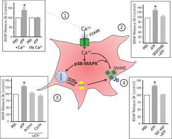
Major advances have been made in identifying the essential molecular components that regulate P2X4 receptor expression and the cellular machinery that drives release of microglial BDNF, a critical substrate for neuropathic pain (Fig. 2). These discoveries have built a complete framework for understanding microglia-to-neuron signaling in neuropathic pain, positioning the P2X4 receptor as a core signaling pathway necessary for ongoing expression of tactile allodynia following nerve injury.
ATP activation of P2X4 receptors causes the synthesis and release of BDNF from microglia through a Ca2+and p38-MAPK-dependent signaling cascade. Activation of P2X4 receptors expressed on the cell surface instigates a series of conformational changes that allow cations, such as Ca2+ and Na+, entry into the cell through a non-selective channel. In a recent study, we reported ATP stimulation causes a biphasic release of BDNF and a concomitant increase in BDNF protein level within primary cultured microglia (Trang et al., Reference Trang, Beggs, Wan and Salter2009). Although drug treatment affected both BDNF accumulation and release, points 1–4 only illustrate the effects on the peak BDNF release response at 60 min post ATP stimulation: (1) in the presence of extracellular Ca2+, activating P2X4 receptors with ATP causes the release of BDNF; (2) treatment with the p38-MAPK inhibitor SB203580 prevents BDNF release; (3) inhibiting transcription or translation with actinomycin-D or cycloheximide, respectively, prevents BDNF release; and (4) microglia treated with TAT-NSF, which interferes with NSF hexamerization necessary for SNARE-dependent exocytosis, abolishes BDNF release. The current model is that influx of Ca2+ through P2X4 receptors drives the activation of p38-MAPK that leads to increase in the SNARE-dependent release per se of BDNF and to an increase in BDNF synthesis in microglia. Modified from Trang et al. (Reference Trang, Beggs, Wan and Salter2009).

A key cellular intermediary in the P2X4 receptor signaling pathway is p38-MAPK. Activation of this kinase is a point of convergence for involvement of microglial P2X7 and P2Y12 receptors in neuropathic pain, and for macrophage P2X4 receptor-mediate inflammatory pain. However, in contrast to the P2X4 receptor signaling pathway that has been systematically dissected, the upstream and downstream components of the P2X7 and P2Y12 receptor pathways have yet to be uncovered. Thus, the next major challenges are to determine the significance of this convergence in signaling for microglia function and how the components of P2X7 and P2Y12 receptor signaling contribute to aberrant spinal nociceptive processing in neuropathic pain.
ACKNOWLEDGEMENTS
This work was supported by grants from the Canadian Institutes of Health Research (CIHR; grant number MT-11219), the Krembil Foundation and the Ontario Research Fund Research Excellence Program. M.W.S. holds a Canada Research Chair (Tier I) in Neuroplasticity and Pain, and is the Anne and Max Tanenbaum Chair in Molecular Medicine at the Hospital for Sick Children. T.T. was supported by a CIHR fellowship.
Statement of interest
None