Implications
Maize and soybean are two of the principle ingredients in animal feed, and as such zearalenone (ZEA) and soybean isoflavones (ISOs) are often found together in maize–soybean meal-based feed. Our study shows that diets containing ISO may accelerate the biotransformation and degradation of ZEA and its metabolites, and thus reduce the residues of ZEA in liver and muscle tissues. These findings suggest a potential means of mitigating the negative effects of high concentration of ZEA contamination in feed.
Introduction
Zearalenone (ZEA) belongs to a group of mycotoxins with strong estrogenic effects. It is produced as a secondary metabolite by a number of Fusarium species, including Fusarium culmorum and Fusarium graminearum (Hestbjerg et al., Reference Hestbjerg, Nielsen, Thrane and Elmholt2002; Glenn, Reference Glenn2007), and is common in maize and wheat (Yamashita et al., Reference Yamashita, Yoshizawa, Aiura, Sanchez, Dizon and Arim1995; Binder et al., Reference Binder, Tan, Chin, Handl and Richard2007; Krska et al., Reference Krska, Welzig and Boudra2007). Among domestic animals and poultry, prepubertal female pigs appear to be the most sensitive to ZEA (Fink-Gremmels and Malekinejad, Reference Fink-Gremmels and Malekinejad2007). ZEA is rapidly absorbed after oral administration, and edema and reddening of the vulva are external clinical signs of ZEA intoxication in pigs (Zinedine et al., Reference Zinedine, Soriano, Molto and Manes2007). Malekinejad et al. (Reference Malekinejad, Maas-Bakker and Fink-Gremmels2006) reported that in pigs ZEA was converted into α-zearalenol (α-ZEA), predominantly catalyzed by 3α-hydroxysteroid dehydrogenase (3α-HSD). Soybean isoflavones (ISOs) are a subgroup of flavonoids found predominantly in soybean (Reinli and Block, Reference Reinli and Block1996) at concentrations ranging from 0.01% to 0.5% (Wang and Murphy, Reference Wang and Murphy1994). ISO is primarily active as an estrogen in low-estrogen environments and as an estrogen competitor in high-estrogen environments (Hwang et al., Reference Hwang, Kwak, Lim, Lee, Kang, Choe, Hur and Han2006). In addition, ISO has been shown to have antioxidant effects (Hsieh et al., Reference Hsieh, Wu and Hu2009).
Maize and soybean are two of the principle ingredients in animal feed, and therefore ZEA and ISO often coexist in maize–soybean meal-based feed, where they may interact. It is generally believed that ZEA has negative effects on animal production and human health (Zinedine et al., Reference Zinedine, Soriano, Molto and Manes2007), whereas, conversely, ISO has potentially beneficial effects on animal production(Greiner et al., Reference Greiner, Stahly and Stabel2001; Payne et al., Reference Payne, Bidner, Southern and Geaghan2001). Therefore, the present study focused on whether ISO could increase or reduce ZEA residues in muscle and liver tissues of prepubertal gilts. It was hoped that the study would suggest means of mitigating the negative effects of ZEA contamination of feed.
Material and methods
Animals and experimental design
The experiment was approved by the Institutional Animal Care and Use Committee at Huazhong Agricultural University, Wuhan, China, and conducted in accordance with the National Institute of Health guidelines for the care and use of experimental animals.
Seventy 75-day-old, prepubertal, female pigs (Duroc ×Landrace × Yorkshire) with an average body weight of 26.50 ± 0.60 kg were allocated to seven diet treatment groups and treated for 21 days as follows: (1) control group, fed the basal diet, (2) control+0.5 mg ZEA/kg, (3) control+0.5 mg ZEA/kg+300 mg/kg ISO, (4) control+0.5 mg ZEA/kg+600mg/kg ISO, (5) control+2.0 mg/kg ZEA, (6) control+2.0 mg/kg ZEA+300 mg/kg ISO and (7) control+2.0 mg/kg ZEA+600 mg/kg ISO. The concentration of ISO (300 or 600 mg/kg) corresponds to either 15% or 30% soybean in the diet, respectively. The concentration of ZEA (0.5 or 2.0 mg/kg) corresponds to either a low or high degree of ZEA contamination in the feed. Each treatment was replicated five times with two pigs per replicate. The pigs were housed in concrete floored indoor pens (two pigs of one replicate per pen), and all pigs had free access to feed and water throughout the 21-day study.
Diet preparation
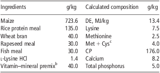
Soybean ISO (extracted from soybean, purity >99%) was obtained from Chengdu Tianyuan Natural Product Co., Ltd (Chengdu, China), and its composition was as follows: daidzein 531.1 mg/g, genistein 321 mg/g, daidzin 90.1 mg/g, glycitin 45.2 mg/g, genistin 17.1 mg/g and glycitein 2.1 mg/g, as determined according to Heimler et al. (Reference Heimler, Vignolini, Galardi, Pinelli and Romani2004). ZEA (purity >99%), obtained from Sigma-Aldrich (St. Louis, MO, USA) was diluted gradually to 1 mg/g before use with defatted rice bran. The basal diet was formulated to meet National Research Council (NRC, 1998) nutrient requirements, and did not contain soybean meal. The concentration of ISO (300 or 600 mg/kg) corresponds to 15% or 30% soybean in the diet, respectively. The ingredients and chemical composition of the basal diets fed to the control group is listed in Table 1. The concentration of mycotoxin in basal diet was determined according to Gimeno (Reference Gimeno1979) and Xu et al. (Reference Xu, Zhang and Chu1988).
Ingredients and chemical composition of the basal dietFootnote a
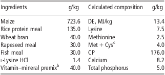
DE = digestible energy.
a Mycotoxin levels in the basal diet (per kg feed): zearalenone, 50 μg; aflatoxin B1, 2.5 μg; deoxynivalenol, 200 μg.
b The vitamin–mineral premix provided (per kg feed): vitamin A, 5000 IU; vitamin D3, 1000 IU; vitamin E, 10 IU; vitamin K3, 1 mg; vitamin B1, 1.50 mg; vitamin B2, 4.00 mg; vitamin B6, 1 mg; vitamin B12, 0.02 mg; niacin, 20 mg; d-pantothenic acid, 8 mg; biotin, 0.1 mg; folic acid, 0.2 mg; Fe, 120 mg; Cu, 160 mg; Mn, 32 mg; Zn, 200 mg; I, 0.2 mg; Se, 0.15 mg; and chloride choline, 0.2 mg.
c Met + Cys stands for the total content of methionine and cystine.
Tissue collection and determination of ZEA
At the end of the experiment, five pigs were selected from each treatment group (one pig at random from each replicate), fasted for 24 h, stunned electrically and exsanguinated. The pigs’ bodies were dissected, and the excised liver and longissimus dorsi muscle were stored at −20°C for subsequent determination of ZEA concentration. Partial samples of liver were excised, frozen in liquid nitrogen and stored at −70°C for subsequent protein extraction.
The residues of ZEA in liver and muscle tissues were measured using an enzyme immunoassay mix kit (R-Biopharm, R1401, Darmatadt, Germany), according to the manufacturer's protocol.
Western blot analysis
For protein extraction, ∼50 mg of liver tissue from five pigs from each treatment (one pig at random from each replicated) were homogenized on ice with a cold buffer containing proteinase, sonicated and then centrifuged at 12 000 × g for 10 min at 4°C. Supernatant was collected and stored at −20°C. The protein concentration was measured using the Bradford dye-binding procedure (Bradford, Reference Bradford1976) with bovine serum albumin as a standard. Protein lysate samples (50 μg) from the different treatment groups were boiled for 5 min in sample buffer, separated in 10% SDS–PAGE and transferred onto nitrocellulose membrane. After blocking in a 5% nonfat dry milk solution in washing buffer containing 10 mM Tris (pH = 7.5), 150 mM NaCl and 0.05% Tween 20, the membranes were incubated overnight at 4°C with a mouse-derived monoclonal antibody against 3α/3β-HSD (Abcam, USA, ab54606 and ab55268) at a dilution of 1 : 200, washed and then incubated for 2 h with a secondary antibody linked to the horseradish-peroxidase-labeled goat anti-mouse IgG (Wuhan Boster Biological Technology, Ltd, China, BA1050) at room temperature. Reactive protein was detected using an ECL chemiluminescence system (Amersham Pharmacia Biotech, UK). To measure the intensities of the bands, the electrophoresis films were scanned into a computer, and Photoshop software was used to convert the color into black/white image. Gray values of the bands (i.e. light intensities) were then measured using the image analysis software, AlphaEastFC.
Statistical analysis
The ANOVA analyses were performed on the premise of the homogeneities of variance by using SAS software. The results of the six experimental treatments were analyzed as a 2 × 3 factorial design using the ANOVA procedure of the SAS software (SAS Institute Inc., 1990), with ZEA and ISO supplementation levels and their interactions as main effects. These groups were then compared with each other using Tukey's multiple comparisons from the SAS program. The data are expressed as mean ± s.e. Differences were considered significant at the P < 0.05 level.
Results
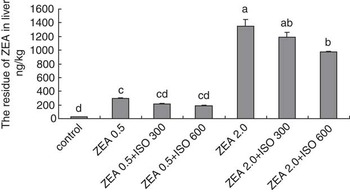
The homogeneity of variance test showed no significant differences among the overall variances in any of the following ANOVA analyses. As shown in Figure 1, pigs fed on a diet supplemented with ZEA showed significantly increased ZEA residues in liver tissue (P < 0.05). ZEA levels in the liver tissue of pigs fed on diets supplemented with 0.5 or 2.0 mg/kg ZEA reached 291.14 and 1352.06 ng/kg, respectively, compared with the control group levels of 26.31 ng/kg. Simultaneous addition of 600 mg/kg ISO to the 2.0 mg/kg ZEA-contaminated diet decreased the ZEA level in liver tissue compared with the diet with 2.0 mg/kg ZEA alone (P < 0.05).
ZEA residue levels in liver tissue of prepubertal gilts. The data are expressed as means ± s.e., n = 5. P-values of the main effects among the six experimental groups are: ZEA, P < 0.0001; ISO, P = 0.0014; ZEA × ISO, P = 0.0437. Mean values with different letter superscripts are significantly different from each other (P < 0.05). ZEA = zearalenone; ISO = isoflavone.

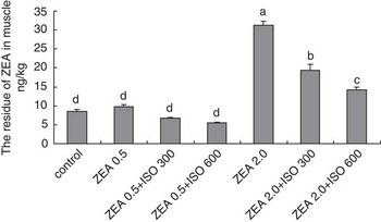
As shown in Figure 2, pigs fed on a diet supplemented with 2.0 mg/kg ZEA showed increased ZEA residue levels in muscle tissue (P < 0.05). Levels of ZEA in the muscle tissue of pigs fed the diet with 0.5 or 2.0 mg/kg ZEA added reached 9.71 ng/kg and 31.30 ng/kg, respectively, compared with the control group level of 8.51 ng/kg. Addition of 300 or 600 mg/kg ISO to 2.0 mg/kg ZEA-contaminated diet decreased residue level of ZEA in muscle tissue compared with the diet with 2.0 mg/kg ZEA added alone (P < 0.05).
ZEA residue levels in muscle tissue of prepubertal gilts. The data are expressed as means ± s.e., n = 5. P-values of the main effects among the six experimental groups are: ZEA, P < 0.0001; ISO, P < 0.0001; ZEA × ISO, P < 0.0001. Mean values with different letter superscripts are significantly different from each other (P < 0.05). ZEA = zearalenone; ISO = isoflavone.

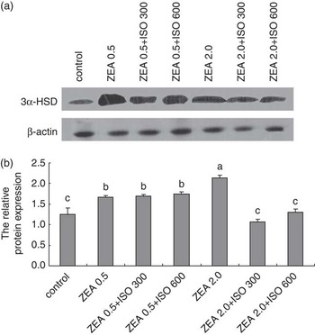
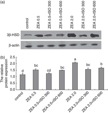
Figures 3 and 4 demonstrated that the diet supplemented with ZEA increased the protein expression of 3α/3β-HSD in liver tissue compared with the control group (P < 0.05). Diets with both 0.5 mg/kg ZEA and either 300 or 600 mg/kg ISO had no significant effect on the expression of 3α/3β-HSD compared with the diets containing 0.5 mg/kg ZEA alone (P > 0.05). Pigs offered ISO and 2.0 mg/kg ZEA showed significantly lower protein expression of 3α/3β-HSD than those offered diets with 2.0 mg/kg ZEA alone (P < 0.05).
Western blot analysis of the expression of 3α-HSD protein in liver tissue of prepubertal gilts. (a) Western blot analysis of 3α-HSD. (b) The data show the relative levels of 3α-HSD protein in liver tissue of prepubertal gilts. Protein expression was expressed relative to β-actin bands. The data are expressed as means ± s.e., n = 5. P-values of the main effects among the six experimental groups are: ZEA, P = 0.0074; ISO, P < 0.0001; ZEA × ISO, P < 0.0001. Mean values with different letter superscripts are significantly different from each other (P < 0.05). HSD = hydroxysteroid dehydrogenase; ZEA = zearalenone; ISO = isoflavone.

Western blot analysis of the expression of 3β-HSD protein in liver tissue of prepubertal gilts. (a) Western blot analysis of 3β-HSD. (b) The data show the relative level of 3β-HSD protein in liver tissue of prepubertal gilts. Protein expression was expressed relative to β-actin bands. The data are expressed as means ± s.e., n = 5. P-values of the main effects among the six experimental groups are: ZEA, P = 0.0006; ISO, P = 0.0038; ZEA × ISO, P = 0.1334. Mean values with different letter superscripts are significantly different from each other (P < 0.05). HSD = hydroxysteroid dehydrogenase; ZEA = zearalenone; ISO = isoflavone.

Discussion
ZEA is absorbed rapidly after oral administration, and the degree of absorption is difficult to measure owing to extensive biliary excretion. The uptake by a pig after a single oral dose of 10 mg/kg BW has been estimated at 80% to 85%, and significant serum levels have been measured in pigs within 30 min of feeding (Biehl et al., Reference Biehl, Prelusky, Koritz, Hartin, Buck and Trenholm1993). After absorption, ZEA is converted into α-ZEA or β-ZEA in the liver, predominantly by 3α/3β-HSD (Olsen et al., Reference Olsen, Pettersson and Kiessling1981). Malekinejad et al. (Reference Malekinejad, Maas-Bakker and Fink-Gremmels2006) reported differences among species in the hepatic biotransformation of ZEA, with α-ZEA being the main metabolite in the liver of the pig. In the present study, we determined ZEA residue levels in muscle tissue as representative of the edible organs of a pig. In addition, we measured ZEA residue levels in liver tissue, where ZEA is converted. In pigs fed the diet containing ZEA at 2.0 mg/kg for 21 days, residue levels of ZEA in liver and muscle tissues reached 1.35 and 0.031 μg/kg, respectively. James and Smith (Reference James and Smith1982) found that, after 4 weeks of feeding ZEA at 40 mg/kg of feed, residues in the liver varied between 78 and 128 μg/kg. Chickens fed with contaminated feed also showed ZEA residues in muscle and liver tissues (Mirocha et al., Reference Mirocha, Robison, Pawlosky and Allen1982). ZEA was detected at a mean concentration of 2.1 μg/kg BW in the livers of hens fed contaminated maize (1580 μg ZEA/kg maize; Danicke et al., Reference Danicke, Ueberschar, Halle, Matthes, Valenta and Flachowsky2002). All these studies demonstrate that ZEA residues accumulate in muscle and liver tissue when animals are fed diets contaminated with high levels of ZEA, and that these concentrations of ZEA increased with increasing amounts of ZEA in the feed.
ISO may act primarily as an estrogen in low-estrogen environments and as an estrogen competitor in high-estrogen environments (Hwang et al., Reference Hwang, Kwak, Lim, Lee, Kang, Choe, Hur and Han2006). In addition, ISO has been shown to elicit antioxidant effects (Hsieh et al., Reference Hsieh, Wu and Hu2009). We show that pigs offered ISO and 2.0 mg/kg ZEA simultaneously exhibited significantly lower protein expression of 3α/3β-HSD than those offered diets with 2.0 mg/kg ZEA alone. Zollner et al. (Reference Zollner, Jodlbaure, Kleinova, Kahlbacher, Kuhn, Hochsteiner and Lindner2002) reported finding large amounts of α-ZEA but only trace amounts of ZEA in muscle tissues of pigs fed on a diet of ZEA-contaminated oats. α-ZEA displays a stronger estrogenic effect than ZEA (Kuiper-Goodman et al., Reference Kuiper-Goodman, Scott and Watanbe1987). Olsen et al. (Reference Olsen, Pettersson and Kiessling1981) reported another biotransformation pathway for ZEA in animals, whereby ZEA and its reduced metabolites can conjugate with glucuronic acid, which is catalyzed by uridine diphosphate glucuronyl transferases (UDPGT). Following metabolism, these conjugates are excreted in the urine and bile fluid (Mirocha et al., Reference Mirocha, Pathre and Robison1981). Omoni and Aluko (Reference Omoni and Aluko2005) reported that UDPGT activity increased in various tissues in rats fed a high-ISO diet (0.81 mg/g) for 2 weeks. Our prior study indicated that ISO can counteract the estrogenic influence of a high dose of ZEA (2.0 mg/kg) in the diet of prepubertal gilts (Wang et al., Reference Wang, Zhang, Peng and Qi2010). Metabolism may accelerate the biotransformation and degradation of ZEA and its metabolites, and then decrease the estrogenic toxicity of ZEA and its metabolites.
In conclusion, we demonstrated here that muscle and liver tissues retained ZEA residues when pigs are fed on a diet contaminated with high concentrations of ZEA, and that the concentrations of ZEA in muscle and liver tissue increase with increasing amounts of ZEA in the feed. When diets are contaminated with high levels of ZEA, the addition of ISO may accelerate the biotransformation and degradation of ZEA and its metabolites, and reduce the levels of ZEA residues in liver and muscle tissue of prepubertal gilts.
Acknowledgments
This work was supported by the National Natural Science Foundation (Grant No.30771561/C120104), China, and by a National Nonprofit Institute Research Grant from CATAS-TCGRI (Grant No. 1630032012021).