Introduction
Due to the huge amount of waste discharged by industry and agriculture in recent decades, water pollution has become a serious global problem (Karimifard & Alavi Moghaddam, Reference Karimifard and Alavi Moghaddam2018). Contaminants such as heavy metals, volatile organic compounds, personal care products, pharmaceuticals, dyes, and spilled oils impair water quality to a significant extent (Foroutan et al., Reference Foroutan, Mohammadi, Razeghi and Ramavandi2019, Reference Foroutan, Mohammadi, Sohrabi, Sahebi, Farjadfard, Esvandi and Ramavandi2020). Many pollutants derived from dyes are applied widely in the fields of plastic, cosmetics, paper, rubber, textiles, and food processing. Each year ~7×10⁵ tons of dye effluent are produced (Thakur et al., Reference Thakur, Pandey and Arotiba2016).
Methylene blue (MB, C16H18N3SC·3H2O), an organic cationic dye, is a heterocyclic compound and a small amount of it in water can block sunlight penetration and inhibit the photosynthesis of organisms (Liu et al., Reference Liu, Omer and Ouyang2018). Moreover, MB is toxic, mutagenic, and carcinogenic (Kang et al., Reference Kang, Zhao, Wang, Zhang, Chen, Yi, Rao and Song2018). Long-term exposure to MB can cause serious problems for human health. Moreover, due to its stable and complex structure, MB is non-biodegradable and can last for a long time. Remediation of dye-contaminated effluents is essential, therefore.
Several techniques for remediation of dye effluent have been applied, such as chemical oxidation, ion-exchange, coagulation, electrolysis, membrane filtration, adsorption, and reverse osmosis (Astuti et al., Reference Astuti, Sulistyaningsih, Kusumastuti, Thomas and Kusnadi2019; Li et al., Reference Li, Zhao, Zhou and Yang2021). Among them, adsorption has proven to be the most favored because of cost-effectiveness and easy scalability (Peng et al., Reference Peng, Li, Liu and Song2017a; Zhu et al., Reference Zhu, Fan, Zhou and Li2019). Various kinds of adsorbents such as activated carbon (AC), zeolites, clay minerals, graphene, etc., have been used (Bujdák et al., Reference Bujdák, Jurečeková, Bujdáková, Lang and Šeršeň2009; Foroutan et al., Reference Foroutan, Khoo, Ramavandi and Abbasi2017, Reference Foroutan, Zareipour and Mohammadi2018; Uddin, Reference Uddin2017; Viglašová et al., Reference Viglašová, Galamboš, Danková, Krivosudský, Lengauer, Hood-Nowotny, Soja, Rompel, Matík and Briančin2018).
Montmorillonite (Mnt) is a typical layered aluminosilicate mineral with a 2:1 phyllosilicate structure composed of two silica tetrahedral sheets sandwiching one alumina octahedral sheet (Pauling, Reference Pauling1930; Marshall & Caldwell, Reference Marshall and Caldwell1946). Because of low-valence substitution in the octahedral and tetrahedral sheets, Mnt has a large cation exchange capacity and is used commonly to adsorb heavy metal ions and cationic dye pollutants (Tombácz & Szekeres, Reference Tombácz and Szekeres2004; Yan et al., Reference Yan, Wang, Yu, Wei, Du and Shan2007; Yi et al., Reference Yi, Ai, Zhao, Zhang and Song2020). The small interlayer spacing of natural montmorillonite is <1 nm. To increase the interlayer spacing, inorganic and organic pillar-bearing methods are often adopted to enhance the adsorption capacity by enlarging the interlayer spacing (Choudary et al., Reference Choudary, Durgaprasad and Valli1990; Ahenach et al., Reference Ahenach, Cool, Impens and Vansant2000). Because of weak interlayer interactions, sodium montmorillonite can be exfoliated easily into 2D montmorillonite nanolayers with thickness <2 nm and transverse dimension <200 nm through ultrasonic treatment to expose almost all the negatively charged surface and active sites (Wang et al., Reference Wang, Zhao, Yi, Chen, Kang, Li and Song2018; Chen et al., Reference Chen, Yuan, Zhao, Rao and Song2019b). The 2D Mnt should be able to adsorb a large amount of MB due to the strong electrostatic interaction between Mnt and dye cations. The separation of 2D Mnt from water is difficult, however.
Preparation of 3D composites composed of 2D Mnt is an effective strategy for solid–liquid separation. 3D Mnt/polymer hydrogels such as Mnt/chitosan, Mnt/carboxymethyl cellulose, and Mnt/polyacrylamide have been synthesized successfully for pollutant adsorption (Weian et al., Reference Weian, Wei and Yue'E2005; Youssef et al., Reference Youssef, Malhat, Abdel Hakim and Dekany2017; Kang et al., Reference Kang, Zhao, Wang, Zhang, Chen, Yi, Rao and Song2018; Wang et al., Reference Wang, Ni, Chen, Ai, Zhao and Song2020a, Reference Wang, Wang, Zhao, Bai, Huang, Zhang and Song2020b). 2D Mnt nanolayers are embedded in the 3D polymer hydrogel through hydrogen bonding and electrostatic interaction (Wang et al., Reference Wang, Wang, Ai, Li, Li, Peng, Zhao and Song2021). Graphene oxide and reduced graphene oxide are fascinating carbon materials and have been researched widely as adsorbents for a variety of wastewaters. Due to the large specific surface area, low density, and good chemical stability, composite aerogels of graphene oxide/Mnt(Yang et al., Reference Yang, Yu, He, Yu, Chen, Lu, Shu, Cui, Zhang and Jin2019) and reduced graphene oxide/Mnt(Yan et al., Reference Yan, Hu, Guo, Cai, Huang, Xiong and Tan2016) have been prepared for purification of wastewater polluted by metal ions and organic materials. The Mnt particles serve as a reinforcing agent to improve the structural stability and mechanical property of the aerogel. Few reports have been published, however, on the composite aerogel composed of 2D montmorillonite nanolayers and reduced graphene oxide for dye removal.
The objective of the present study was to assemble 2D Mnt and graphene oxide sheets into a three-dimensional aerogel (3D Mnt-rGO Gel) via a hydrothermal process to achieve easy solid–liquid separation of 2D Mnt used as adsorbents, and to assess the MB removal performance of Mnt-rGO Gel under various conditions.
EXPERIMENTAL
Materials
Mnt with a purity of >98% was purchased from Chifeng Ningcheng montmorillonite company (Chifeng, Inner Mongolia, China). Hydrochloric acid (HCl), sodium hydroxide (NaOH), methylene blue (MB), and flaky graphite with purity of >99.85% were purchased from Sinopharm Chemical Reagent Co. Ltd (Shanghai, China). All the reagents were of analytical grade, and used without purification. Purified water with a resistance of 18.25 MΩ·cm was obtained from Milli-Q Direct 8 (Millipore, Darmstadt, Germany), and used in all experiments.
Preparation of 2D Mnt
Two-dimensional montmorillonite nanolayers (2D Mnt) were prepared through sodium-modification and ultrasonic exfoliation. First, 50 g of Mnt was dispersed in 1 L of NaCl solution (5 wt.% suspension, 0.85 mole L–1) with magnetic stirring for 24 h at room temperature; then the Mnt was separated from the supernatant by centrifugation, and the supernatant was decanted and discarded. This was repeated two more times. Second, after the third wash with NaCl, the Mnt was washed three times with purified water. Third, purified water was then added to the final centrifugate and resuspended ultrasonically (60% amplitude for 4 min). Then, the Mnt suspension was centrifuged at 15,777×g (12,000 rpm, F15-6×100y) for 2 min to remove large particles. After centrifugation, 10 mL of the obtained suspension was extracted, weighed, and dried to calculate the concentration of 2D Mnt in the suspension. Finally, the 2D Mnt suspension was diluted to 30 mg L–1 for use.
Preparation of Mnt-rGO Aerogels
Graphite oxide (GO) was prepared using the modified Hummers’ method (Peng et al., Reference Peng, Li and Song2017b). Graphene oxide suspension was prepared as follows. First, 0.4 g of GO powder was dispersed in 100 mL of purified water with magnetic stirring for 20 min. Then, the GO suspension was sonicated at an amplitude of 40% for 20 min. Third, the exfoliated GO suspension was centrifuged at 15,777×g (12,000 rpm, F15-6×100y) for 10 min to obtain a graphene oxide suspension. The precipitate was collected and dried to calculate the concentration of the graphene oxide suspension obtained. Finally, the graphene oxide suspension was diluted to 1.5 mg mL–1.
Mnt-rGO hydrogel was prepared via a hydrothermal method. First, a specific amount of 2D Mnt suspension was added to 80 mL of graphene oxide suspension with a concentration of 1.5 mg mL–1. The mixed suspension was stirred for 5 min and then sonicated at 40% amplitude for 2 min. Next, the suspension was transferred to a 100 mL Teflon-lined stainless-steel bomb, which was then sealed and kept at 120°C for 6 h. After the hydrothermal process, the resulting hydrogel was freeze dried at –48°C under low pressure of 24 Pa for 24 h to obtain Mnt-rGO aerogels. The mass ratios of 2D Mnt to graphene oxide were 0.5:1, 1:1, and 1.5:1, respectively, and the corresponding Mnt-rGO aerogels were labeled as Mnt-rGO Gel-1, Mnt-rGO Gel-2, and Mnt-rGO Gel-3, respectively. The rGO hydrogel was synthesized by the same process without the addition of 2D Mnt.
Material Characterization
2D Mnt was characterized by atomic force microscopy (AFM, Multimode8, Bruker, Leipzig, Germany). Fourier-transform infrared spectrometry (FTIR, Nicolet 6700, Thermo Electron Scientific Instruments, Waltham, Massachusetts, USA) was applied to investigate the functional groups of the sample. X-ray diffraction (XRD, D8-Advance, Bruker AXS, Berlin, Germany) was used to analyze the mineral phase. The microstructures of the hydrogel were observed by scanning electron microscopy (SEM, JSM-7100F, Joel, Tokyo, Japan). The surface zeta potentials of the hydrogel were measured by a Zeta Analyzer (ZS90, Malvern, UK). X-ray photoelectron spectroscopy (XPS, Escalab 250Xi, Thermo Fisher Scientific, Waltham, Massachusetts, USA) was performed to analyze the surface atom state of the samples. Thermal stability was performed on a thermal gravimetric analyzer (TGA, SDTA851, Mettler Toldedo, Schwerzenbach, Switzerland) over a temperature range of 50–800°C with a heating rate of 10°C min–1 under a nitrogen atmosphere.
Adsorption Experiment
The adsorption of MB onto Mnt-rGO Gels was investigated by batch experiment. A certain mass of gel adsorbent was added to 200 mL MB solution contained in a 250 mL conical flask. The effects of suspension pH, initial MB concentration, temperature, 2D Mnt content in the aerogel, and adsorbent dosage on the MB removal performance were investigated. The adsorption experiments were conducted using a shaker bath (SHA-B, GuoHua, China) with an oscillating speed of 150 rpm at 25°C. The samples of MB solution were drawn by a syringe at appropriate time intervals and filtered through an organic membrane with a pore diameter of 0.22 μm.
The concentration of MB solution was measured using a UV-Vis spectrophotometer (UV-Orion AquaMate 8000, Waltham, Massachusetts, USA) at a wavelength of 664 nm. All the tests were repeated three times. The removal (R, %) and adsorption capacity (q, mg g–1) were calculated by the following equations:


where C 0 (mg L–1) and C t (mg L–1) are the initial and residual concentrations of MB in the suspension, respectively. V(L) and m(g) are the volume of MB solution and mass of the adsorbent, respectively. As a control group, MB adsorption experiments without adsorbent were conducted through the same process at various pH, MB concentrations, and temperature conditions. Because of stable chemical properties, the removal of MB under these control conditions was 0%.
Reusability
400 mg of Mnt-rGO Gel-3 was added to 200 mL of MB aqueous solution with an initial concentration of 25 mg L–1. After shaking for 9 h at 150 rpm and 25°C, the MB concentration in the supernatant was measured. The Mnt-rGO Gel-3 used in water was filtered with qualitative filter paper and regenerated by soaking in 20 mL of H2O2 (15%) solution. Then, the eluted Mnt-rGO Gel-3 was washed with deionized water and applied to MB adsorption. The above procedure was repeated four more times.
RESULTS AND DISCUSSION
Characterization of 2D Mnt and Mnt-rGO Gel
Atomic force microscopy (Fig. 1) was used to measure the size of exfoliated Mnt. Large aggregates composed of many small layers were observed from the 2D AFM image (Fig. 1a). The small layers were irregular in shape, and the length and width ranged from tens to hundreds of nanometers. The 3D AFM image (Fig. 1b) showed that the heights of all the 2D Mnt were <2 nm. The thickness distribution of the selected area marked by the red line (Fig. 1c) demonstrated that the thickness of the nanolayers was also <2 nm. Due to the theoretical layer spacing of Mnt being in the range of 0.96–1.5 nm, the AFM characterization suggested that the Mnt had been exfoliated fully into monolayers. Because of the positive charge on the edge and permanent negative charge on the surface of Mnt in water, the formation of agglomerates might be caused by the electrostatic attraction between different nanolayers (Chen et al., Reference Chen, Zhao, Chen, Bai, Zhang, Li, An and Song2019a).
AFM results of 2D Mnt: a 2D and b 3D images of Mnt; c thickness distribution of the selected area marked by the red line

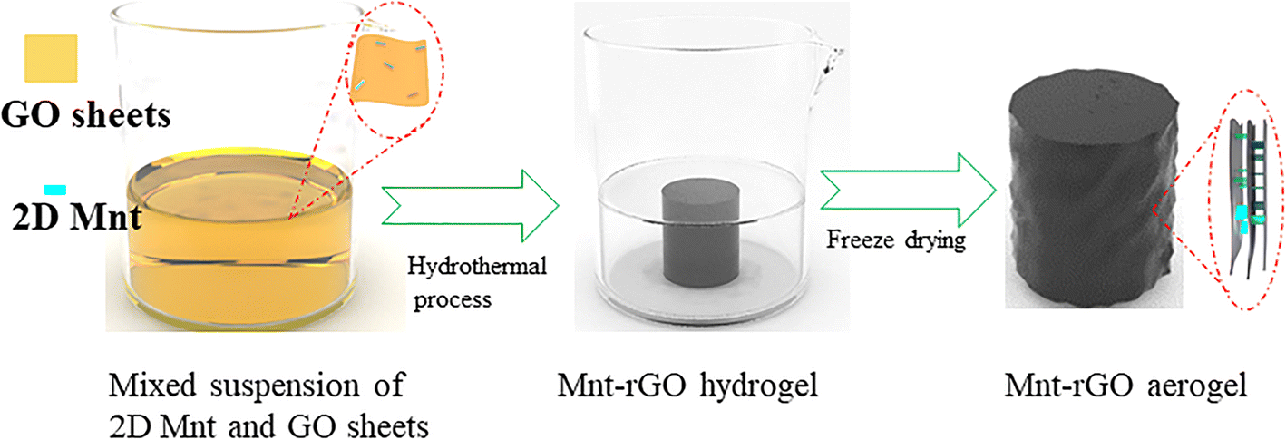
Mnt-rGO Gels were prepared through a hydrothermal process (Fig. 2). The pH of GO suspension was 3.50. In an acidic solution, the surfaces of the GO sheet and Mnt layer were negatively charged, and the edge of the Mnt layer was charged positively. Thus, in the mixed suspension of Mnt and GO, electrostatic repulsion existed between the surfaces of Mnt and GO, and electrostatic attraction existed between the Mnt edges and the surface of the GO sheets. Thus, due to the characteristics of surface potential and the small size of Mnt, Mnt may adsorb normally on the surfaces of the GO sheets to form spacers. During the hydrothermal process, GO sheets were reduced to have increased hydrophobicity. Driven by this hydrophobic force, the reduced GO sheets could approach each other and assemble into a 3D hydrogel. The Mnt adsorbed on the GO surfaces acted as spacers to prevent serious stacking of the graphene sheets.
Schematic illustration of the preparation process of Mnt-rGO aerogel

The photographs (Fig. 3a) of rGO Gel and Mnt-rGO Gels with various Mnt contents demonstrated that the rGO Gel and Mnt-rGO Gels had been prepared successfully through the hydrothermal process. With the increase in Mnt content, the volume of Mnt-rGO Gel (diameter and height) increased significantly, and the Mnt-rGO Gel-3 had the largest volume. The significant increase in volume indicated that the Mnt and graphene oxide sheets were assembled into a 3D structure during the hydrothermal process, which was beneficial for the separation of Mnt from water. X-ray diffraction (Fig. 3b) was used to characterize the Mnt, rGO Gel, and Mnt-rGO Gel with various Mnt contents. The diffraction peaks located at 7.15, 19.83, 28.43, 35.16, and 61.76°2θ corresponded to the (001), (100), (005), (110), and (300) crystal planes, respectively, of Mnt (Temuujin et al., Reference Temuujin, Jadambaa, Burmaa, Erdenechimeg, Amarsanaa and MacKenzie2004; Kang et al., Reference Kang, Zhao, Wang, Zhang, Chen, Yi, Rao and Song2018). The broad diffraction peak at 24°2θ was the characteristic peak of rGO, indicating the reduction of graphene oxide during the hydrothermal process (Alizadeh et al., Reference Alizadeh, Hosseini, Pourjavadi and Bahramian2020). In the patterns of Mnt-rGO Gels, diffraction peaks of both rGO and Mnt were observed, demonstrating the successful incorporation of Mnt into rGO Gel. As the 2D Mnt content increased, the peak intensity of rGO decreased gradually, and the peak intensities of Mnt increased gradually. Compared to that of rGO, the characteristic peaks of rGO in Mnt-rGO Gels became broad, suggesting a more disordered stacking of reduced graphene oxide sheets (Zhao et al., Reference Zhao, Liu, Wang, Sha, Li, Zheng, Amjadipour, MacLeod and Motta2017). The Mnt could, therefore, reduce the degree of restacking of the reduced graphene sheets.
a Images of rGO Gel and Mnt-rGO Gels with various Mnt contents, b XRD patterns of Mnt, rGO, and Mnt-rGO Gels, c FTIR curves of rGO, Mnt, and Mnt-rGO Gel, and d TGA curves of Mnt-rGO Gel

The FTIR measurements were used to characterize rGO Gel, Mnt, and Mnt-rGO Gel. The characteristic peaks of rGO Gel and Mnt (Fig. 3c) were noted on the XRD pattern for Mnt-rGO Gel. The peaks found at 1725, 1571, and 1226 cm–1 were assigned to stretching vibrations of the carboxyl groups, the conjugated carbon backbone, and the C–O–C or C–O–H groups, respectively (Das & Sharma, Reference Das and Sharma2020; Li et al., Reference Li, Sztukowska, Liu, Dong, Małaszkiewicz, Zhang and Wang2020; Wei et al., Reference Wei, Xiang, Ren, Cui, Zhou, Xiao, Jiang, Yao, Shen and Guo2020). The peaks found at 1033, 519, and 466 cm–1 were attributed to the bending vibrations of Si–O, Si–O–Al, and Si–O–Si, respectively (Temuujin et al., Reference Temuujin, Jadambaa, Burmaa, Erdenechimeg, Amarsanaa and MacKenzie2004; Zhao et al., Reference Zhao, Kang, Qin, Wang, Zhang, Song and Komarneni2020). The FTIR results further confirmed the formation of 3D Mnt-rGO composite Gel.
The thermal stability of Mnt-rGO Gel was evaluated using TGA (Fig. 3d). In the heating process, the Mnt-rGO Gel had two general stages of weight loss. The first stage, from 30 to 150°C, was the most abrupt and was attributed to the evaporation of physically adsorbed water molecules in Mnt-rGO Gel (Soares et al., Reference Soares, Nascimento, Menezes and Batista2004). The weight loss beyond 150°C was more gradual and probably was due to a combination of decomposition of oxygen-containing functional groups in rGO and of Mnt dehydroxylation (Lv et al., Reference Lv, Li, Jiang, Chang and Liao2015; Coros et al., Reference Coros, Pogacean, Turza, Dan, Berghian-Grosan, Pana and Pruneanu2020).
The SEM measurements (Fig. 4) were applied to characterize structures of rGO Gel and Mnt-rGO Gels with different Mnt contents. The rGO Gel was composed of a 3D reduced graphene oxide network with interconnected openings (Fig. 4a). The morphologies of Mnt-rGO Gel-1, Mnt-rGO Gel-2, and Mnt-rGO Gel-3 (Fig. 4b–d) also revealed a porous structure, which was not obviously different from the structure of rGO.
SEM images of a rGO Gel and b–dMnt-rGO Gels

MB-Removal Performance
Effect of pH on the removal performance of MB
The influence of pH on the removal of MB by Mnt-rGO Gel-3 was investigated at pH values of 2, 4, 6, 8, and 10, respectively. The concentrations of MB and Mnt-rGO Gel-3 were 25 and 200 mg L–1, respectively. MB removal at various pH values increased rapidly over the first 240 min (Fig. 5a), and then slowed to reach 100% in 540 min, suggesting the slow adsorption process of MB onto Mnt-rGO Gel-3. As the pH increased from 2 to 10, the rate of MB removal increased slightly, which suggested that the suspension pH had little or no influence on the adsorption of MB onto the Mnt-rGO Gel. The zeta potential of Mnt-rGO Gel-3 was negative in the pH range of 2–10(Fig. 6). As MB is a cationic dye, the removal of MB by Mnt-rGO Gel may involve electrostatic adsorption (Liu et al., Reference Liu, Omer and Ouyang2018). When the pH increased from 2.5 to 9.8, the zeta potential of Mnt-rGO Gel decreased slowly from –18 to –28 mV, which was consistent with the trend of MB removal increasing slightly with increasing pH (Fig. 5a). The Mnt-rGO Gel can be applied in a wide pH rage of 2–10, which is wider than polymer-Mnt Gel (Roufegari-Nejhad et al., Reference Roufegari-Nejhad, Sirousazar, Abbasi-Chiyaneh and Kheiri2019) and GO-Mnt/sodium alginate Gel (E et al., Reference Das, Ma, Yang and Hao2020).
Removal of MB with key parameters varied: a pH values, b 2D Mnt content; c initial MB concentrations; d adsorbent dosage; e temperature; and f reusability of Mnt-rGO Gel-3

Zeta potential of Mnt-rGO Gel and Mnt as a function of pH values

Effect of Mnt content on the removal performance
The effect of Mnt content in Mnt-rGO Gel on MB removal (Fig. 5b) was investigated at pH 8, and the concentrations of initial MB and adsorbent were 25 and 200 mg L–1, respectively. The removal of MB by rGO Gel increased slowly with time and reached 80% in 540 min. Compared with rGO Gel, Mnt-rGO Gel-1 removed MB more quickly in the first 240 min, and then the removal increased slowly to 95% in 540 min. In the same contact time, the performances of Mnt-rGO Gel-2 and -3 were improved even further, and complete removal was achieved after 540 min. 2D Mnt enhanced the MB adsorption performance of Mnt-rGO Gel. Due to the slow process of adsorption of MB on rGO, the slow process of removal of MB by Mnt-rGO Gel after 240 min might be caused by the rGO. As shown in Fig. 6, the negatively charged surface of Mnt with zeta potential of <–25 mV promoted the adsorption of electropositive MB through electrostatic interaction (Rakhsh et al., Reference Rakhsh, Golchin, Beheshti Al Agha and Alamdari2017). The inhibition by 2D Mnt of the restacking of graphene sheets might also increase the number of exposed adsorption sites, thereby enhancing the adsorption of MB onto rGO.
Effect of initial MB concentration on the removal performance
The effect of initial concentration of MB on the removal performance of Mnt-rGO Gel-3 (Fig. 5c) was tested at pH 8, with the concentration of Mnt-rGO Gel-3 fixed at 200 mg L–1. At a concentration of 25 mg L–1, the removal of MB reached 100% in 540 min. As the initial concentration of MB increased continuously to 100 mg L–1, the MB removal decreased significantly to 41%. At an initial MB concentration of 25 mg L–1, complete MB removal might be attributed to the notion that active sites of the Mnt-rGO Gel were sufficient to adsorb all the MB molecules. As the initial MB concentration increased, the number of active sites available to adsorb MB were reduced, leading to a continuous decrease in the rate of MB removal.
Effect of adsorbent dosage on MB removal
The removal of MB by Mnt-rGO Gel-3 with various dosages was conducted at pH 8 and MB concentration of 25 mg L–1, and the result is shown in Fig. 5d. The MB removal was 38% at the Mnt-rGO Gel-3 concentration of 50 mg L–1, indicating that the concentration of adsorbent was insufficient, and MB could not be adsorbed completely. As the concentration of Mnt-rGO Gel-3 increased from 50 to 200 mg L–1, the removal of MB showed a significant increase, and 100% MB removal was achieved at the Mnt-rGOGel-3 concentration of 200 mg L–1. As the concentration of Mnt-rGO Gel-3 further increased to 300 mg L–1, complete MB removal was achieved in a shorter contact time. The Mnt-rGO Gel-3 at a concentration of 200 mg L–1 could, therefore, adsorb all the MB molecules.
Effect of temperature on MB removal
The effect of temperature on MB removal was determined at temperatures of 15, 30, 45, and 60°C, respectively (Fig. 5e). The initial MB concentration and Mnt-rGO Gel-3 concentrations were 40 mg L–1 and 200 mg L–1, respectively. At 15°C, the MB removal reached 78% in 540 min. As the temperature increased to 30°C, the removal of MB increased significantly to 88% in 540 min. At 45 and 60°C, the removal increased further to 100% in 240 min. The improved MB removal performance caused by the temperature increase suggested that the adsorption of MB onto Mnt-rGO Gel was an endothermic process. The increase in temperature promoted the diffusion and adsorption process of MB molecules.
Reusability study
Five adsorption-desorption cycles for MB onto Mnt-rGO-3 were conducted at pH 8 with an initial MB concentration of 25 mg L–1 and a solution volume of 200 mL (Fig. 5f). The MB removal performance was stable for five cycles. After repeated use (four times), MB removal of Mnt-rGO Gel-3 was still as high as 97%, indicating good reusability.
Adsorption and Mechanism
Adsorption kinetics
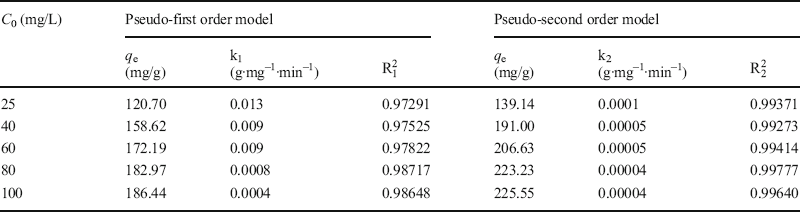
The adsorption capacity of MB removed by Mnt-rGO Gel-3 as a function of contact time was studied at pH 8 and 25°C (Fig. 7a). At an MB concentration of 25 mg L–1, the adsorption capacity increased rapidly in the first 240 min. After 240 min, the adsorption capacity increased slowly. Therefore, the adsorption of MB by Mnt-rGO Gel was a slow process. As the initial concentration of MB increased, the MB adsorption capacity did not increase significantly over the first 60 min. After that, the difference in adsorption capacity appeared gradually. At 540 min, the curve with an initial concentration of 25 mg L–1 was close to adsorption equilibrium, while the other curves were far from adsorption equilibrium, proving further that the adsorption process was slow. Pseudo-first order and pseudo-second order models were used to fit the experimental data to obtain a greater understanding of the adsorption kinetics (He et al., Reference He, Jiang, Chen, Jiang and Zhang2018).
a Adsorption kinetics and b adsorption isotherm of MB removed by Mnt-rGO Gel

Pseudo-first order model:

Pseudo-second order model:

where q t (mg g–1) and q e (mg g–1) are the adsorption capacity of MB at time t(min) and at equilibrium time, respectively, and k (g·mg–1·min–1) is the adsorption rate constant. The fitting results are presented in Fig. 7a and the parameters obtained from the two models given in Table 1. The correlation coefficient R2 of the pseudo-second order model was greater than that of the pseudo-first order. Therefore, the pseudo-second order could describe the adsorption process better. The q e was close to the experiment data, verifying the rationality of the fitting results.
Adsorption kinetic parameters for adsorption of MB onto Mnt-rGO Gel

Adsorption isotherm
The adsorption capacity of Mnt-rGO Gel as a function of equilibrium concentration was investigated at pH 8, 25°C, and the concentration of Mnt-rGO Gel-3 was 200 mg L–1. The MB adsorption capacity reached 196 mg L–1 at a MB equilibrium concentration of 19 mg L–1. Then, as the equilibrium concentration was increased to 78 mg L–1, the MB adsorption capacity of Mnt-rGO Gel-3 increased to 207 mg g–1, which was much greater than that of Go-Mnt/sodium alginate beads (141 .02 mg g–1) (E et al., Reference Das, Ma, Yang and Hao2020) and carboxymethyl cellulose/carboxylated GO composite beads (180.32 mg g–1) (Eltaweil et al., Reference Eltaweil, Elgarhy, El-Subruiti and Omer2020). The large adsorption capacity suggested the superior MB removal performance of Mnt-rGO Gel-3, which might be attributed to the adsorption of MB onto 2D Mnt and the increased adsorption surface of rGO resulting from the reduced restacking of graphene sheets.
The Freundlich and Langmuir models were used to fit the experimental data of the adsorption isotherm for a better understanding of the adsorption process (Foo & Hameed, Reference Foo and Hameed2010; Chen et al., Reference Chen, Chen, Bai and Li2013).
Langmuir isotherm model:

Freundlich isotherm model:

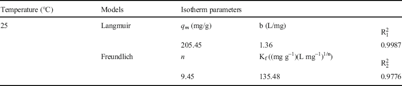
where q e (mg g–1) is the adsorption capacity at equilibrium, q m (mg g–1) is the maximum adsorption capacity, b (L mg–1) is the Langmuir constant, Kf ((mg g–1)(L mg–1)1/n ) is the Freundlich constant, n is the heterogeneity factor, and C e (mg L–1) is the concentration of adsorbates. The fitting isotherms for the adsorption of MB onto Mnt-rGO Gel-3 are shown in Fig. 7b and the parameters obtained from the fitting results are listed in Table 2. The Langmuir isotherm showed better fitting with a greater correlation coefficient, which suggested monolayer adsorption of MB onto the Mnt-rGO gel-3. The maximum adsorption capacity of MB obtained by the Langmuir model was 205 mg g–1, which was close to the experimental data.
Adsorption isotherm parameters for adsorption of MB onto Mnt-rGO Gel

Adsorption mechanism
The Mnt-rGO Gel showed improved adsorption performance for MB. In order to reveal the adsorption mechanism of Mnt-rGO Gel toward MB molecular, EDS, FTIR, and XPS were carried out. The distributions of hydrogel elements after MB adsorption (Fig. 8) suggested that the distributions of Si and Al elements were even, demonstrating the uniform distribution of 2D Mnt in the rGO Gel. The presence of a uniformly distributed N element proved the uniform adsorption of MB onto Mnt-rGO Gel surface.
SEM image of Mnt-rGO Gel after adsorption of MB; EDS maps of various elements (C, O, Si, Al, and N)

FTIR measurements were used to characterize MB and Mnt-rGO Gel before and after MB adsorption (Fig. 9a). The characteristic adsorption peaks of MB at 1602, 1396, 1252, 887, and 669 cm–1 corresponded to the v(=N+(CH3)2), δ(CH3), δ(C–H), γ(C–H), and v(C–S–C), respectively (Ovchinnikov et al., Reference Ovchinnikov, Evtukhova, Kondratenko, Smirnov, Khokhlov and Erina2016; Pang et al., Reference Pang, Fu, Ding, Lu, Li and Tang2017). The characteristic peaks of MB could be found on the spectra of Mnt-rGO Gel after adsorption, suggesting the successful adsorption of MB onto the Mnt-rGO Gel.
a FTIR spectra of MB, Mnt-rGO Gel before and after adsorption; XPS spectra: b survey scan and narrow spectra of c C1s, and d O1s

Compared with the XPS survey scan of Mnt-rGO Gel before MB adsorption, the presence of a weak peak of nitrogen on the scan after MB adsorption further confirmed the adsorption of MB on the hydrogel surface (Fig. 9b). As to the spectrum of C1s before MB adsorption (Fig. 9c), the peaks at 284.63, 286.28, 288.78, 290.11, and 291.70 eV were assigned to the C–C/C=C, C–OH, C–O––C, C=O, and O=C–OH bonds, respectively (Wei et al., Reference Wei, Chai, Cao and Jia2018). After MB adsorption, the peak at 285.25 eV was attributed to C–NH from MB (Zhou et al., Reference Zhou, Meng, Yi, Shu, Han, Zhu, Gao, Liu, Li, Yang and Yi2020). The peaks of C=C/C–C, C–OH, C–O–C, C=O, and O=C–OH had distinctly negative shifts. Because MB and graphene have the same aromatic rings, the negative shift of C=C/C–C indicated that MB molecules might interact with the π electrons of aromatic rings of graphene via π–π electron coupling. The negative shifts of C–OH, C–O–C, C=O, and O=C–OH suggested the interactions between oxygen containing functional groups and MB molecules. The peak intensities of C–O–C, C=O, and O=C–OH were weak, suggesting that the contents of these groups on reduced graphene oxide were small. The groups of C–O–C, C=O, and O=C–OH were not the main active sites for adsorption. On the O1s spectra (Fig. 9d), the peaks at 531.03, 531.83, 532.56, and 533.46 eV on the O1s spectra before MB adsorption were assigned to the Al–O, C–OH, Si–O, and –OH bonds, respectively (E et al., Reference Das, Ma, Yang and Hao2020). After MB adsorption, the peaks of C–OH and –OH had negative shifts, further confirming the interactions between MB and oxygen-containing groups of rGO and Mnt (Yan et al., Reference Yan, Hu, Guo, Cai, Huang, Xiong and Tan2016; Molla et al., Reference Molla, Li, Mandal, Kang, Hur and Chung2019). As for the negatively charged surfaces of rGO and Mnt, the positively charged MB might be adsorbed on rGO and Mnt via electrostatic interaction. Due to the nitrogen groups on MB molecules and oxygen-containing groups of rGO and Mnt, MB molecules might also adsorb onto rGO and Mnt via hydrogen bonding (Fig. 10).
Possible adsorption mechanism of MB adsorbed on Mnt-rGO Gel

Conclusions
A composite aerogel of reduced graphene oxide supported by 2D montmorillonite nanolayers was synthesized successfully via a hydrothermal method for easy solid–liquid separation. 2D montmorillonite nanolayers acted as spacers to inhibit the restacking of graphene sheets, which enhanced the performance of Mnt-rGO Gel for MB removal with a large adsorption capacity of 207 mg g–1. The adsorption mechanism of MB onto Mnt-rGO Gel might be attributed to the π–π interactions between aromatic rings of MB and graphene, hydrogen bonding, and electrostatic interactions between the nitrogen groups on the MB and oxygen-containing groups on the Mnt-rGO Gel.
ACKNOWLEDGMENTS
Financial support from the Fundamental Research Funds for the Central Universities (WUT: 2020IVA082) is acknowledged gratefully.
Funding
Funding sources are as stated in the Acknowledgments.
Declarations
Conflict of Interest
The authors declare that they have no conflict of interest.