Introduction
Phthalates are a group of colorless, odorless liquids which are used as plasticizers or softeners. Their principal use is to soften polyvinyl chloride (PVC). Plasticized PVC is used in a wide range of commercial products, including food packaging, PVC tubing, medical tubing and blood bags, toys, and cosmetics (Wormuth et al. Reference Wormuth, Scheringer, Vollenweider and Hungerbuhler2006). Phthalates can leach into foods heated in plastic containers. Mouthing of toys containing phthalates can also result in phthalate exposure. Individuals receiving feedings or transfusions through medical tubing containing phthalates are likely to be exposed to phthalates (U.S. EPA, 2007). Di(2-ethylhexyl) phthalate (DEHP), di-n-butyl phthalate (DBP) and benzyl-butyl phthalate (BBP) are now classified as reprotoxic substances [i.e. category 1B according to Regulation (EC), category 2 according to Directive 67/548/EEC], and the use of these phthalates is prohibited in the production of toys and childcare articles [Registration, Evaluation, Authorization and Restriction of Chemicals (REACH) Annex XVII, 51]. Since 2004, the use of DEHP, DBP, and BBP has been prohibited in cosmetics (Directive 2004/93/EC) and in materials to come into contact with food since 2007 (Directive 2007/19/EC). Di-iso-nonyl phthalate (DINP), di-iso-decyl phthalate (DIDP), and di(n-octyl) phthalate (DNOP) are restricted only in toys that can be placed into the mouth.
Phase 1 bio-transformations rapidly metabolize phthalates to their monoesters. Phthalate monoesters can be further metabolized by oxidation of the lipophilic aliphatic side-chain. Phthalate metabolites are excreted unchanged in urine and feces or as glucuronide conjugates in urine. Secondary oxidized DEHP metabolites like mono-2-ethylhexyl phthalate (MEHP), mono(2-ethyl-5-oxohexyl)phthalate (MEOP), and mono-2-ethyl-5-hydroxyhexyl phthalate (MEHHP) are most valuable biomarkers of DEHP exposure. For DBP, the simple monoester, mono-n-butyl phthalate (MBP), is the major metabolite excreted in urine and used as a biomarker of DBP exposure (Latini, Reference Latini2005). Elimination half-lives of the oxidized metabolites are longer than those of the monoesters. Koch and colleagues estimated an excretion half-life of 5 h for MEHP and 10–24 h for their oxidized metabolites (Koch et al. Reference Koch, Preuss and Angerer2006) and 3.3 h for MBP and 4.5–10.3 h for their oxidized metabolites (Koch et al. Reference Koch, Christensen, Harth, Lorber and Bruning2012).
Previous animal studies have reported that phthalate compounds might cause hyperactivity and impulsivity in rats (Masuo et al. Reference Masuo, Morita, Oka and Ishido2004). In humans, Engel et al. (Reference Engel, Miodovnik, Canfield, Zhu, Silva, Calafat and Wolff2010) reported that prenatal phthalate exposure was associated with problems with aggression, conduct, and attention, as well as with poor executive functioning and emotional control. Previously, we have reported an association between urine DEHP or DBP metabolite levels and attention deficit hyperactivity disorder (ADHD) symptoms (Kim et al. Reference Kim, Cho, Kim, Shin, Yoo, Kim, Yang, Kim, Bhang and Hong2009) and low intelligence (Cho et al. Reference Cho, Bhang, Hong, Shin, Kim, Kim, Yoo, Cho and Kim2010) in a cross-sectional study of community-dwelling Korean children. However, no studies have been conducted in children who have a confirmed diagnosis of ADHD obtained through meticulous diagnostic testing.
At this point, additional research is needed to investigate the contribution of phthalates to the pathophysiological mechanisms leading to ADHD, related externalizing behaviors, and cognitive problems. First, case-control studies are needed to examine whether phthalate metabolite concentrations are higher in children with ADHD than in healthy controls. Second, studies of clinical sample of ADHD should be conducted to investigate which phenotypes of ADHD were best attributed to the phthalates. Third, neurobiological studies are required to analyze how phthalates affect the neural system, leading to cognitive and/or behavioral changes. Recent animal toxicity studies have indicated that exposure to phthalates has a negative impact on the development of the hippocampus in male rats (Smith et al. Reference Smith, Macdonald and Holahan2011); however, the effects of phthalates on human brain development have not been established.
In this study, three levels of analyses were conducted. First, we conducted a case-control study comparing DEHP or DBP metabolite levels between children with and without ADHD. Second, we investigated urine DEHP or DBP metabolite concentrations in relation to the clinical subtype of ADHD diagnosed or each of the individual behavioral measures conducted in a sample of children with ADHD. Third, we investigated the correlation between urine phthalate metabolite concentration and cortical thickness (CT). Based on the results of previous studies (Kim et al. Reference Kim, Cho, Kim, Shin, Yoo, Kim, Yang, Kim, Bhang and Hong2009; Engel et al. Reference Engel, Miodovnik, Canfield, Zhu, Silva, Calafat and Wolff2010), we hypothesized that DEHP or DBP metabolites in urine would be higher in children with ADHD than in those without ADHD and that these metabolites would be associated with symptom severity. We also hypothesized that phthalate metabolite levels would be negatively correlated with CT in the right frontal and temporal lobes, which are the cortical regions most closely related to aggression and externalizing behaviors (Wahlund & Kristiansson, Reference Wahlund and Kristiansson2009; Fahim et al. Reference Fahim, He, Yoon, Chen, Evans and Perusse2011) and where CT has been found to be reduced in ADHD patients (Seidman et al. Reference Seidman, Valera and Makris2005; Makris et al. Reference Makris, Biederman, Valera, Bush, Kaiser, Kennedy, Caviness, Faraone and Seidman2007; Almeida Montes et al. Reference Almeida Montes, Prado Alcantara, Martinez Garcia, De La Torre, Avila Acosta and Duarte2013).
Materials and method
Participants and procedures
We recruited 180 medication-naïve children with ADHD from the child psychiatric clinic of the Seoul National University Hospital in South Korea. The recruited children were aged between 6 and 15 years and had been diagnosed with ADHD according to DSM-IV criteria, as ascertained by a child psychiatrist and a semi-structured interview. To diagnose ADHD and any co-morbid disorder, we used the Korean Kiddie-Schedule for Affective Disorders and Schizophrenia – Present and Lifetime Version (K-SADS-PL; Kim et al. Reference Kim, Cheon, Kim, Chang, Yoo, Kim, Cho, Seo, Bae, So, Noh, Koh, McBurnett and Leventhal2004). Exclusion criteria included the following: (1) a history of pervasive developmental disorder, mental retardation, bipolar disorder, psychotic disorder, obsessive compulsive disorder, or Tourette's syndrome; (2) a history of organic brain disease, seizure disorder, or other neurological disorder; (3) an IQ < 70; (4) the presence of learning disabilities or language disorders; (5) the presence of major depressive disorder, anxiety disorder, or tic disorder requiring drug therapy; and (6) any previous course of methylphenidate treatment lasting more than 1 year or occurring within the last 4 weeks.
The comparison group included participants of a research project entitled ‘The effects of pollution on neurobehavioral development and future policies to protect our children’, who were aged 8–11 years and recruited from ordinary primary schools in Korea from 2008 to 2009 (Cho et al. Reference Cho, Bhang, Hong, Shin, Kim, Kim, Yoo, Cho and Kim2010). Phthalate metabolites have been known to be influenced by sex and age (CDC, U.S. Department of Health and Human Services, 2005). Because the patient and control groups differed with respect to age ranges and gender ratios, we used a selection method to match the age and gender distributions. First, among the 180 children with ADHD, 87 who were aged 8–11 years were selected. Among these 87 children with ADHD, only nine were female. Consequently, we excluded female patients entirely and compared the remaining 78 male patients with male controls. Among 473 boys from the control group who had participated in the diagnostic interview, 35 boys met DSM-IV criteria for ADHD (21 inattentive type, nine hyperactive-impulsive type, five combined type). These boys were excluded from the control group and were used as a non-referred ADHD group. The remaining 438 boys comprised the non-ADHD group.
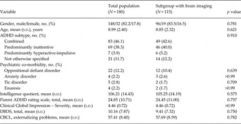
The parents completed the Korean version of the ADHD Rating Scale-IV (ADHD-RS; So et al. Reference So, Noh, Kim, Ko and Koh2002), the Disruptive Behavioral Disorder Rating Scale according to DSM-IV (DBDS; Silva et al. Reference Silva, Alpert, Pouget, Silva, Trosper, Reyes and Dummit2005), and the Children's Behavior Checklist (CBCL; Oh et al. Reference Oh, Lee, Hong and Ha1997). Certified child and adolescent psychiatrists who had completed inter-rater reliability training courses conducted the clinical assessments of children with ADHD using the Clinical Global Severity (CGI) scale (National Institute of Mental Health, 1970). The children with ADHD were administered the Korean version of the computer-based Continuous Performance Test (CPT; Greenberg & Waldman, Reference Greenberg and Waldman1993), named ADHD Diagnostic System (Shin et al. Reference Shin, Cho, Chun and Hong2000). Raw data on the CPT variables were converted to aged-adjusted T scores. Higher T scores indicated poorer test performance. Among 180 children with ADHD, a subset of 115 participants completed all assessments, including magnetic resonance imaging (MRI). This subset did not differ from the original participant pool in age, sex, or clinical characteristics (Table 1).
Demographic and clinical characteristics of total population of ADHD (N = 180) and subgroup of ADHD with brain imaging (N = 115)

ADHD, Attention deficit hyperactivity disorder; DBDS, Disruptive Behavioral Disorder Rating Scale according to DSM-IV; CBCL, Child Behavior Checklist.
We provided detailed information about the study to the parents and children and then obtained written informed consent before any child entered the study. The study protocol was approved by the institutional review board of Seoul National University Hospital.
Phthalate level measurements
The spot urine samples were collected in 50 ml sterile specimen containers at the outpatient clinics in the morning and were refrigerated at −20 °C immediately. Refrigerated specimens were transported to the laboratory within 2 h. The metabolites measured in this study included one primary metabolite of DBP, mono-n-butyl phthalate (MBP), and two secondary metabolites of DEHP, mono-2-ethylhexyl phthalate (MEHP) and mono(2-ethyl-5-oxohexyl)phthalate (MEOP). The urine phthalate metabolite concentrations were measured with high-performance liquid chromatography tandem mass spectrometry (Agilent 6410 triple Quad LCMS; Agilent, USA). Five hundred microliters of urine were buffered with 30 μl of 2.0 mol/l sodium acetate (pH 5.0) and then spiked with a mixture of isotope phthalate monoester standards (100 ng/ml) and 10 μl of β-glucuronidase. The sample was incubated at 37 °C for 3 h to deconjugate the glucuronidated phthalate metabolites. After incubation, 100 μl of 2 nmol/l hydrogen chloride was added to collect phthalate monoester. The extract was dried with nitrogen gas and reconstituted with 1 ml of high-performance liquid chromatography-grade water in a 2-ml glass vial. One blank and one quality control (QC) sample were included in each batch of samples. The QC sample was spiked with pooled urine and a mixture of phthalate monoester standard (100 ng/ml). The supernatants were purified by solid phase extraction with disposable Agilent C18 1.8 μm (2.1 × 50 mm). The mobile phase was 0.1% acetic acid water: 0.01% acetic acid acetonitrile (90:10, v/v) at a flow rate of 0.25 ml/min, and the eluates were monitored at target masses of 221, 293, and 291 and internal standard masses of 225, 297, and 95. We used the value (μg/l)/creatinine (g/l) for dilution correction in the analyses. Because the concentrations were not normally distributed, we used their natural log-transformed values.
Image acquisition and processing
Whole-brain structural MRI was acquired with a T1-weighted magnetization-prepared rapid acquisition gradient echo (MPRAGE) scan on a 3 T Siemens scanner (Siemens Magnetom Trio Tim Syngo MR B17, Germany). Images were acquired with the following parameters: TR = 1900 ms, TE = 3.13 ms, inversion time 900 ms, flip angle = 9°, voxel size 0.9 mm3, FOV = 230 mm, slices 176.
T1-weighted images were registered in the ICBM 152 average template using a linear transformation and corrected for intensity non-uniformity artifacts. The images were then classified into white matter (WM), gray matter (GM), cerebrospinal fluid (CSF) and background using an advanced neural net classifier. Hemispheric cortical surfaces were automatically extracted from each T1-weighted image using the Constrained Laplacian-based Automated Segmentation with Proximities (CLASP) algorithm, which reconstructed the inner cortical surface by deforming a spherical mesh onto the WM/GM boundary and then expanding the deformable model to the GM/CSF boundary (MacDonald et al. Reference MacDonald, Kabani, Avis and Evans2000). The reconstructed hemispheric cortical surfaces consisted of 40 962 vertices, each forming high-resolution meshes. The inner and outer cortical surfaces had the same number of vertices, and there was a close correspondence between the counterpart vertices of the inner and outer cortical surfaces. CT was defined using the t-link method, which captures the Euclidean distance between these linked vertices (MacDonald et al. Reference MacDonald, Kabani, Avis and Evans2000). For group analysis, each individual thickness map was transformed to a surface group template using a 2-dimensional (2D) surface-based registration that aligns variable sulcal folding patterns through sphere-to-sphere warping (Lerch & Evans, Reference Lerch and Evans2005).
Statistical analysis
We conducted three principal sets of statistical analyses.
First, we compared the urine phthalate metabolite (e.g. MEOP, MEHP, MBP) concentrations between clinic-referred boys with ADHD and boys without ADHD using independent-sample t tests and analysis of covariance (ANCOVA). In the ANCOVA, years of paternal and maternal education, both of which were significantly different between groups, were used as covariates. We also compared the urine phthalate metabolite concentrations between non-referred boys with ADHD and boys without ADHD.
Second, we conducted a comparison of the urine phthalate metabolite concentrations among the children with ADHD according to subtype. Because only seven children were hyperactive-impulsive subtype, we grouped the hyperactive-impulsive and combined subtypes together for the purpose of statistical analysis; these two subtypes of ADHD are known to have similar phenomenology and pathophysiology (Grizenko et al. Reference Grizenko, Paci and Joober2010).Twenty-one children with ADHD, not otherwise specified were excluded from this analysis. We also conducted linear regression analyses to elucidate the associations of urine phthalate metabolite concentrations with ADHD symptoms, externalizing behaviors, and attentional deficits among children with ADHD. Regression analyses were performed using a set of covariates based on established predictors of children's cognitive and behavioral functioning: age, gender, years of parental education, yearly income, subtype of ADHD, and IQ.
The statistical analyses above were performed using SPSS v. 21.0 (SPSS Inc., USA), with statistical significance defined as alpha level < 0.05.
Third, to investigate the correlation between brain CT and phthalate metabolite concentrations, multiple regression analyses were performed with urine phthalate metabolite concentrations, age, sex, and intracranial volume (ICV) as independent variables and each of the vertices of CT as a dependent variable. All the 81 924 vertices (40 962 per hemisphere) were used in statistical analysis. We utilized the SurfStat (K. Worsley, http://www.math.mcgill.ca/keith/surfstat/), which is a MATLAB toolbox (MathWorks Inc., USA) for the statistical analysis of univariate and multivariate surface data using linear mixed-effects models. The regression equation was defined as follows:
 $$Y{\sim}b_0 + b_1 \,{\rm urine}\,{\rm phthalate}\,{\rm metabolite}\,{\rm concentrations} + b_2 \,{\rm Age} + b_3 \,{\rm Sex} + b_4 \,{\rm ICV}$$
where Y is the CT, b 0 is the Y intercept, b 1–4 are the regression coefficients, and ε is the residual error.
Then, we included the ADHD-RS score and the DBDS score as well as age, sex, and ICV as covariates. The regression equation was defined as follows:
 $$ Y{\sim}b_0 + b_1 \,{\rm urine}\,{\rm phthalate}\,{\rm metabolite}\,{\rm concentrations} + b_2 \,{\rm Age} + b_3 \,{\rm Sex \,+\,} b_4 \,{\rm ICV} + b_5 \,{\rm ADHD} - {\rm RS \,+\,} b_6 \,{\rm DBDS} + \varepsilon $$
The results were thresholded at an uncorrected p value of 0.001 and at a false discovery rate (FDR)-corrected p value of 0.05.
Results
Characteristics and urine phthalate metabolite concentrations among children with ADHD
Table 1 shows the demographic and clinical characteristics of 180 clinic-referred children with ADHD (148 males and 32 females, age 6–15 years, mean age 8.99 ± 2.40 years). The combined type (46.1%) was the most common in our sample, followed by the inattentive type (38.3%). The ADHD, hyperactive-impulsive type and the ADHD, not otherwise specified (NOS) type were identified in 3.9% and 11.7% of the sample, respectively. The most common co-morbidity was oppositional defiant disorder (ODD) (12.2%), followed by tic disorder (2.8%). Anxiety disorder and tic disorder were found in 2.2% each of the sample.
The geometric mean (ln) concentrations of creatinine-corrected MEHP, MEOP, and MBP were 45.60 μg/g [geometric s.d. (GSD) = 2.41, range 2.95–892.65], 43.82 μg/g (GSD = 2.18, range 3.70–785.44), and 68.03 μg/g (GSD = 2.10, range 91.90–109.76), respectively.
Phthalate metabolite concentration differences between boys with and without ADHD
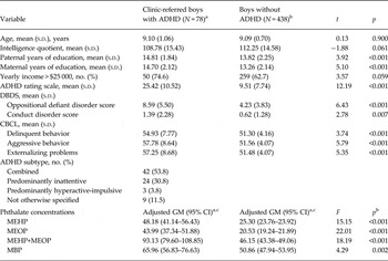
Table 2 shows comparisons between the clinic-referred boys with ADHD (age 8–11 years) and the age-matched control boys without ADHD. The geometric mean concentrations of MEHP (44.46 ± 2.14 v. 25.49 ± 2.41), MEOP (43.65 ± 1.89 v. 20.71 ± 1.94), and MBP (66.00 ± 1.93 v. 50.31 ± 1.79) were significantly higher in boys with ADHD than in boys without ADHD (p < 0.001 each). ANCOVA revealed that all significant group differences that were found in the t tests persisted even after adjusting for years of paternal and maternal education, both of which were significantly different between groups (Table 2). To cross-validate these findings, we compared the urine phthalate metabolite concentrations between non-referred boys with ADHD and boys without ADHD. The geometric mean concentrations of MEHP (29.68 ± 1.72 v. 25.49 ± 2.41, p = 0.158) and MEOP (23.79 ± 1.83 v. 20.71 ± 1.94, p = 0.230) were not significantly different between two groups. The geometric mean concentrations of MBP (60.13 ± 1.98 v. 50.31 ± 1.79, p = 0.085) were higher, although not statistically significant, in non-referred boys with ADHD than in boys without ADHD.
Comparison of boys with ADHD and boys without ADHD (age 8–11 years)

ADHD, Attention deficit hyperactivity disorder; DBDS, Disruptive Behavioral Disorder Rating Scale according to DSM-IV; CBCL, Child Behavior Checklist; GM, geometric mean; GSD, geometric standard deviation; MEHP, mono-2-ethylhexyl phthalate; MEOP, mono(2-ethyl-5-oxohexyl)phthalate; MBP, mono-n-butyl phthalate.
a Presented as loge-transformed creatinine-corrected concentrations (μg/g).
b Results of analysis of covariance (ANCOVA) test.
c Adjusted for years of paternal and maternal education.
Phthalate metabolite concentrations according to the subtype of ADHD
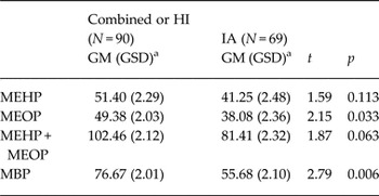
Table 3 shows comparison of phthalate metabolite concentrations according to the subtype of ADHD. We found that the children with the combined or hyperactive-impulsive subtype of ADHD exhibited higher MEOP and MBP concentrations compared to the children with the inattentive subtype (GM ± GSD: 49.38 ± 2.03 v. 38.08 ± 2.36 for MEOP, p = 0.033 and 76.67 ± 2.01 v. 55.68 ± 2.10 for MBP, p = 0.006, respectively).
Comparison of the urine phthalate metabolite concentrations between children with inattentive subtype of ADHD and children with combined or hyperactive-impulsive subtype of ADHD

ADHD, Attention deficit hyperactivity disorder; IA, predominantly inattentive; HI, predominantly hyperactive-impulsive; NOS, not otherwise specified; GM, geometric mean; GSD, geometric standard deviation; MEHP, mono-2-ethylhexyl phthalate; MEOP, mono(2-ethyl-5-oxohexyl)phthalate; MBP, mono-n-butyl phthalate.
Twenty-one children with ADHD, not otherwise specified, were excluded from this analysis.
a Presented as loge-transformed creatinine-corrected concentrations
Associations between phthalate metabolite concentrations and ADHD symptoms, externalizing problems, and CPT scores
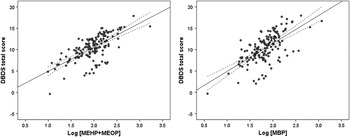
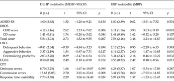
Table 4 shows associations between phthalate metabolite concentrations and clinical and neuropsychological variables among 180 children with ADHD. After adjusting for age, gender, years of paternal and maternal education, yearly income, subtype, and IQ, significant associations were found between urine DEHP or DBP metabolite concentrations and the CGI-S [B = 0.50, 95% confidence interval (CI) −0.10 to 0.90 for DEHP and B = 0.53, 95% CI 0.10–0.96 for DBP], ODD (B = 4.12, 95% CI 1.23–7.02 for DEHP and B = 6.11, 95% CI 3.03–9.10 for DBP), and total DBDS scores (B = 5.54, 95% CI 1.52–9.55 for DEHP and B = 7.56, 95% CI 3.24–11.87 for DBP). Urine DBP metabolite concentrations were also significantly associated with the aggressive behavior (B = 6.16, 95% CI 1.47–10.85) and externalizing problem (B = 2.16, 95% CI 0.46–10.22) scores. Urine DEHP metabolite concentrations were significantly associated with commission error (B = 13.63, 95% CI 3.65–23.61) and response time variability (B = 7.73, 95% CI 1.06–14.40) scores. We presented the relationships between the urine DEHP or DBP metabolites and the total scores on DBDS in Fig. 1.
Distribution of the DBDS total scores according to the natural-log transformed concentrations of urine DEHP and DBP metabolites after adjustment for age, gender, years of paternal and maternal education, yearly income, subtype, and IQ. DBDS, Disruptive Behavioral Disorder Rating Scale according to DSM-IV; DEHP, di(2-ethylhexyl) phthalate; DBP, di-n-butyl phthalate.

Associations [B coefficients (95% CIs)] between natural log-transformed urine phthalate metabolite concentration and the behavioral problem scales among children with ADHD (N = 180)

ADHD, Attention-deficit hyperactivity disorder; DEHP, di(2-ethylhexyl) phthalate; MEHP, mono-2-ethylhexyl phthalate; MEOP, mono(2-ethyl-5-oxohexyl)phthalate; DBP, di-n-butyl phthalate; MBP, mono-n-butyl phthalate; ADHD-RS, ADHD rating scale; CGI-S, Clinical Global Impression-Severity; DBDS, Disruptive Behavioral Disorder Rating Scale according to DSM-IV; ODD, oppositional defiant disorder; CD, conduct disorder; CBCL, Child Behavior Checklist, CPT, Continuous Performance Test.
B coefficients are with 1-unit increase in log-transformed urine DEHP or DBP metabolite concentrations.
Models were adjusted for age, gender, years of paternal and maternal education, yearly income, subtype, and intelligence quotient.
Correlations between phthalate metabolite concentrations and CT
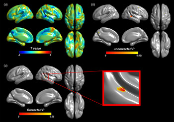
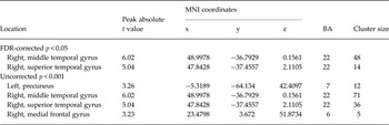
Among the 115 participants with imaging data (96 males, 19 females), a negative correlation was observed between urine DEHP metabolite concentrations and CT in right middle and superior temporal gyri at an FDR-corrected p < 0.05, after adjusting for age, sex, and ICV (Fig. 2, Table 5). This correlation remained even after adjusting for ADHD-RS and DBDS scores (online Supplementary Fig. S1, Supplementary Table S1). No significant correlation was found between CT in any brain area and urine DBP metabolite concentrations at an uncorrected p < 0.001.
Correlation analysis between CT and urine DEHP metabolite concentrations in 115 patients with ADHD, controlling for age, sex, and intracranial volume. (a) Statistical t map with t value range of −6.0 to 6.0. Negative correlations are marked as blue and positive correlations are marked as red. Negative correlations were observed in the right superior and middle temporal gyri at (b) an uncorrected p < 0.001 and at (c) a FDR-corrected p < 0.05. CT, cortical thickness; DEHP, di(2-ethylhexyl) phthalate; ADHD, attention deficit hyperactivity disorder.

Brain areas of negative correlation of CT with urine DEHP metabolite concentrations in 115 patients with ADHD, controlling for age, sex, and intracranial volume

CT, Cortical thickness; DEHP, di(2-ethylhexyl) phthalate; ADHD, attention-deficit hyperactivity disorder; MNI, Montreal neurological institute; BA, Brodmann area.
All MNI coordinates of peak absolute t values are selected in the significant regions.
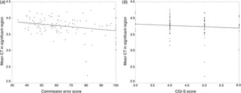
To further clarify the relationships between DEHP, CT in brain regions that were significantly correlated with DEHP metabolite concentrations (significant regions), and ADHD symptoms, we conducted multiple regression analysis using the ADHD-RS, DBDS, CGI-S, and CPT scores as a main predictive variable and mean CT in significant regions as a dependent variable. After controlling for age, sex, and ICV, mean CT in significant regions showed negative and significant correlations with commission error scores (T = −3.00, df = 111, p = 0.003) and negative, but not significant correlations with CGI-S scores (T = −1.42, df = 111, p = 0.157) (Fig. 3). No significant correlation was found between CT in significant regions and ADHD-RS (T = −0.66, df = 111, p = 0.511) or DBDS (T = −0.66, df = 111, p = 0.511) scores.
Correlations between mean CT in significant regions (right middle and superior temporal gyri) and commission error scores (a) and CGI-S scores (b) in 115 patients with ADHD, after controlling for age, sex, and intracranial volume. CT, cortical thickness; CGI-S, Clinical Global Improvement – Severity; ADHD, attention deficit hyperactivity disorder.

Discussion
In this study, three levels of analyses were conducted. First, we found that the concentrations of phthalates were significantly higher in clinic-referred boys with ADHD than in boys without ADHD. Second, we found that the concentrations of phthalates, especially the DBP metabolite concentrations, were significantly higher in the combined or hyperactive-impulsive subtypes of ADHD compared to the inattentive subtype and were associated with more severe externalizing symptoms. Third, we found that concentrations of urine DEHP metabolites were negatively correlated with CT in the right middle and superior temporal regions of the brains of ADHD patients.
To our knowledge, this is the first case-control study that has identified an association between phthalates and the diagnosis of ADHD obtained through meticulous diagnostic testing. Our results are consistent with previous studies that reported an association between prenatal maternal phthalate levels in urine and childhood externalizing problems (Engel et al. Reference Engel, Miodovnik, Canfield, Zhu, Silva, Calafat and Wolff2010) and between urine phthalate levels and ADHD symptoms in children (Kim et al. Reference Kim, Cho, Kim, Shin, Yoo, Kim, Yang, Kim, Bhang and Hong2009), suggesting the importance of both prenatal and postnatal phthalate exposure for childhood externalizing behaviors including ADHD. However, we did not find significant differences in the concentrations of urine phthalate metabolites between non-referred boys with ADHD and boys without ADHD, both of which were recruited from the school community and were ascertained by the same diagnostic instruments. These results raise a concern that observed differences in the concentrations of urine phthalate metabolites between clinic-referred boys with ADHD and boys without ADHD may be due to, at least in part, the discrepant sample selection (e.g. clinics v. community schools, K-SADS-PL v. DISC-IV).
However, there are some possible explanations for these discrepant results (significantly higher urine phthalate metabolite concentrations in the clinic-referred boys with ADHD, but not in the non-referred boys with ADHD, compared to boys without ADHD). It is notable that the concentrations of the urine DBP metabolite were significantly higher for the clinic-referred boys with ADHD than for the community-dwelling boys without ADHD and that the concentrations of the urine DBP metabolites for the non-referred boys with ADHD were between the other two groups. Also notable was that urine phthalate metabolite concentrations were higher in the combined or hyperactive-impulsive subtypes than the inattentive subtype of ADHD. Children with the combined subtype of ADHD exhibit more externalizing behaviors than those with the inattentive subtype, which influences treatment-seeking behaviors (Faraone et al. Reference Faraone, Biederman, Weber and Russell1998). In the present study, referred boys with ADHD are more likely to have combined subtype than non-referred boys with ADHD (53.8% v. 14.3%) and non-referred boys with ADHD are more likely to have inattentive subtype than referred boys with ADHD (60.0% v. 30.8%). Such different subtype composition between referred and non-referred boys with ADHD may have contributed, at least in part, to group differences in the concentrations of urine phthalate metabolites. In addition, children with ADHD who are referred to clinics usually suffer from a severe form of the disorder and many of them suffer from other co-mobid conditions, such as ODD (Gadow et al. 2007). From our data, it is possible to speculate that urine phthalate concentrations are associated with severity as well as the diagnosis of ADHD. The influence of phthalate metabolites on symptom severity and externalizing behaviors is further supported by the observed finding that there is a positive association between the behavioral problem scale scores and the urine DEHP or DBP metabolite concentrations.
Several studies have reported structural abnormalities in individuals with ADHD, such as a decrease in the volume of various encephalic structures or in CT in several brain regions, including frontal and temporal cortices (Seidman et al. Reference Seidman, Valera and Makris2005; Makris et al. Reference Makris, Biederman, Valera, Bush, Kaiser, Kennedy, Caviness, Faraone and Seidman2007; Shaw et al. Reference Shaw, Eckstrand, Sharp, Blumenthal, Lerch, Greenstein, Clasen, Evans, Giedd and Rapoport2007; Konrad & Eickhoff, Reference Konrad and Eickhoff2010; Almeida Montes et al. Reference Almeida Montes, Prado Alcantara, Martinez Garcia, De La Torre, Avila Acosta and Duarte2013). Our results find negative correlations between DEHP metabolite concentrations and CT in right middle and superior temporal gyri and suggest that DEHP may play an additional role in developmental delays or abnormal development in these brain regions in children. In addition, the urine DEHP metabolite concentrations were positively correlated with commission error scores, which were negatively correlated with mean CT in the right middle and superior temporal gyri. Commission errors are indicators of a deficit in response inhibition, which is considered to be a core deficit in ADHD (Aron & Poldrack, Reference Aron and Poldrack2005). Therefore, our results suggest that DEHP may have a negative impact on temporal maturation, possibly leading to deficits in response inhibition and increasing the risk of ADHD. However, there were no significant correlations between mean CT in significant regions and ADHD and externalizing symptom scores, suggesting that the association between DEHP levels and externalizing symptom severity may not be mediated by cortical change.
There were several limitations to this study. First, because phthalates have short biological half-lives and do not accumulate in the tissues of living organisms (Fennell et al. Reference Fennell, Krol, Sumner and Snyder2004; Koch et al. Reference Koch, Bolt and Angerer2004), the fact that only a single sample was acquired per participant limits our ability to estimate the effects of long-term exposure to these chemicals; however, several studies have reported moderate consistency of phthalate measurements over time (Hoppin et al. Reference Hoppin, Brock, Davis and Baird2002; Fromme et al. Reference Fromme, Bolte, Koch, Angerer, Boehmer, Drexler, Mayer and Liebl2007). When sources or exposure patterns are consistent, we can assume that a single measurement reflects a typical measurement. Even so, caution should be exercised when expanding our results to other phthalates because the vulnerability of the developing brain to chemicals reportedly depends on the duration of exposure (Tanida et al. Reference Tanida, Warita, Ishihara, Fukui, Mitsuhashi, Sugawara, Tabuchi, Nanmori, Qi, Inamoto, Yokoyama, Kitagawa and Hoshi2009). Second, in our case-control comparison, only boys within a specific age range were included; therefore, these results cannot be generalized to a broader population of children. Third, we only applied the parent-rated form of the ARS. This approach may have contributed to the non-significant association found between the phthalate metabolite concentrations and the ARS scores because the parent-rated form of the ARS has been reported to be less reliable and less valid than the teacher-rated form of the ARS in a Korean epidemiological study (Yang et al. Reference Yang, Kim, Kim, Cho and Kim2008). Fourth, there was no control group in the imaging analyses. To determine whether the association between the DEHP metabolite concentrations and the CT in the temporal region was specific to ADHD patients, we should have obtained both phthalate levels and imaging data from the healthy controls. Finally, because our data were cross-sectional and correlational, no inferences about causality are possible at this time. Therefore, further research using prospective designs is needed to investigate causality.
Conclusions
The results of this study suggest that DEHP or DBP metabolites can induce brain changes and externalizing disorders in children. This study is the first to reveal the impact of phthalate metabolites on the brains of children. Further neuroimaging and epigenetic studies are required to understand how phthalates affect the nervous system and to investigate the contribution of phthalates to the epigenetic pathophysiological mechanisms leading to cognitive and behavioral changes.
Supplementary material
For supplementary material accompanying this paper visit http://dx.doi.org/10.1017/S0033291714002694.
Acknowledgements
This study was supported by a grant from the Korean Health Technology R&D Project, Ministry of Health & Welfare, Republic of Korea (A120013 to B.-N.K.) and by the Basic Science Program through the National Research Foundation of Korea (NRF) grant funded by the Korean government (MEST) (2010-0002283 to J.-W.K. and 2011-0028333 to J.-M.L.).
Declaration of Interest
None.