Introduction
The feasibility of mammalian oocyte cryopreservation was first demonstrated about 35 years ago (Whittingham, Reference Whittingham1977) and despite significant recent progress over the past 20 years, the efficiency of oocyte cryopreservation is still very low.
The cryopreserved-warmed oocyte normally shows different alterations in the ultrastructural components such as disorganization of chromosomes, microtubules and actin microfilaments (Rojas et al., Reference Rojas, Palomo, Albarracı ´n and Mogas2004), disorganization of the spindle apparatus with the consequent risk of chromosomal loss and aneuploidy (Liu et al., Reference Liu, Sun, Li, Jiao and Wang2003), nuclear fragmentation (Men et al., Reference Men, Monson, Parrish and Rutledge2003), diminished plasma membrane selective permeability, microvilli loss, extensive ooplasm disorganization (Diez et al., Reference Diez, Duque, Gomez, Hidalgo, Tamargo, Rodriguez, Fernandez, de la Varga, Fernandez, Facal and Carbajo2005), and changes in the zona pellucida (Ghetler et al., Reference Ghetler, Skutelsky, Ben Nun, Bendor, Anihai and Shalgi2006).
With access to a reliable method for cryopreservation of mammalian oocytes beside its advantages on domestic animal breeding by genetic selection programs (Vajta, Reference Vajta2000) and its contribution on the maintenance of biodiversity through wildlife species conservation (Pukazhenthi & Wildt, Reference Pukazhenthi and Wildt2004), would increase the availability of materials for basic research in the field of reproductive biology.
The most encouraging results in domestic animals have been obtained in cattle, where vitrification of immature and mature oocytes has resulted in healthy offspring after in vitro fertilization and culture (Papis et al., Reference Papis, Shimizu and Izaike2000; Vieira et al., Reference Vieira, Mezzalira, Barbieri, Lehmkuhl, Rubin and Vajta2002). Although, the infrequent studies on vitrification of small ruminants’ oocytes, especially in ovine, have resulted to the poor developmental rates after vitrification of immature (Al-Aghbari & Menino Jr, Reference Al-Aghbari and Menino2002; Silvestre et al., Reference Silvestre, Yaniz, Salvador, Santolaria and Lopez-Gatius2006) and mature oocytes (Kelly et al., Reference Kelly, Kleemann, Kuwayama and Walker2006; Succu et al., Reference Succu, Leoni, Berlinguer, Mossa, Galioto and Naitana2007).
Many attempts have been conducted to improve oocyte cryopreservation protocols such as increased cooling and warming rates, using different types and concentrations of cryoprotectants, supplementation of vitrification solutions with cytoskeleton stabilizer (Vajta & Kuwayama, Reference Vajta and Kuwayama2006; Ledda et al., Reference Ledda, Bogliolo, Succu, Ariu, Bebbere, Leoni and Naitana2007), using different macromolecules in vitrification solution, using different cryodevice, and using oocytes at different developmental stage in the presence or absence of cumulus cells.
It is also known that the developmental stage at which the oocytes are frozen greatly influence the post-thaw survival rate, as such the in vitro matured oocytes are more permeable to cryoprotectants than immature freshly collected oocytes (Le Gal et al., Reference Le Gal, Basil and Vallet1993; Men et al., Reference Men, Monson and Rutledge2002). There are also controversial reports regarding the effects of cytoskeleton stabilizer on post-vitrification development of mammalian oocytes (Isachenko et al., Reference Isachenko, Soler, Isachenko, Perez-Sanchez and Grishchenko1998; Mezzalira et al., Reference Mezzalira, Vieira, Barbieri, Machado, Thaler Neto, Bernardi, Silva and Rubin2002; Vieira et al., Reference Vieira, Mezzalira, Barbieri, Lehmkuhl, Rubin and Vajta2002; Fujihira et al., Reference Fujihira, Kishida and Fukui2004; Silvestre et al., Reference Silvestre, Yaniz, Salvador, Santolaria and Lopez-Gatius2006; Bogliolo et al., Reference Bogliolo, Ariu, Fois, Rosati, Zedda, Leoni, Succu, Pau and Ledda2007; Zhang et al., Reference Zhang, Nedambale, Yang and Li2009).
To date, only a few papers have reported some progress on vitrification of ovine oocytes (Isachenko et al., Reference Isachenko, Alabart, Nawroth, Isachenko, Vajta and Folch2001; Silvestre et al., Reference Silvestre, Yaniz, Salvador, Santolaria and Lopez-Gatius2006) in which the best result has been reported by Succu et al. (Reference Succu, Bebbere, Bogliolo, Ariu, Fois, Leoni, Berlinguer, Naitana and Ledda2008). The present study was aimed to assess the effects of: (i) FBS concentration in vitrification solutions; (ii) equilibration time; and (iii) pre-treatment of oocytes with cytochalasin B on post-vitrification development of ovine immature (GVCOCs) and mature (MII) oocytes with (MIICOCs) or without cumulus cells (MIIDOs).
Materials and methods
Except when otherwise indicated, all chemicals were obtained from Sigma (St. Louis, MO, USA).
Oocyte collection
Sheep ovaries were collected from a local slaughterhouse (latitude: 32′17″ N; longitude 50′51″ E; altitude 2049 m) and transported to the laboratory in saline (30–35°C) in a thermos flask, within 1 to 3 h following collection. Ovaries were washed three times with prewarmed fresh saline (37°C), and all visible follicles with a diameter of 2–6 mm were aspirated using gentle vacuum (30 mmHg) via a 20-gauge short beveled needle connected to a vacuum pump. Prior to aspiration, the collecting tube was filled with 2 ml preincubated HEPES-buffered TCM, supplemented with 50 IU/ml heparin.
In vitro maturation
After aspiration, only oocytes surrounded by more than three layers of unexpanded cumulus cells (COCs: cumulus–oocyte complexes) were recovered and selected for in vitro maturation (IVM). Before culturing, oocytes were washed in HEPES-buffered TCM199 (H-TCM199) supplemented with 10% FBS, and 2 mM glutamine. The oocyte culture medium (OCM) consisted of bicarbonate-buffered TCM199 with 2 mM l-glutamine supplemented with 0.1 IU/ml FSH, 100 μl/ml penicillin, 100 μg/ml streptomycin, 10% FBS, and 0.2 mM Na-pyruvate. The medium osmolarity was adjusted to 280 mOsm. The COCs were distributed randomly in maturation droplets (10 oocytes in 50 μl) and covered by sterile paraffin oil in a 60-mm Petri dish (Falcon 1008; Becton Dickinson, Lincoln Park, NJ, USA) and were then incubated under an atmosphere of 5% CO2 –95% air with 100% humidity at 39°C for 22 h.
Experimental design
In all experiments the effects of cryopreservation on developmental competence of vitrified-warmed sheep oocytes were evaluated in terms of survival, cleavage, and blastocyst rates in both immatured (GVCOCs) and matured (MII) oocytes with (MIICOCs) or without cumulus cells (MIIDOs).
Prior to vitrification, the immature and mature oocytes were first cultured in IVM medium for 2 h and 22 h, respectively. Before fertilization, the vitrified-warmed GV and MII oocytes were incubated in IVM medium for 22 h and 2 h, respectively.
Experiment I: The effect of FBS concentration in base medium on post-vitrification development of oocytes
In this experiment, in total 2633 COCs with a compact cumulus investment were used to compare the effect of two concentrations of FBS in vitrification solutions (10 and 20%) on the ability of sheep oocytes to withstand vitrification and warming procedure. Four hundred and thirty-one COCs were considered as non-vitrified controls to establish the normal cleavage and blastocyst rates. As the only two blastocysts were achieved in the presence of 10% FBS, in the rest of experiment the 10% FBS was applied.
Experiment II: The effect of equilibration time on post-vitrification development of oocytes
In this experiment, a total of 3008 COCs with a compact cumulus investment was used. The oocytes were subjected randomly to the equilibration solution for 5, 7, or 10 min prior to vitrification. The effect of equilibration time on post-vitrification development was then assessed. Based on the results of this experiment 10 min (MIICOCs) and 7 min (GVCOCs and MIIDOs) equilibration time was considered for the next experiment.
Experiment III: The effect of pre-treatment of oocytes with cytochalasin-B on post-vitrification development of oocytes
In this experiment, a total of 3190 COCs with a compact cumulus investment was used.
Before vitrification, the oocytes in corresponding groups were distributed randomly in two groups and incubated in maturation medium with or without 7.5 μg/ml CCB for 30 min (Fujihira et al., Reference Fujihira, Kishida and Fukui2004). The effect of oocyte pre-treatment with CCB on post-vitrification development was then assessed.
Vitrification and warming procedures
All vitrification procedures were carried out at room temperature (25°C). The oocytes (GVCOCs, MIICOCs, and MIIDOs), before being exposed to the equilibration and vitrification solutions, were first transferred to the base medium for 1 min. The base medium for preparation of all vitrification solutions was prepared in HEPES-buffered TCM199 supplemented with antibiotics and 10% or 20% (v/v) FBS. One-step equilibration was performed, COCs or denuded oocytes were equilibrated (5, 7, or 10 min) in 200 μl of equilibration solution (7.5% EG plus 7.5% DMSO prepared in base medium). Vitrification solution (VS) was composed of 15% EG (2.76 M) plus 15% dimethyl sulphoxide (DMSO; 1.9 M) supplemented with 0.5 M sucrose (Vieira et al., Reference Vieira, Mezzalira, Barbieri, Lehmkuhl, Rubin and Vajta2002; Men et al., Reference Men, Monson, Parrish and Rutledge2003). The COCs were then transferred to the VS and loaded with a fine bore pasture pipette onto the inner surface of the tip of sharpened 0.25 ml straw (the straw was cut at an angle with the scalpel blade to resemble the tip of long beveled injection needle) with a the minimum volume (<0.1 μl) of vitrification medium (Shirazi et al., Reference Shirazi, Soleimani, Karimi, Nazari, Ahmadi and Heidari2010). After loading, almost all the solution was removed with a fine bore pasture pipette and the straw was quickly immersed in liquid nitrogen.
The time limit form the moment of oocyte exposure to VS and loading to the tip of straw before plunging into liquid nitrogen was less than 45 s. Oocytes were kept in liquid nitrogen for at least 1 week. Recovered oocytes were rinsed from cryoprotectants in warming solution (0.5M sucrose in base medium) for 5 min. The oocytes were then washed twice in base medium for 5 min, and finally the GV and MII oocytes, before being fertilized, were incubated in maturation medium for 22 h and 2 h, respectively.
Toxicity test
In order to assess the toxicity of the cryoprotectants and cytochalasin B (CCB) employed in different groups; the oocytes were assigned randomly to three toxicity groups, corresponding to the vitrification groups (GVCOCs, MIICOCs, and MIIDOs). In each group the oocytes were first exposed to the either CCB and VSs or vitrification solutions alone and then directly processed through the warming solutions. For all groups, following exposure to the warming solutions, the GV and MII oocytes were washed in H199 medium and transferred into the IVM drops for 22 h and 2 h, respectively and IVF (in vitro fertilization) was then performed. The non-treated oocytes were directly fertilized and cultured in vitro as control. The experiment was at least repeated three times.
Preparation of sperm and in vitro fertilization
Before transfer to fertilization drops, the oocytes were washed four times in H-SOF (HEPES-Synthetic Oviduct Fluid) and once in fertilization medium. The H-SOF and fertilization medium were the same as used by Tervit et al. (Reference Tervit, Whittingham and Rowson1972) with some modification as such for preparing H-SOF, 20 mM of NaHCO3 was substituted with 20 mM HEPES (10 mM free acid plus 10 mM Na salt). Both media were supplemented with antibiotics.
Fresh semen was collected from a Lori-Bakhtiari breed ram of proven fertility. For swim up, 80–100 μl of semen was kept under 1 ml of BSA-HSOF in a 15 ml conical Falcon tube at 39°C for up to 45 min. After swim up, the 700–800 μl of the supernatant was added to 3 ml of BSA-HSOF, centrifuged twice at 200 g for 3 min and the final pellet was resuspended with BSA-HSOF. Insemination was carried out by adding 1.0 × 10⁶ sperm/ml to the fertilization medium. The fertilization medium was SOF enriched with 20% heated inactivated estrous sheep serum. A 5-μl aliquot of sperm suspension, containing 1 × 10⁶ sperm/ml, was added into the fertilization drop (10 oocytes per 45 μl fertilization drop). Fertilization was carried out by co-incubation of sperm and oocytes in an atmosphere of 5% CO2 in humidified air at 39 °C for 22 h.
In vitro culture
After IVF, presumptive zygotes were vortexed for 2–3 min to remove the cumulus cells and then washed in H-SOF to remove spermatozoa and cellular debris. They were then allocated to 20 μl culture drops (five to six embryos/drop) consisted of SOF supplemented with 2% (v/v) BME-essential amino acids, 1% (v/v) MEM-nonessential amino acids, 1 mM glutamine and 8 mg/ml fatty acid free bovine serum albumin (BSA). The incubation condition was humidified 7% O2, 5%CO2, and 88% N2 at 39°C. On the third and fifth day of culture (day 0 defined as the day of fertilization) 10% charcoal stripped FBS was added to the medium. The culture was continued until 8 days post-fertilization. The osmolarity was maintained at 270 mOsm.
Statistical analysis
Data were collected over at least five replicates. The post-vitrification development of vitrified oocytes was analyzed using one-way ANOVA (analysis of variance). When ANOVA revealed a significant effect, the experimental groups were compared by Tukey method. When equal variance test was failed the treatments were compared by Student–Newman–Keuls method. When normality test failed the Kruskal–Wallis One-Way Analysis of Variance on Ranks was applied. A P-value < 0.05 level was considered to be significant (Sigma Stat Ver.2).
Results
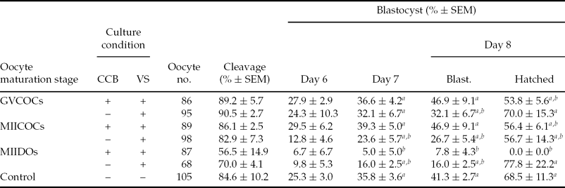
The results of the toxicity test revealed no significant differences in the rates of cleavage and day 6 blastocysts between toxicity test groups and control group. Although, in MIIDOs group when the oocytes were exposed to both CCB and VS, the rates of days 7 and 8 blastocysts and hatched blastocysts were decreased (P < 0.01) compared with the control and all other toxicity test groups (Table 1).
Developmental competence of ovine GV and MII stage oocytes after pre-treatment with cytochalasin B and cryoprotectant (cytotoxicity test)

a,bNumbers with different superscript letters in the same column differ significantly; P < 0.01.
GVCOCs: GV stage oocyte enclosed with cumulus cells; MIICOCs: MII stage oocyte enclosed with cumulus cells; MIIDOs: Denuded MII stage oocyte; CCB: cytochalasin B; VS: vitrification solution.
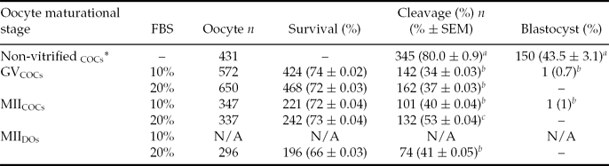
Despite the insignificant difference in survival rate of oocytes vitrified-warmed at different maturational stages using two different concentrations of FBS, the cleavage rate was significantly higher (P < 0.05) in MIICOCs oocytes when exposed to VSs containing 20% FBS compared with the vitrified-warmed oocytes in other groups (Table 2).
Post-warming development of ovine oocytes vitrified at GV and MII stages using two different concentrations of fetal calf serum (FBS)
a,b,c Numbers with different superscript letters in the same column differ significantly (P < 0.05).
*In this group the non-vitrified oocytes were subjected to in vitro maturation, in vitro fertilization, and in vitro culture.
GVCOCs: GV stage oocyte enclosed with cumulus cells; MIICOCs: MII stage oocyte enclosed with cumulus cells; MIIDos: Denuded MII stage oocyte.
As shown the rate of blastocyst was quite low in vitrified-warmed oocytes irrespective of oocyte nuclear maturational stage and FBS concentration. The only two blastocysts, however, both derived from those oocytes vitrified in the presence of 10% FBS in VSs. Moreover, the two blastocysts were derived from the GV and MII oocytes that had been enclosed with cumulus cells (Table 2). The rates of cleavage and blastocyst in vitrified-warmed oocytes were significantly lower than non-vitrified oocytes (Table 2).
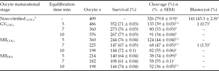
There was no significant difference in post-warming survival rate of ovine oocytes vitrified at GV and MII (with or without cumulus cells) stages with different equilibration times. Among vitrified-warmed oocytes, the highest cleavage rates were achieved in MIICOCs and MIIDOs oocytes (P < 0.05) which had been equilibrated for 10 and 7 min, respectively (Table 3). Although, the two resulting blastocysts had been obtained from GVCOCs and MIICOCs oocytes which had been equilibrated for 5 and 7 min, respectively. The rates of cleavage and blastocyst were obviously higher in non-vitrified oocytes compared with the vitrified oocytes (Table 3).
Effect of equilibration time on post-warming development of ovine oocytes vitrified at GV and MII stages
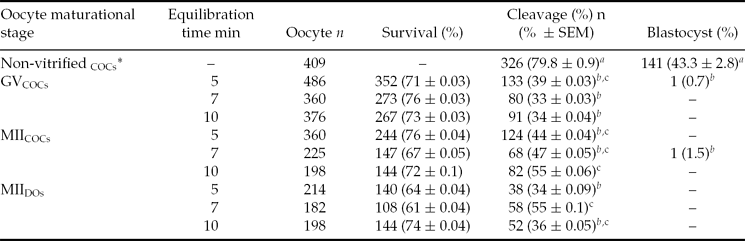
a,b,cNumbers with different superscript letters in the same column differ significantly (P< 0.05).
*In this group the non-vitrified oocytes were subjected to in vitro maturation, in vitro fertilization, and in vitro culture.
GVCOCs: GV stage oocyte enclosed with cumulus cells; MIICOCs: MII stage oocyte enclosed with cumulus cells; MIIDos: Denuded MII stage oocyte.
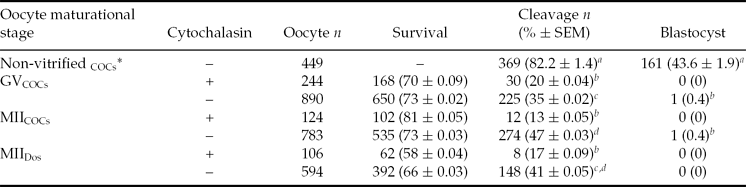
The survival rate of vitrified-warmed oocytes irrespective of oocyte maturational stage and the presence or absence of cumulus cells was not influenced by oocyte pre-treatment with CCB. The cleavage rate, however, was negatively influenced (P < 0.001) by the presence of CCB in all experimental groups (GVCOCs and MIICOCs, and MIIDOs). Additionally, the two blastocysts were obtained from those vitrified-warmed oocytes which had not been exposed to CCB (Table 4).
Effect of the presence of cytochalasin B on post-warming development of ovine oocytes vitrified at GV and MII stages
a–dNumbers with different superscript letters in the same column differ significantly (P < 0.01).
*In this group the non-vitrified oocytes were subjected to in vitro maturation, in vitro fertilization, and in vitro culture.
GVCOCs: GV stage oocyte enclosed with cumulus cells; MIICOCs: MII stage oocyte enclosed with cumulus cells; MIIDos: Denuded MII stage oocyte.
Discussion
It is known that cryotolerance of oocytes is complicated by their structural complexity, low surface area:volume ratio and relatively low plasma membrane permeability to water and/or cryoprotectants (Agca et al., Reference Agca, Liu, Peter, Critser and Critser1998; Leibo, Reference Leibo1980). Additionally, there are many contributing factors that influence the oocyte cryosurvival such as oocyte maturational stage, which has been reported to affect the quality and developmental competence of cryopreserved oocytes (Hochi et al., Reference Hochi, Ito, Hirabayashi, Ueda, Kimura and Hanada1998; Palasz and Mapletoft, Reference Palasz and Mapletoft1996).
In the current study, despite the lack of difference in post-vitrification survival rate between ovine oocytes vitrified at MII and GV stages, in vitro matured ovine oocytes were found to be more resistant to vitrification injury than immature oocytes in term of cleavage rate, particularly, when MIICOCs oocytes were vitrified in VS containing 20% FBS (53 versus 37% in MIICOCs and GVCOCs oocytes, respectively). This finding was in agreement with the reports in human (Boiso et al., Reference Boiso, Marti, Santalo, Ponsa, Barri and Veiga2002), cattle (Albarracin et al., Reference Albarracin, Morato, Izquirerdo and Mogas2005), porcine (Rojas et al., Reference Rojas, Palomo, Albarracı ´n and Mogas2004), and equine (Hurtt et al., Reference Hurtt, Landim-Alvarenga, Seidel and Squires2000; Tharasanit et al., Reference Tharasanit, Colenbrander and Stout2006a). Although, in another study in equine, the COCs vitrified at GV stage yielded higher cleavage rate than those vitrified after IVM (34 and 16%, respectively). Moreover, the vitrified-warmed GV oocytes enclosed with compacted cumulus cells did offer the best chance of having normal spindle and the potential for fertilization compared with GV oocytes with expanded cumulus cells (Tharasanit et al., Reference Tharasanit, Colleonione, Lazzarione, Colenbrander, Galli and Stout2006b). This controversy may vary depending on species involved and on cryopreservation procedures utilized. In various species, the difference in membrane lipid composition may affect the oocyte's ability to withstand chilling injuries, through acting on membrane fluidity (Zeron et al., Reference Zeron, Tomczak, Crowe and Arav2002).
Nevertheless, some possible causes for the lower developmental potential of GV oocytes compared with MII oocytes may include irreversible structural damage to the oocyte membrane (Arav et al., Reference Arav, Zeron, Leslie, Behboodi, Anderson and Crowe1996) and impaired intercellular communication between the oocyte and cumulus cell compartments. Indeed, the extensive cumulus cell membrane damage and reduced oocyte–cumulus cell communications after vitrification will impair oocyte maturation and its subsequent development (Gilchrist et al., Reference Gilchrist, Ritter and Armstrong2004; Li et al., Reference Li, Bunch, White, Rickords and Session2006).
In the current study the presence of cumulus cells during oocyte vitrification had a positive effect on subsequent embryo development in term of cleavage rate (53 and 41% in MIICOCs and MIIDOs oocytes, respectively; Table 2). Moreover, in all three experiments (Tables 2–4), the resulting blastocysts were obtained from those vitrified oocytes which had been enclosed with cumulus cells. Since in vitrification procedure the oocytes could adversely be affected by the osmotic stress caused by the rapid concentration or dilution of cryoprotectants, the higher developmental potential of vitrified-warmed MIICOCS compared with the MIIDOs could be related to the protective effect of cumulus cells against the osmotic stress (Imoedemhe & Sigue, Reference Imoedemhe and Sigue1992). Additionally, the cumulus cells could stabilize the oocyte structure and prevent the morphological damages caused by the vitrification–warming procedure (Park et al., Reference Park, Chung, Cha, Hwang, Lee and Lim2001). This finding, however, was in contrast to what reported by Zhang et al. (Reference Zhang, Nedambale, Yang and Li2009), indicating that the presence of cumulus cells had no effect on post-warming survival and subsequent cleavage and blastocyst rates of vitrified ovine matured oocytes.
It has been also shown that the presence of cumulus cells may minimize the release of cortical granules and prevent premature zona reaction; thereby it improves the fertilization rates after oocyte cryopreservation (Vincent et al., Reference Vincent, Pickering and Johnson1990). Conversely, the presence of cumulus cells interferes with the permeability of cryoprotectants to porcine oocytes (Fujihira et al., Reference Fujihira, Nagai and Fukui2005). In this context, the vitrified bovine matured oocytes without cumulus cells had a higher post-warming survival rate and embryo development, up to the 8-cell stage, compared with the cumulus-enclosed MII oocytes (Chian et al., Reference Chian, Kuwayama, Tan, Tan, Kato and Nagai2004). These controversies regarding to the effect of cumulus cells on post-vitrification development of oocytes need to be more elucidated.
The presence of macromolecules such as FBS alters the temperature at which the solution vitrifies (Shaw et al., Reference Shaw, Kuleshova, MacFarlane and Trounson1997) and thereby reduce the amount of intracellular cryoprotectants required to achieve vitrification (Kuleshova et al., Reference Kuleshova, Shaw and Trounson2001; Shaw et al., Reference Shaw, Oranratnachai, Trounson, Trounson and Gardner2000), which in turn reduce the toxicity of the solution. It also prevents the conversion of the zona pellucida proteins to a state known as zona hardening (Carroll et al., Reference Carroll, Depypere and Matthews1990 George et al., Reference George, Johnson and Vincent1992) and preserves the condition for normal fertilization after oocyte cryopreservation.
In the current study the 20% FBS concentrations in VSs was more protective than 10% FBS in oocyte cryopreservation, as illustrated by the higher cleavage rate in MIICOCs oocytes compared with other groups (Table 2). However, the resulting blastocysts were obtained in oocytes which had been vitrified in the presence of 10% FBS. It seems the higher concentration of FBS in oocyte VS had a detrimental effect on blastocyst formation. In bovine, the high concentration of FAF-BSA, as a macromolecule, in oocyte VSs has adversely affected the subsequent embryo development to blastocyst (Checura & Seidel Jr, Reference Checura and Seidel2007).
In the current study the speed of embryo development following fertilization of vitrified-warmed oocytes was slightly lower than non-vitrified oocytes (data not shown), which was in accordance with vitrified bovine oocytes (Checura & Seidel, Reference Checura and Seidel2007). Apart from all possibilities the two main reasons for retarded embryo development following vitrification-warming procedures might be attributed to the decrease in total content of inherited maternal transcripts related to the several cell functions and developmental competence (Succu et al., Reference Succu, Bebbere, Bogliolo, Ariu, Fois, Leoni, Berlinguer, Naitana and Ledda2008) as well as impaired mitochondrial activity (Fu et al., Reference Fu, Shi, Zhang, Zhao, Yan, Hou, Zhou, Fan, Suo and Zhu2009; Nagai et al., Reference Nagai, Mabuchi, Hirata, Shoda, Kasai, Yokota, Shitara, Yonekawa and Hoshi2006). Beside mitochondrial fragmentation and its impaired activity, the extent of microtubule damage due to vitrification-warming procedure can negatively affect the normal distribution of mitochondria in vitrified oocytes (Van Blerkom, Reference Van Blerkom1991; Sun et al., Reference Sun, Wu, Lai, Park, Day, Prather and Schatten2001; Zhou and Li, Reference Zhou and Li2009).
It has been shown that the effect of equilibration time on the rate of morphologically normal porcine oocytes after vitrification is dependent on the presence or absence of cumulus cells. The oocytes enclosed with cumulus cells are needed more equilibration time compared with denuded oocytes (Fujihira et al., Reference Fujihira, Nagai and Fukui2005). In the current study there was no difference in survival rates of vitrified-warmed oocytes equilibrated at different times. Although in MIICOCs group there was a trend of an increased cleavage rate as the equilibration time was increased (Table 3). Moreover, the highest cleavage rate in MIICOCs (55%) and MIIDOs (55%) groups were achieved after 10 and 7 min equilibration, respectively. It seems that the oocytes enclosed with cumulus cells have needed more equilibration time compared with the cumulus-free oocytes, which confirm what reported by Fujihira et al. (Reference Fujihira, Nagai and Fukui2005). In denuded oocytes, however, the cleavage rate was negatively affected, although insignificant, as the equilibration time was increased (10 min). This finding was, probably, due to the increased exposure time to cryoprotectant and its toxic consequences (Table 3).
Cytochalasin B pre-treatment is used to reduce cryoinjury to oocytes and embryos during vitrification (Isachenko et al., Reference Isachenko, Soler, Isachenko, Perez-Sanchez and Grishchenko1998; Dobrinsky et al., Reference Dobrinsky, Pursel, Long and Johnson2000; Fujihira et al., Reference Fujihira, Kishida and Fukui2004). The effects of CCB on developmental potential of vitrified-warmed oocytes, however, are controversial and are dependent on the animal species and the method of vitrification used. Some works have found that CCB increases oocyte survival after vitrification of immature porcine oocytes (Isachenko et al., Reference Isachenko, Soler, Isachenko, Perez-Sanchez and Grishchenko1998; Fujihira et al., Reference Fujihira, Kishida and Fukui2004); while, in bovine CCB had no effect on both immature and mature vitrified bovine oocytes (Mezzalira et al., Reference Mezzalira, Vieira, Barbieri, Machado, Thaler Neto, Bernardi, Silva and Rubin2002; Vieira et al., Reference Vieira, Mezzalira, Barbieri, Lehmkuhl, Rubin and Vajta2002).
In agreement with Bogliolo et al. (Reference Bogliolo, Ariu, Fois, Rosati, Zedda, Leoni, Succu, Pau and Ledda2007), in the current study ovine oocytes pre-treatment with CCB had no effect on survival rates of vitrified-warmed oocytes. Although, the cleavage rate was negatively affected by CCB pre-treatment of oocytes. Moreover, the resulting blastocysts were exclusively obtained in oocytes vitrified in the absence of CCB (Table 4). This finding was in contrary to the report indicating the higher blastocyst development rate in oocytes treated with 7.5 or 10 μg/ml CCB compared with those treated with lower CCB concentration (Zhang et al., Reference Zhang, Nedambale, Yang and Li2009).
In toxicity test, oocyte exposure to the VS with or without CCB had no detrimental effect on rates of cleavage, blastocyst, and hatched blastocyst except for the MIIDOs group in which the rates of blastocyst, and hatched blastocyst was negatively influenced by the presence of CCB (Table 1). The question that why the post-warming development of vitrified oocytes in all treatment groups (GVCOCs, MIICOCs and MIIDOs) was negatively influenced by CCB pre-treatment need to be further investigated. How extent the inhibitory effect of CCB on oocyte's microtubules assembling, after vitrification-warming procedure, is reversible and thereby how the normal cleavage can be affected by delayed reassembling of microtubules in CCB pretreated groups may be the probable reason.
In conclusion, in our study condition the post warming development of vitrified ovine oocytes was quite low and partially influenced by the presence of cumulus cells, FBS concentration, and CCB pre-treatment.
Acknowledgements
The authors would like to acknowledge the Research Institute of Animal Embryo Technology for technical and financial supports, Shahrekord University and Shahrekord's slaughterhouse staff for their cooperation.