INTRODUCTION
The morphological diversity of spermatozoa in gastropods has been considered a guide to understanding phylogenetic and taxonomic relationships within the molluscs. The Muricidae are an important component of marine communities around the world, with more than 1150 species grouped into eight subfamilies (Vokes, Reference Vokes1996), and it is among the many groups of marine gastropods that need a comprehensive revision (Pastorino, Reference Pastorino2005). The subfamily Trophoninae is one of the most conspicuous groups of marine gastropods living currently around the southern tip of South America (Pastorino, Reference Pastorino2005). The genus Trophon Monfort, 1810, includes at least 15 species (Pastorino, Reference Pastorino2005). In despite of the importance of this genus, biological knowledge about many species is extremely poor (Pastorino, Reference Pastorino2005). Trophon geversianus (Pallas 1774) shows the widest geographic distributional range and inhabits both rocky intertidal and shallow subtidal zones. In the south-western Atlantic this species is reported from 35°8′S to 56°8′S latitude, including the Malvinas (Falkland) Islands, while on the south-eastern Pacific coast it ranges from 42°8′S to 56°8′S (Castellanos & Landoni, Reference Castellanos and Landoni1993; Griffin & Pastorino, Reference Griffin and Pastorino2005; Pastorino, Reference Pastorino2005). Taxonomy and anatomy were the focus in the most of the studies on T. geversianus in the south-western Atlantic (Pastorino, Reference Pastorino2005), as well as some reproductive aspects including spawn characteristics, hatching modality and reproductive seasonality (Zaixso, Reference Zaixso1973; Penchaszadeh, Reference Penchaszadeh1976; Cumplido et al., Reference Cumplido, Averbuj and Bigatti2010).
Ultrastructural study of the spermatogenesis and sperm (euspermatozoa and paraspematozoa) morphology in T. geversianus is reported here for the first time; the eusperm, the typical and fertile sperm, and the parasperm, atypical sperm or non-fertile sperm (Healy & Jamieson, Reference Healy and Jamieson1981). Alternative theories have been advanced in an attempt to answer the role of paraspermatozoa. Buckland-Nicks & Chia (Reference Buckland-Nicks and Chia1976) consider the function of nutrition and/or stimulation of euspermatozoa by the products of paraspermatozoan breakdown. Fretter & Graham (Reference Fretter and Graham1962) suggest the ‘transportation’ of euspermatozoa during and after sperm transfers from male to female, via mobile associations of euspermatozoa. Buckland-Nicks (Reference Buckland-Nicks1998) suggest paraspermatozoa may be involved in creating a hostile pre-fertilization environment for rival sperm.
Comparisons are made with other caenogastropods, in particular with other neogastropod taxa, to identify possible diagnostic sperm features of the Muricidae and to assess the relationships with volutids and other neogastropods. These new ultrastructural descriptions may contribute to resolving some of the relationships of the Muricidae.
MATERIALS AND METHODS
A total of twenty Trophon geversianus were collected manually at depths of 5 m at low tide, in June and December of 2010, from Golfo Nuevo, Puerto Madryn (42°43′S 65°01′W), Argentina.
For light microscopy, gonadal tissues from ten individuals were fixed in Bouin's aqueous solution, dehydrated in a graded ethanol series, and embedded in methacrylate using standard procedures. Serial sections (5 µm thick) were stained with haematoxylin/eosin and with periodic acid-Schiff (PAS) for the demonstration of neutral glycoconjugates.
The different cell structures were identified and recorded by viewing and photographing tissue sections using a Zeiss Axiostar light microscope.
For transmission electron microscopy (TEM), the gonads of ten individuals were examined. After removal from the shell, pieces of the testis (9 mm3) or sperm duct were fixed in 2.5% glutaraldehyde in 0.1 M sodium phosphate buffer containing filtered seawater for 4 h at 4°C, and washed thoroughly either in sucrose-adjusted cacodylate buffer or in CaCl2-adjusted phosphate buffer. Subsequently the tissue pieces were placed in a 1% solution of osmium tetroxide (in 0.1 M cacodylate or phosphate buffer) for 1.5 h, and again washed in buffer. Tissues were dehydrated using an ascending series of ethanol (from 20% to absolute ethanol), then placed first in a 1:1 ethanol:propylene oxide solution for 15 min, and finally embedded in Araldite resin. Ultrathin sections (1 µm) were cut using either a Reichert or an LKB IV ultramicrotome and stained with uranyl acetate and lead citrate. All sections were examined and photographed using Philips EM 301 transmission electron microscopes operated at 75– 80 kV.
RESULTS
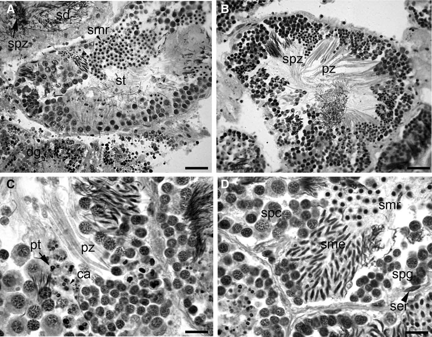
The testis of Trophon geversianus was in contact with the digestive gland, and both were surrounded by dense connective tissue. The testis consisted of numerous spermatogenic tubules and spermatic ducts separated from each other by connective tissue (Figure 1A). Trophon geversianus exhibits two main types of reproductive mature cells: eusperm and parasperm (Figure 1B).
Light microscope sections of the testis of Trophon geversianus stained with haematoxylin/eosin: (A) transverse section of a spermatogenic tubule (st) surrounded by connective tissue, digestive gland (dg), and spermatogenic duct (sd) filled with mature spermatozoa (spz); (B) detail of different spermatogenic stages. Note both spermatozoa types in same tubule, spermatozoa and paraspermatozoa (pz); (C) view of a cluster of euspermatogonia lying adjacent to the basal membrane. An early paraspermatid is seen with caryomerites inside the cytoplasm, as well as a cluster of mature paraspermatozoa. Note their vermiform shape; (D) note the different stages of spermatogonia, euspermatocytes, and developmental stages of euspermatids. Note also the rounded shape euspermatid, the elongated shape euspermatid, and the ovoid shape of Sertoli cell nucleus. ca, caryomerites; sme, elongated euspermatid; smr, ring euspermatid; pt, paraspermatid; ser, Sertoli cell; spg, spermatogonia; spc, euspermatocytes. Scale bars: A, B, 50 µm; C, D, 20 µm.

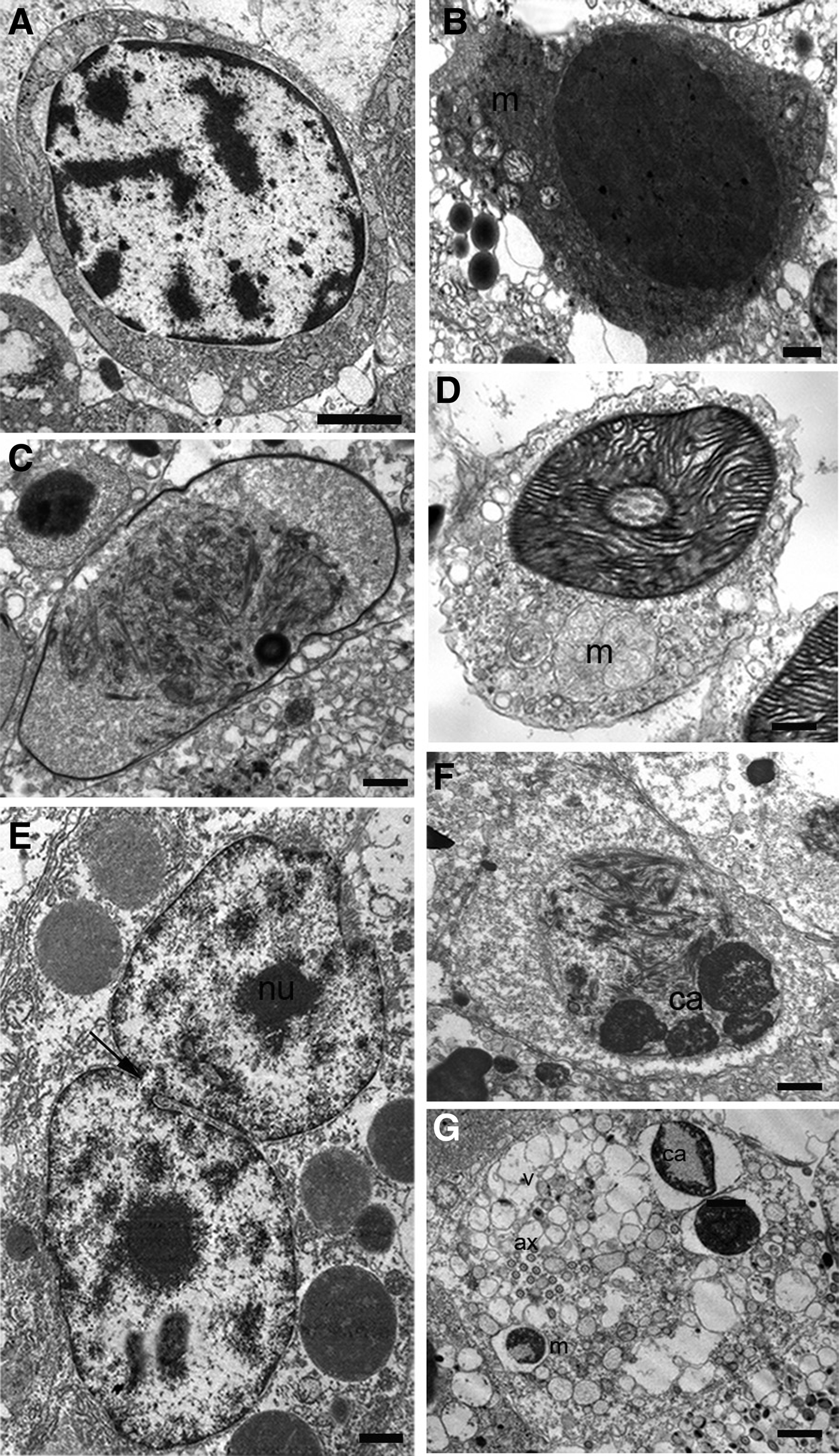
During the spermatogenesis, euspermatogenic cells were generally clustered in small groups in the same maturation phase, and were joined by a cytoplasmic continuity. These groups of cells were distributed throughout the tubule (Figure 1A, B). Once the euspermiogenesis process was complete, the mature euspermatozoa generally filled the lumen of the tubule (Figure 1C); Sertoli cells were characterized by an ovoid nucleus with euchromatin. Small clumps of electron-dense chromatin were loosely distributed in the nucleoplasm, giving the nucleus a patchy appearance. Late euspermatocytes have the chromatin dispersed in patches throughout the nucleoplasm, the mitochondrias are located in one pole of the cell (Figure 2A).
Stages of spermatogenesis of Trophon geversianus, viewed by transmission electron microscopy: (A) late euspermatocyte; (B) euspermatid in granular stage; (C) euspermatid in fibrilar stage; (D) transverse section through lamellar euspermatid stage of nucleus showing distribution of chromatin in lamellae. A well-developed Golgi complex and five rounded mitichondrias; (E) nuclei of paraspermatocyte with nucleoli, note the synaptonemal complex in the chromatin. Note the cytoplasmic bridges (arrowhead); (F) early paraspermatid showing numerous caryomerites; (G) late paraspermatid. Note the vesicle excreted from a cell (arrowhead). Note nuclear vesicles, which are pale and similar in density to the many clear vesicles in cytoplasm. ca, caryomerites; g, Golgi complex; m, mitichondrias; n, nuclei; nu, nucleoli; syc, synaptonemal complex. Scale bars: A, 2 µm; B, C, D, F, 0.5 µm; E, G, 1 µm.
The early euspermatids are roughly spherical and characterized by the condensation of the nuclear granular chromatin. The mitochondria is aggregate at the posterior pole of the cell (Figure 2B). Concomitantly, the condensation of the entire nucleus began, as the nuclear granular chromatin formed electron-dense fibrils (Figure 2C). The next step of nuclear condensation was characterized by a thickening of the chromatin fibrils, and the formation of a lamellar arrangement showing a helical organization (Figure 2D). The condensation was clearly correlated with nuclear elongation and a middle piece composed of five mitochondrial elements arranged around the axoneme (Figure 2D).
The parasperm cells were observed in clusters distributed throughout the tubule (Figure 1B, C). As seen by optical microscopy, paraspermatid development was characterized by elongation of the cell, concurrent with the appearance of a cytoplasmic elongation at the apex of the cell and the breakdown of the nucleus into small round fragments (caryomerites) (Figure 1D).
Paraspermatocytes are connected by cytoplasmic bridges, the nuecleoli are visible and synaptonemal complex are present in this nucleus (Figure 2E). The ultrastructure of the paraspermatid show an early stage as well as the final stage characterized by the fragmentation of the nucleus into caryomerites, the presence of mitochondria, and internal and central axonemes and vesicles (Figure 2F, G).
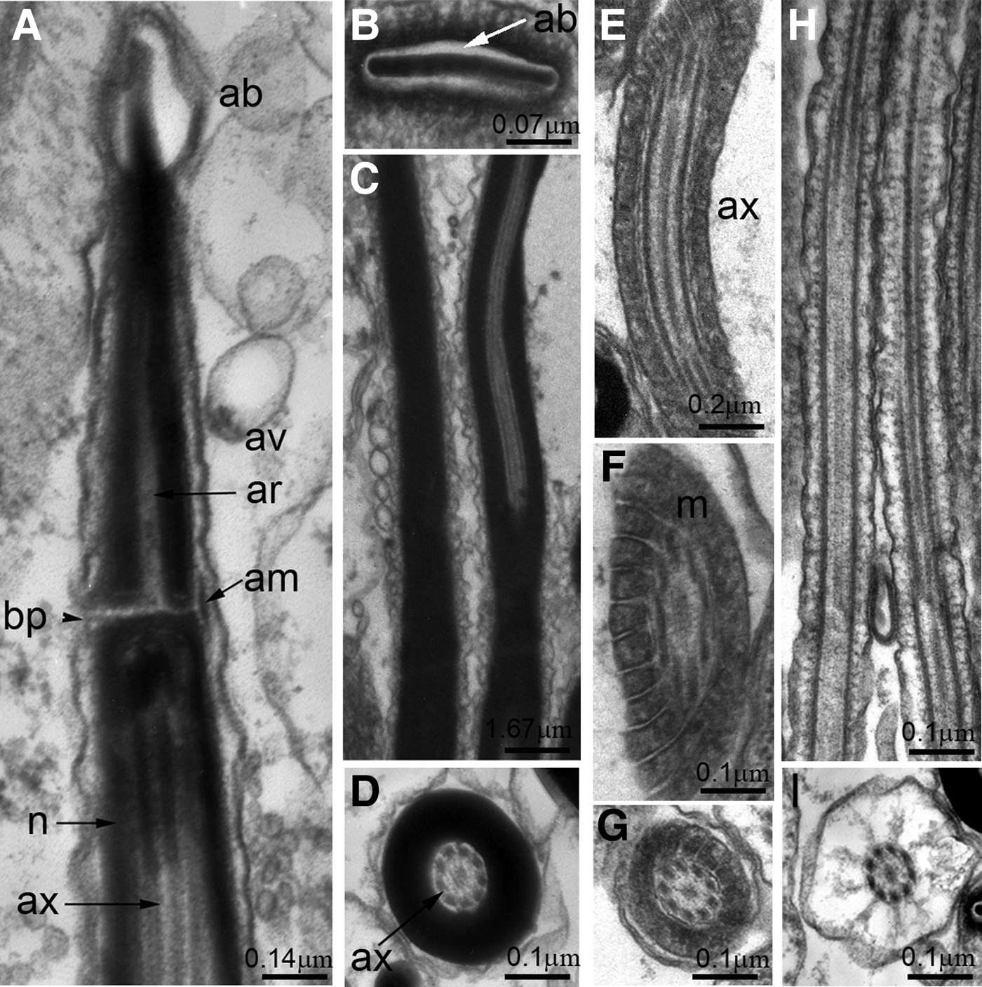
The ultrastructure of the testis shows that T. geversianus possesses euspermatozoa: fertile sperm composed of an acrosomal complex, nucleus, midpiece, glycogen piece and end piece, with a single incorporated axoneme. The acrosomal complex consists of a tall, conical, membrane-bound acrosomal vesicle (Figure 3A). The acrosomal vesicle is approximately N = 7 1.14 ± 0.08 µm long. Apically the vesicle membrane separates from the vesicle contents and lies close to the plasma membrane to form an electron-lucent, balloon-like space, called the apical bleb (Figure 3A). In transverse section the acrosome is compressed laterally showing in the anterior region the flat apical bleb (Figure 3B). The acrosomal vesicle has a very deep invagination (length 0.71 ± 0.04 µm) within which is subacrosomal material (Figure 3B). An accessory membrane is present in the basal part of the acrosome (Figure 3B).
Euspermatozoa of Trophon geversianus: (A) longitudinal section (LS) through acrosomal complex showing the apical bleb; subacrosomal material; acrosomal vesicle separated by the basal plate with the anterior portion of nucleus. The axoneme is continuously surrounded peripherically by nuclear material. Note the dense accessory membrane associated with the plasma membrane in the basal portion of the acrosome; (B) transverse section (TS) of the acrosomal complex from apical bleb region of the acrosomal vesicle; (C) LS of the nucleus. Note the internal axoneme (ax); (D) TS of the nucleus. Note the disposion 9 + 2 of the axoneme; (E) LS of the midpiece showing spiralling of mitochondria around the axoneme; (F) oblique section of the midpiece showing spiralling of mitochondria; (G) TS of the midpiece. Note the disposion 9 + 2 of the axoneme; (H) LS of the glycogen piece showing radiating, longitudinal rows of putative glycogen granules; (I) TS of the glycogen piece showing the internal axoneme. ab, apical bleb; am, accessory membrane; av, acrosomal vesicle; ax, axonemes; m, mitochondria; n, nucleus; sam, subacrosomal material. Scale bars: A, 0.14 µm; B, 0.07 µm; C, 1.67 µm; E, 0.2 µm; D, F, G, H, I, 0.1 µm.
The nucleus is filiform with a complete and long invagination and contains a centriolar derivative (Figure 3B, C). In transverse sections of the nucleus it is apparent that the axoneme has a 9 + 2 arrangement of microtubules (Figure 3D). Posterior to the nucleus the axoneme is enclosed in a mitochondrial sheath, forming the midpiece region (Figure 3E–G).
Beyond the midpiece the axoneme is associated with the glycogen piece (Figure 3H),
Transversal sectioning shows nine longitudinal and radiating tracts of dense granules (one tract per axonemal doublet) (Figure 3I). The glycogen composition of these granules has been demonstrated cytochemically, with a PAS positive result.
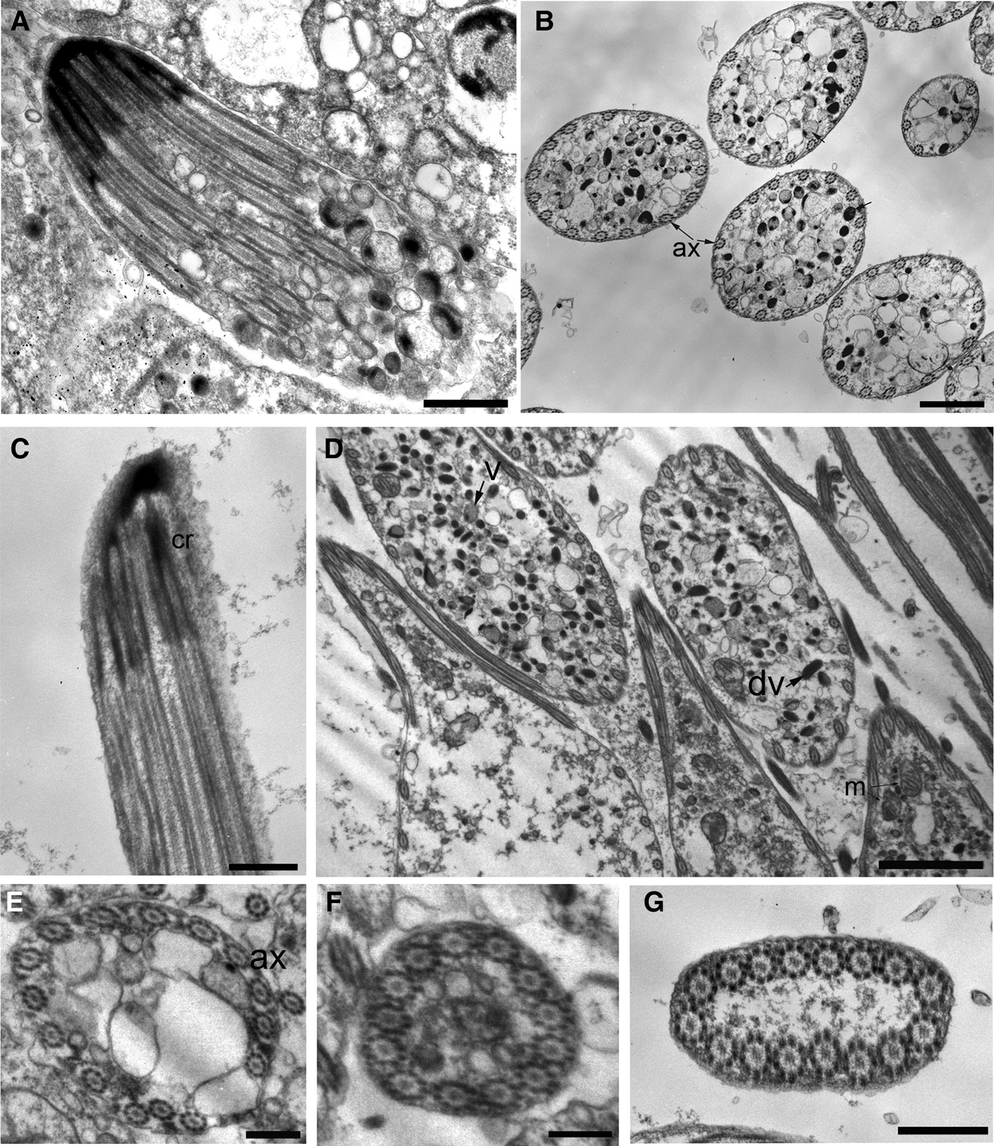
The ultrastructure of the testis shows that Trophon geversianus exhibits a type of paraspermatozoa that is vermiform with multiple incorporated axonemes. The paraspermatozoa of T. geversianus (N = 24, length 60 ± 4 µm) have tapered anterior and posterior extremities and contain no discernible nucleus or nuclear derivative (Figure 4A, C). Observed in the main body region of the paraspermatozoa are: (1) 12–16 (14 ± 2, N = 21) peripherally distributed axonemes lying close to or in contact with the inner surface of the plasma membrane (with axonemes approximately equidistant from each other); (2) numerous very electron-dense vesicles; (3) occasional, large round vesicles of low to moderate electron-density (putative lipid vesicles); (4) numerous small vesicles (possible mucoid deposits) with low electron density; and (5) occasional mitochondria (Figure 4B, D).
Paraspermatozoa of Trophon geversianus: (A) longitudinal section (LS) through apex showing showing sheath of granular material enclosing axonemes and basal body/centriolar complexes with some dense vesicles; (B) transverse section (TS) of the main body region of a paraspermatozoon, showing 14–15 peripheral axonemes closely adherent to the plasma membrane, dense vesicles and less dense vesicles; (C) LS through apex showing cap of granular material enclosing axonemes and basal body/centriolar complexes; (D) LS and TS of the body region of the paraspermatozoid showing axonemes peripherally; (E–G) TS of the apex region showing a part of the peripheral axonemes. ax, axonemes; dv, dense vesicles; v, less dense vesicles. Scale bars: A, 0.54 µm; B, 1 µm; C, 0.35 µm; E, 0.2 µm; D, 2 µm; E, F, G, 0.5 µm.
In the apex of the parasperm only the axonemes and some cytoplasm persist, where each axoneme attaches to a granular deposit (Figure 4A, C). Present near the extremities of the paraspermatozoa only are the peripheral axonemes (Figure 4E). Transverse sections toward the apex of the parasperm show a diminishing diameter and a flattened shape; in both cases no vesicles or mitochondria are present (Figure 4F, G).
DISCUSSION
Features of Trophon geversianus euspermatozoa can be observed in many other neogastropods (Buckland-Nicks, Reference Buckland-Nicks1973; Healy, Reference Healy1982). These shared characters include: (1) the morphology of the acrosomal complex (apical bleb, accessory membrane, shape of the acrosomal vesicle and existence of the axial rod material); (2) a midpiece containing numerous helically coiled mitochondria; and (3) a glycogen piece with nine tracts of granules associated with the axonemal doublets. The morphology of the nucleus varies widely among the caenogastropods (see Healy, Reference Healy and Ponder1988). In many groups the nucleus is short, rod-shaped and solid, with a short basal invagination (e.g. some Littorinoidea, some Rissooidea, Loxonematoidea, Vermetoidea, Cypraeoidea and some Conoidea). In some Muricoidea (especially many Volutidae) the short basal invagination of the axoneme is present (Giménez et al., Reference Giménez, Healy, Hermida, Lo Nostro and Penchaszadeh2008; Zabala et al., Reference Zabala, Hermida and Giménez2009; Arrighetti & Giménez, Reference Arrighetti and Giménez2010; Giménez, Reference Giménez2011), as well as in some Olividae (Giménez et al., Reference Giménez, Arrighetti, Teso, Hermida, Zabala and Penchaszadeh2009; Teso & Giménez, Reference Teso and Giménez2013). This study demonstrates that the nucleus of T. geversianus is long and almost totally penetrated by the axonemal insertion, as seen in some other groups, e.g. some Littorinoidea (Littorininae, Provannidae), some Rissooidea, Muricoidea, Nassarinae, and many Conoidea. (Gallardo & Garrido, Reference Gallardo and Garrido1989; Healy, Reference Healy1990; Hodgson, Reference Hodgson1993; Ropstorf et al., Reference Ropstorf, Healy, Riedel and Sitnikova2002). The presence of continuous axoneme along the nucleus may prove to be a diagnostic feature of the Muricoidea.
Euspermatogenesis in T. geversianus includes many features that are common in other caenogastropods (Walker & MacGregor, Reference Walker and MacGregor1968; Buckland-Nicks & Chia, Reference Buckland-Nicks and Chia1976). These include substructural changes in the nucleus. The pattern of nuclear condensation in T. geversianus (with granular, fibrillar and lamellar phases) described by Henley (Reference Henley1973) has been reported in other caenogastropods (Healy, Reference Healy1982; Buckland-Nicks et al., Reference Buckland-Nicks, Williams, Chia and Fontaine1983; Suwanjarat & Klepal, Reference Suwanjarat and Klepal2001; Zabala et al., Reference Zabala, Hermida and Giménez2012).
The morphology of the paraspermatozoa of T. geversianus categorizes them as the Type 5 paraspermatozoa of Nishiwaki (Reference Nishiwaki1964) (vermiform paraspermatozoa, exhibiting multiple peripheral axonemes enclosed at maturity and bunched anteriorly, scattered mitochondria and small dense vesicles and total absence of nuclear material). Vermiform paraspermatozoa are present in most of the Neotaenioglossa and Neogastropoda (Melone et al., Reference Melone, Lora Lamia Donin and Cotelli1980; Healy, Reference Healy and Ponder1988; Hodgson, Reference Hodgson1997; Buckland-Nicks, Reference Buckland-Nicks1998).
The vermiform parasperm of T. geversianus consist of a conical region where the axonemes decrease in number and are embedded in electron-dense material. This structure has been observed in other Muricoidea (Giménez et al., Reference Giménez, Healy, Hermida, Lo Nostro and Penchaszadeh2008; Arrighetti & Giménez, Reference Arrighetti and Giménez2010; Zabala et al., Reference Zabala, Hermida and Giménez2009; Giménez, Reference Giménez2011), and other caenogastropods such as the Fusitriton and Ceratostoma (Buckland-Nicks, Reference Buckland-Nicks1998). In other caenogastopods, paraspermatozoa are clustered in cohorts of either four or eight parasperm linked by cytoplasmic bridges (Buckland-Nicks, Reference Buckland-Nicks1998). In T. geversianus the same clustering of parasperm was seen, but the cytoplasmic bridges were not observed.
ACKNOWLEDGEMENTS
The author wishes to thank Gregorio Bigatti and Carlos Sanchez Antelo for their generous help with collection of the samples, Natalio de Vicenzo for his help with the TEM, and Jennifer Antonides for her help with the English version and improvement of this manuscript.
FINANCIAL SUPPORT
Financial support for this project was provided by UBACyT X117 and CONICET PIP 2788.