Hypoxic–ischaemic encephalopathy was described by Amiel-Tison in 1969.Reference Amiel-Tison1 Hypoxic–ischaemic cerebral injury that occurs during the perinatal period is one of the most commonly recognised cause of severe, long-term neurologic deficits, which is known as cerebral palsy in children.Reference Perlman2 The intensity of the initial insult may determine the mode of death, with severe injury resulting in necrosis and milder insults resulting in apoptosis.Reference Bonfold, Krainc, Ankarcrona, Nicotera and Lipton3 These infants must be identified as soon as possible – 2 to 6 hours – after delivery to facilitate the implementation of early interventions.Reference Perlman2 The Sarnat grades of encephalopathy are commonly used, but there are some differences in opinion about the criteria defining moderate and severe encephalopathy.Reference MacLennan4, 5 Infants with severe encephalopathy frequently have an adverse outcome, whereas the outcome of those with moderate encephalopathy is less certain.Reference MacLennan4 Acute hypoxia usually affects all the vital organs and not just the brain. Hypoxic–ischaemic encephalopathy, may be responsible for multisystem involvement such as acute bowel necrosis, renal failure, hepatic injury, cardiac damage, respiratory complications, and haematological insult.Reference MacLennan4, 5 This requires testing in the early neonatal period, that is, within 24 hours. The purpose of our study was to evaluate the importance of myocardial performance index – Tei index – in order to distinguish newborn babies with moderate and severe hypoxic–ischaemic encephalopathy according to the Sarnat criteria as an additive criterion.Reference Sarnat and Sarnat6
Methods
Subjects
Our study group consisted of 20 newborn babies with hypoxic–ischaemic encephalopathy, who were referred to our Newborn Intensive Care Unit between July, 2008 and 2009, and 50 healthy term newborn babies – control group – born in our obstetrics clinics. None of the subjects had congenital heart disease, rhythm abnormalities, syndromes, chromosomal abnormalities, or other abnormalities such as prematurity, which can influence ventricular functions. Infants with hypoxic–ischaemic encephalopathy were diagnosed according to modified American College of Obstetric and Gynecology clinic and laboratory criteria.Reference MacLennan4, 5, Reference Shah, Riphagen, Beyene and Perlman7 Using the Sarnat grades, 20 babies with hypoxic–ischaemic encephalopathy were scored as severe (four babies), moderate (six babies), and mild (10 babies).Reference Sarnat and Sarnat6 All subjects were examined serially on the first day and thereafter in the 1, 3–4, 6–7, and 11–12 months.
Echocardiographic investigations
Echocardiographic investigations were performed using Sonos 5500 (Hewlett-Packard, Philips, Holland) with 5–12 megahertz transducers in our Pediatric Cardiology Echocardiographic Laboratory. Echocardiograms were recorded on a 1/2-inch VHS videotape and were subsequently analysed directly from the stored images. Ejection fraction and fractional shortening of the left ventricle are investigated by M-Mode echocardiography. Myocardial performance index for the left and right ventricles were determined by pulsed Doppler technique. The sample volume was placed just distal to the mitral and tricuspid annuli at the tips of the leaflets at an angle that was as optimally parallel to flow as possible in the apical four-chamber view. Ejection time for the left and right ventricles was measured from the apical long-axis view and the parasternal short-axis view with Doppler sample volume positioned just below the aortic or the pulmonary valve.Reference Sugiuro, Suziki, Hussein, Kato and Togari8, Reference Tsutsumi, Ishii, Eto, Hota and Kato9
Myocardial performance index calculation
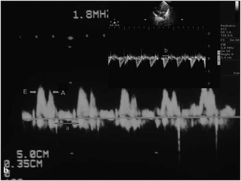
Myocardial performance index is defined as the sum of isovolumic contraction and relaxation times divided by the ejection time, which requires measurement of the time interval between the end and the onset of mitral or tricuspid inflow (the “a” interval) and the ejection time of the left and right ventricles’ outflow (the “b” interval).Reference Tei, Ling and Hodge10–Reference Eto, Ishii, Tei, Tsutsumi, Akagi and Kato15 For the left ventricle, “b” interval was measured from the aortic valve opening to the aortic valve closure, and for the right ventricle from the pulmonary valve opening to the pulmonary valve closure Reference Cui, Roberson and Lawn16 (Fig 1). We calculated the Tei index – myocardial performance index – using the formula (a − b)/b. Each time interval was measured on three consecutive cardiac cycles and subsequently averaged.
Myocardial performance index measurement by pulsed Doppler method.

Statistics
Results are expressed as mean ± standard deviation. All data analyses were performed with a commercially available statistical analysis software package (SPSS for Windows, version 13.0). The Mann–Whitney U-test was used to compare the groups. Values of p less than or equal to 0.05 were considered statistically significant. Correlations between groups and the repeated measurements were determined by variance analysis. If statistical significance was reached (p ≤ 0.05), further analysis was carried out by post hoc Wilcoxon test with Bonferroni corrections.
Results
Left ventricular systolic functions, such as ejection fraction and fractional shortening obtained by M-Mode echocardiography techniques for mature healthy babies and babies with hypoxic–ischaemic encephalopathy, were in the normal ranges and there was no significant difference between the two groups (p > 0.05, all). Ejection fraction and fractional shortening increased with advancing age for both groups, but this increment was not statistically significant (p > 0.05, all; Table 1).
Mean values of EF and FS for control and severe HIE babies.

EF = ejection fraction; FS = fractional shortening; HIE = hypoxic–ischaemic encephalopathy
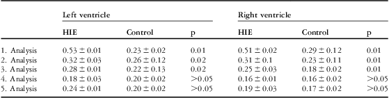
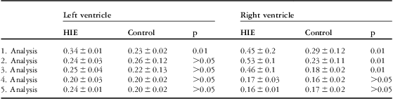
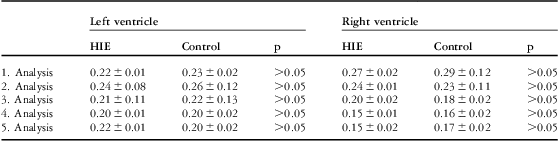
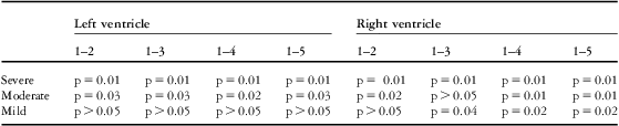
Myocardial performance indexes of the left ventricle (p = 0.01, p = 0.02, and p = 0.02, respectively) and right ventricle (p = 0.01, all) were significantly higher in the severe hypoxic–ischaemic encephalopathy group than in the control group during the first, second, and third analyses. On the other hand, there were no significant differences between the two groups with regard to left and right ventricle myocardial performance indexes during the fourth and fifth analyses (p > 0.05, all; Table 2). Myocardial performance index of the left ventricle only during the first analysis (p = 0.01) and myocardial performance index of the right ventricle during the first, second, and third analyses (p = 0.01, all) were significantly higher in the moderate hypoxic–ischaemic encephalopathy group than in the control group (Table 3). During the entire analysis, myocardial performance indexes of the left and right ventricles in the mild hypoxic–ischaemic encephalopathy group were not statistically different from the control group (p > 0.05, all; Table 4). The myocardial performance index of the left ventricle did not change significantly with time in the mild hypoxic–ischaemic encephalopathy group (p > 0.05, all). However, it decreased significantly during the second, third, fourth, and fifth analyses in both severe (p = 0.01, all) and moderate (p = 0.03, p = 0.03, p = 0.02, p = 0.03, respectively) hypoxic–ischaemic encephalopathy groups when compared with the first analysis results. When compared with the first analysis, myocardial performance index of the right ventricle decreased significantly during the second, third, fourth, and fifth analyses (p = 0.01, all) in the severe hypoxic–ischaemic encephalopathy group; during the second, fourth, and fifth analyses (p = 0.02, p = 0.01, p = 0.01, respectively) in the moderate hypoxic–ischaemic encephalopathy group; and during the third, fourth, and fifth analyses (p = 0.04, p = 0.02, p = 0.02, respectively) in the mild hypoxic–ischaemic encephalopathy group (Table 5).
Left and right ventricular MPI for control and severe HIE babies.
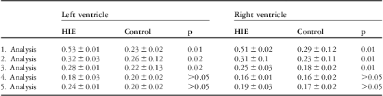
HIE = hypoxic–ischaemic encephalopathy; MPI = myocardial performance index
Left and right ventricular MPI for control and moderate HIE babies.
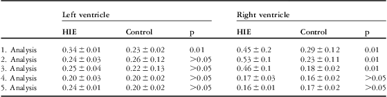
HIE = hypoxic–ischaemic encephalopathy; MPI = myocardial performance index
Left and right ventricular MPI for control and mild HIE babies.
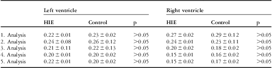
HIE = hypoxic–ischaemic encephalopathy; MPI = myocardial performance index
Statistical changes of left and right ventricular MPI for severe, moderate, and mild HIE groups.
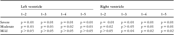
HIE = hypoxic–ischaemic encephalopathy; MPI = myocardial performance index
Discussion
Neonatal asphyxia may induce myocardial dysfunction.Reference Williams, Mallard, Tan and Gluckman17, Reference Borke, Munkeby and Halversen18 The clinical picture varies from bradycardia or tachycardia to cardiogenic shock and the transient abnormalities such as hypotension, myocardial ischaemia, mitral and/or tricuspid regurgitation, and pulmonary hypertension.Reference Walther, Siashi, Ramadan and Wu19–Reference Rowe and Hoffman22 Doppler echocardiographic studies in adults and children with heart disease have shown that impairment of ventricular function leads first to diastolic dysfunction and then to combined diastolic and systolic dysfunction.Reference Thomas and Weyman23, Reference Sohn, Kim, Kim, Chun, Park and Choi24 In addition, echocardiographic studies in hypoxic healthy volunteers have shown a preservation of left ventricular contractility, but reported altered mitral flow patterns suggestive of left ventricle diastolic dysfunction.Reference Allemann, Rotter and Hutter25–Reference Oliver, Peacock, Challenor, Fleming and Walter27 In our study, ejection fraction and fractional shortening, which are the most common clinical methods of assessing systolic function, were within the normal ranges for the left ventricle and there were no significant differences between the healthy and hypoxaemic neonatal groups during the entire analysis (p > 0.05, all; Table 1).Reference Colan28 Measurement that combines assessment of systolic and diastolic functions may better reflect global cardiac functions than an isolated evaluation of either ejection or relaxation.Reference Tsutsumi, Ishii, Eto, Hota and Kato9 Myocardial performance index has been reported to be useful for the assessment of global functions of the left and right ventricles independent of ventricular geometry, heart rate, and blood pressure.Reference Tei, Ling and Hodge10–Reference Eto, Ishii, Tei, Tsutsumi, Akagi and Kato15, Reference Eidem, O'Leary, Tei and Seward29, Reference Ishii, Eto and Tei30 In the present study, left ventricular myocardial performance index was significantly higher in the severe hypoxic–ischaemic encephalopathy group than in the control group during the first, second, and third analyses (p = 0.01, p = 0.02, p = 0.02, respectively; Table 2). In addition, in the moderate hypoxic–ischaemic encephalopathy group the myocardial performance index of only the first analysis was significantly higher than the control group (p = 0.01; Table 3). Hypoxia exerts predominant effects on the right ventricle because of increased loading due to hypoxia-induced pulmonary vasoconstriction.Reference Naeije31 In addition, in our study right ventricular myocardial performance index in both severe and moderate hypoxic–ischaemic encephalopathy groups were significantly higher than in the control group during the first, second, and third analyses (p = 0.01, all; Tables 2 and 3). However, right and left ventricular myocardial performance indexes for the mild hypoxic–ischaemic encephalopathy group were not significantly different from the control group (p > 0.05, all; Table 4).
In an animal study, the right ventricle Tei index was higher in hypoxia than in normoxia. In contrast, the left ventricle Tei index in hypoxia was not different from the value in normoxia.Reference Sugiuro, Suziki, Hussein, Kato and Togari8 However, we can mention that in both severe and moderate hypoxic–ischaemic encephalopathy groups right and left ventricular functions are influenced from hypoxia, as myocardial performance indexes for both ventricles were higher than the control group in our study. In addition, we found that in the moderate hypoxic–ischaemic encephalopathy group hypoxia-induced alteration has a longer duration in the right ventricle than in the left ventricle, as myocardial performance index for the right ventricle continues to be higher than the control group during the second and third analyses (Table 3). Ichihashi et alReference Ichihashi, Yada, Takahashi, Honda and Momoi32 reported that in the patients with mild asphyxia, myocardial performance index of the left ventricle became normal in the third week after birth, and they suggested that this change shows mild cardiac dysfunction owing to the fact that mild asphyxia is reversible. However, in our study, we did not find any statistical difference for myocardial performance indexes of the left and right ventricles in the mild hypoxic–ischaemic encephalopathy and control groups during the 1-year period.
The goals of management of a newborn infant who has sustained a hypoxic–ischaemic insult and is at risk for evolving injury should include supportive care to facilitate adequate perfusion and nutrients to the brain and consideration of interventions to ameliorate the processes of ongoing brain injury.Reference Perlman2 The time interval after hypoxia–ischaemia during which interventions might be efficacious in reducing the severity of ultimate brain injury is likely to be short. According to experimental studies, the therapeutic window is estimated to vary from 2 to 6 hours.Reference Perlman2 New technologies available to determine cerebral damage, such as computed tomography scanning, magnetic resonance imaging, cerebral function monitoring, cranial ultrasound, and Doppler ultrasound of the middle cerebral artery, are not available in many neonatal units. However, in many neonatal units echocardiographic investigations can be performed. The most widely used classification of hypoxic–ischaemic encephalopathy is the Sarnat criteria. The decision as to whether an infant falls into the moderate or severe category is at times difficult. We try to evaluate whether myocardial performance index can be used as an additive criterion in order to distinguish newborn babies with moderate and severe encephalopathy. In our study, we demonstrated that babies with severe and moderate hypoxic–ischaemic encephalopathy have higher myocardial performance index values for left and right ventricles especially 24 hours after birth. The biggest problem with regard to this subject is which value will be accepted as pathologic and discriminative between severe and moderately affected groups, because many haemodynamic changes and myocardial maturation take place in this period and normal values for this age group are not precisely determined. However, Cui et alReference Cui, Roberson and Lawn16 suggested that the upper normal limit for myocardial performance index was less than 0.5. As seen in Tables 2 and 3, only in the severe hypoxic–ischaemic encephalopathy group myocardial performance index was greater than or equal to 0.5 for the left and right ventricles during the first analysis. We suggested that a myocardial performance index greater than or equal to 0.5 can be used as a discriminative additive criterion in order to distinguish between moderately and severely ill hypoxic–ischaemic encephalopathy babies according to Sarnat grades. However, we need further investigations upon this subject.
Acknowledgement
We wish to thank Lütfi Saltuk Demir M.D, Department of Public Health, Selcuk University, Meram School of Medicine Hospital, for his help in statistical analysis.