INTRODUCTION
Strongyloidiasis is a parasitic infection caused by Strongyloides stercoralis, a widely distributed nematode, particularly in the tropical areas (Olsen et al. Reference Olsen, van Lieshout, Marti, Polderman, Polman, Steinmann, Stothard, Thybo, Verweij and Magnussen2009). The definitive diagnosis of this disease relies on the visualization of larvae, especially in the stool, being considered of low sensitivity. Therefore, serological techniques have been used as complementary alternatives to parasitological diagnosis (Levenhagen and Costa-Cruz, Reference Levenhagen and Costa-Cruz2014).
The serological techniques include enzyme linked immunosorbent assay (ELISA) and Western blotting (WB), the former being the most extensively used for the diagnosis of strongylodiasis, especially for its practicality in analysing large numbers of serum samples (Requena-Mendez et al. Reference Requena-Mendez, Chiodini, Bisoffi, Buonfrate, Gotuzzo and Munoz2013; Levenhagen and Costa-Cruz, Reference Levenhagen and Costa-Cruz2014; Buonfrate et al. Reference Buonfrate, Formenti, Perandin and Bisoffi2015). In turn, WB has been a useful tool for many immunological approaches in various diseases, but few studies have reported its use in immunodiagnosis of strongyloidiasis (Sato et al. Reference Sato, Inoue, Matsuyama and Shiroma1990; Conway et al. Reference Conway, Bailey, Lindo, Robinson, Bundy and Bianco1993; Lindo et al. Reference Lindo, Conway, Atkins, Bianco, Robinson and Bundy1994; Ravi et al. Reference Ravi, Ramachandran, Thompson, Andersen and Neva2002). In this context, molecular mass ranges of proteins have been described as immunodominants (Conway et al. Reference Conway, Bailey, Lindo, Robinson, Bundy and Bianco1993) from one-dimensional (1D) gel electrophoresis, but the exact identities of the proteins remain to be determined.
A number of genome and transcriptome sequencing initiatives have allowed the identification of parasite proteins using proteomic approaches, including those devoted to the Strongyloides genus (Marcilla et al. Reference Marcilla, Sotillo, Perez-Garcia, Igual-Adell, Luz Valero, Sanchez-Pino, Bernal, Munoz-Antoli, Trelis, Toledo and Esteban2010; Hunt et al. Reference Hunt, Tsai, Coghlan, Reid, Holroyd, Foth, Tracey, Cotton, Stanley, Beasley, Bennett, Brooks, Harsha, Kajitani, Kulkarni, Harbecke, Nagayasu, Nichol, Ogura, Quail, Randle, Xia, Brattig, Soblik, Ribeiro, Sanchez-Flores, Hayashi, Itoh, Denver, Grant, Stoltzfus, Lok, Murayama, Wastling, Streit, Kikuchi, Viney and Berriman2016). In addition, providing antisera are available, imunoreactive proteins can be detected and selected for protein identification, through the technique collectively termed imunoproteomics. This approach has been shown to be highly promising for the diagnosis of various infectious agents (Yan et al. Reference Yan, Xu, Liu, Yan, Song and Li2010; Zhan et al. Reference Zhan, Ajmera, Geiger, Goncalves, Liu, Wei, Wilkins, Fujiwara, Gazzinelli-Guimaraes, Bottazzi and Hotez2015). As for the diagnosis of human strongyloidiasis, heterologous antigens from Strongyloides ratti and Strongyloides venezuelensis can be easily obtained, and therefore they represent valuable sources of antigens to be explored (Levenhagen and Costa-Cruz, Reference Levenhagen and Costa-Cruz2014). In this study, using a combination of 1D WB and mass spectrometry, we revealed the protein constituents (from filariform larvae of S. venezuelensis) mostly recognized by the antisera of individuals with strongyloidiasis.
MATERIAL AND METHODS
Serum samples
A total of 46 serum samples from individuals registered at the ‘Hospital das Clínicas da Faculdade de Medicina da Universidade de São Paulo (HCFMUSP)’ were divided into two groups: group I consisted of 20 patients who were shedding S. stercoralis larvae only in their feces; group II included 26 patients infected with other parasites, including those presenting monoinfections: hookworm (n = 4); Ascaris lumbricoides (n = 2); Enterobius vermicularis (n = 1); Giardia intestinalis (n = 3); Hymenolepis nana (n = 1); Schistosoma mansoni (n = 9); and those exhibiting polyinfections: hookworm, H. nana (n = 1); S. mansoni, A. lumbricoides, Entamoeba coli, Blastocystis spp., Endolimax nana (n = 1); G. intestinalis, E. nana (n = 1); A. lumbricoides, Blastocystis spp. (n = 1); E. nana, S. mansoni, Blastocystis spp. (n = 1); hookworm, E. coli, Entamoeba dispar/histolytica; S. mansoni (n = 1). Stool samples from all individuals were analysed by: (1) spontaneous sedimentation, (2) modified Baermann (Garcia, Reference Garcia2001) and (3) the agar plate culture (Koga et al. Reference Koga, Kasuya, Khamboonruang, Sukhavat, Ieda, Takatsuka, Kita and Ohtomo1991) techniques. Briefly, the spontaneous sedimentation method is based on sedimentation of fecal material using a cup, after homogenization in water followed by filtration. The modified Baermann technique relies on the positive thermotropism and hydrotropism of the larvae; however, it employs a sedimentation cup containing heated water. The study received approval from the Research Ethics Committee of ‘Universidade de São Paulo’, state of São Paulo, Brazil (Protocol 266·046).
Parasites
Strongyloides venezuelensis filariform larvae were obtained from the feces of experimentally infected Rattus norvegicus. Stool samples were collected and homogenized with charcoal and water followed by incubation at 28 °C for 3 days. After this procedure, the larvae were recovered according to the modified Baermann method (Garcia, Reference Garcia2001).
Antigenic fractions
Approximately 200 000 filariform larvae of S. venezuelensis were resuspended in 1 mL of phosphate-buffered saline (PBS) (0·01 m, pH 7·2) or Tris–HCl (25 mm, pH 7·5), containing 1× protease inhibitor cocktail (Sigma-Aldrich, St. Louis, MO, USA). These were sonicated using five cycles of 20 s pulse each, on ice. The larval suspensions were centrifuged at 12 400 g for 30 min at 4 °C and the supernatant was collected as the soluble fraction (SS and TS in PBS, Tris–HCl, respectively). SS was then resuspended in 1% SDS, boiled for 5 min (100 °C), centrifuged at 12 400 g for 30 min at 4 °C and the supernatant was collected (membrane fraction SM). TS pellets were resuspended in 5 m urea, 2 m thiourea and 4% 3-[(3-Cholamidopropyl)dimethylammonio]-1-propanesulfonate (CHAPS) followed by homogenization on ice for 30 min, centrifugation at 12 400 g for 30 min at 4 °C and collection of the supernatant (membrane fraction TM). All fractions were subjected to protein assays according to the method described by Lowry et al. (Reference Lowry, Rosebrough, Farr and Randall1951).
Electrophoresis and electrophoretic transfer
Analysis of antigenic fractions by polyacrylamide gel electrophoresis (SDS–PAGE) was performed as described by Laemmli (Reference Laemmli1970), with some modifications. Briefly, 140 µg of each antigenic fraction was subjected to SDS–PAGE, using a 12% gel. The samples were resuspended in buffer (2 mm Ethylenediaminetetraacetic acid (EDTA), 125 mm Tris–HCl pH 6·8, 2% SDS, 20% glycerol, 50 mm 1,4-Dithiothreitol (DTT), 0·01% bromophenol blue) and heated at 100 °C in a water bath for 5 min. The samples were then loaded on the gel immersed in buffer (25 mm Tris, 192 mm glycine, 3,5 mm SDS) and subjected to electrophoresis at 40 mA and 100 V for approximately 2 h.
Immediately after electrophoresis, the proteins on the gel were transferred to polyvinylidene difluoride (PVDF) membranes (0·2 µ m) (Headquarter Bio-Rad Laboratories, Hercules, CA, USA) in a Mini Trans-blot system using the transfer buffer (25 mm Tris, 192 mm glycine, 20% ethanol, 0·69 mm SDS) at 200 mA and 50 V for approximately 2 h. The membranes were cut into strips of approximately 3 mm and stored until required.
Western blotting
After blocking for an hour in Tris–HCl buffer (1 m, pH 7·5) with 3% Tween-20 and 3% milk, the membrane was washed three times for 5 min in 10 mm Tris–HCl, and then incubated with test sera diluted 1:100 in T buffer (50 mm Tris–HCl, pH 7·5, 100 mm NaCl, 0·1% Tween-20, 5% skimmed milk powder) for 18 h at 4 °C. The secondary antibody (anti-human IgG Fc conjugated with peroxidase, Sigma-Aldrich, St. Louis, MO, USA) was then diluted 1:1000 in T buffer and added to the membrane. The binding was detected using DAB substrate (Sigma-Aldrich) in the dark. The antigenic components were then detected by Luminescent image analyser scanner (Fujifilm, Minato, Tokyo, Japan) and Image Quant LAS 400 version 1.2 (GE, Fairfield, CT, USA). The positivity of the immunogenic bands was determined based on the studies by Silva et al. (Reference Silva, Barcelo, Passos-Lima, Espindola, Campos and Costa-Cruz2003) and Feliciano et al. (Reference Feliciano, Gonzaga, Goncalves-Pires, Ribeiro-Goncalves, Rodrigues, Ueta and Costa-Cruz2010). A cut-off higher than 50% positivity was established to evaluate the bands recognized more frequently by the sera of group I patients, in the four antigenic preparations.
Protein analysis by mass spectrometry
The bands showing the strongest recognition by the sera of infected patients were excised from the gel and immersed in fixative solution until completely destained. After washing in ultrapure water, the bands were digested with trypsin solution, diluted in ammonium bicarbonate (40 mm), for 20 min, followed by incubation for 12 h at 37 °C in ammonium bicarbonate (20 mm). After peptide purification, mass spectrometric experiments were performed on the Q-Exactive Orbitrap (Thermo Scientific®, Waltham, Massachusetts, EUA) instrument.
Mass spectral files were subjected to database searching using SequestHT algorithm underpinned by the Proteome Discoverer software (version 1.4, Thermo Scientific®). The area detector of precursor ions was chosen for quantification by a label-free approach. Search parameters included: (i) enzyme: trypsin/P; (ii) number of missed cleavage sites = 2; (iii) carbamidomethylation, as a static modification; (iv) oxidation of methionine N-terminal and acetylation, as dynamic changes; and (v) a mass tolerance of 10 ppm for parent ions and 0·1 Da for fragments. The Strongyloides protein database available at the UniProt server (www.uniprot.org) containing 12 544 sequences was used for the searches. A protein was considered to be accurately identified, at 1% false discovery rate, when two or more peptides were found.
RESULTS
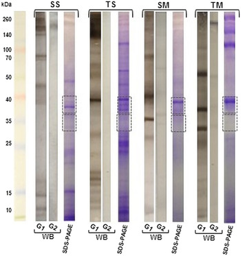
The SDS–PAGE electrophoretic profile of different antigenic fractions revealed similarity in protein migration with molecular masses ranging from 15 to 150 kDa. Nevertheless, distinct protein bands in each lane could be observed by comparing soluble and membrane fractions, attesting for their differential composition.
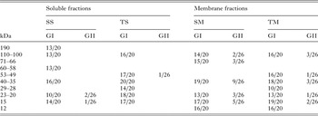
In this study, protein bands exhibiting different antigenic components were recognized by the sera of patients in group I. Anyhow, we considered as immunodominants only those with a frequency of greater than 50% recognition. Positivity of WB technique was determined when the sample presented with two or more immunodominant bands. IgG antibodies present in the sera recognized 11 separate protein bands present in the soluble and membrane antigenic fractions (Table 1). The band at 35–40 kDa was the only one present in all antigenic fractions. Bands of 14–15 kDa were identified in most antigenic fractions, followed by bands of 100–110 kDa. Cross-reactivity in group II was observed in individuals infected with S. mansoni, hookworms, and A. lumbricoides.
Reactive frequency of serum samples in WB using soluble (SS and TS) and membrane (SM and TM) fractions of Strongyloides venezuelensis

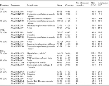
Upon observing the strongest recognition of the 30–40 kDa band, the band was excised and subjected to mass spectrometry (Fig. 1), followed by database searching for protein identification. The identities of the proteins and their abundance are shown in Table 2. The immunoreactive proteins that were identified in the soluble fractions, which exhibited the highest values of abundance, corresponded to metabolic enzymes (i.e. glutamine synthetase/guanido kinase and malate dehydrogenase). In addition, cytoskeletal constituents (i.e. actin and myosin heavy chain) and galectins were also identified as more abundant proteins in the membrane fraction.
WB and SDS–PAGE of soluble (SS and TS) and membrane (SM and TM) fractions of S. venezuelensis filariform larvae. WB of SS, TS, MS, and TM fractions, using serum samples of Groups I (G1) and II (G2). Polyacrylamide gel after SDS–PAGE, stained with Coomassie Brilliant Blue is shown to indicate the excised regions (~30–40 kDa) of the antigenic fraction, by the dotted lines.

Proteins identified from soluble (SS and TS) and membrane (SM and TM) fractions of Strongyloides venezuelensis filariform larvae. For this study, the Strongyloides database available in UniProt (www.uniprot.org) was used, and proteins that showed at least two different peptides with a confidence interval of above 95% were considered as identified

a Mostly abundant component.
DISCUSSION
The use of heterologous antigens from S. ratti or S. venezuelensis applied to immunodiagnosis constitutes a common practice in research laboratories, particularly in Brazil (Silva et al. Reference Silva, Barcelo, Passos-Lima, Espindola, Campos and Costa-Cruz2003; Rigo et al. Reference Rigo, Lescano, de Marchi and Amato Neto2008; Gonzaga et al. Reference Gonzaga, Ribeiro, Feliciano, Manhani, Silva, Ueta and Costa-Cruz2011). Indeed, S. ratti larval extracts are employed routinely as they constitute the basis for the ELISA kits commercially available for the immunodiagnosis of strongyloidiasis (Anderson et al. Reference Anderson, Klein, Dornink, Jespersen, Kubofcik, Nutman, Merrigan, Couturier and Theel2014; Bisoffi et al. Reference Bisoffi, Buonfrate, Sequi, Mejia, Cimino, Krolewiecki, Albonico, Gobbo, Bonafini, Angheben, Requena-Mendez, Muñoz and Nutman2014). In this context, this study is the first to evaluate different antigenic fractions from S. venezuelensis filariform larvae using a proteomics approach for the serological diagnosis of human strongyloidiasis.
Although the WB technique is considered of high specificity, there are few studies in the literature on its application to serological diagnosis of human strongyloidiasis using homologous (Sato et al. Reference Sato, Inoue, Matsuyama and Shiroma1990; Conway et al. Reference Conway, Bailey, Lindo, Robinson, Bundy and Bianco1993; Siddiqui et al. Reference Siddiqui, Koenig, Sinensky and Berk1997) or heterologous antigens (Silva et al. Reference Silva, Barcelo, Passos-Lima, Espindola, Campos and Costa-Cruz2003; Feliciano et al. Reference Feliciano, Gonzaga, Goncalves-Pires, Ribeiro-Goncalves, Rodrigues, Ueta and Costa-Cruz2010; Gonzaga et al. Reference Gonzaga, Ribeiro, Feliciano, Manhani, Silva, Ueta and Costa-Cruz2011). Here we intended to explore this technique further to identify potential protein markers for diagnosis of human strongyloidiasis.
The pattern of protein bands seen on the SDS–PAGE for the different antigenic fractions of S. venezuelensis larvae revealed the integrity of our preparations, with proteins exhibiting molecular masses within the full range 12–190 kDa. Similar profiles have been reported by Feliciano et al. (Reference Feliciano, Gonzaga, Goncalves-Pires, Ribeiro-Goncalves, Rodrigues, Ueta and Costa-Cruz2010) and Gonzaga et al. (Reference Gonzaga, Ribeiro, Feliciano, Manhani, Silva, Ueta and Costa-Cruz2011) in the analysis of saline soluble antigens. In the present paper, we present a novel soluble protein extract made in Tris–HCl buffer. The remaining insolubilized material, composed mostly of membrane fractions, had its protein extracted in a highly denaturing solution. These two fractions were used for the WB experiments. The presence of specific proteins in the membranes and excretory–secretory products of infective S. stercoralis larvae was described by Brindley et al. (Reference Brindley, Gam, Pearce, Poindexter and Neva1988) and Northern and Grove (Reference Northern and Grove1990). According to Zhan et al. (Reference Zhan, Ajmera, Geiger, Goncalves, Liu, Wei, Wilkins, Fujiwara, Gazzinelli-Guimaraes, Bottazzi and Hotez2015) nematode membrane-associated antigens are exposed to the host's immune system and could therefore represent good immunodiagnostic targets. Recently, our research group emphasized the importance of membrane fractions from both the filariform larvae and the female parasite of S. venezuelensis for immunodiagnosis of strongyloidiasis by ELISA [Corral et al. (Reference Corral, Paula, Gottardi, Meisel, Castilho, Gonçalves, Chieffi and Gryschek2015)].
In this study, the fractions analysed by WB showed great diversity of antigenic components, especially in the membrane fractions. Of the bands identified as immunogenic, the 35–40 kDa fraction was more frequently observed in all preparations. Similar results have been reported in several studies independently of which extraction methods were used for recovery of the antigenic fractions (Conway et al. Reference Conway, Bailey, Lindo, Robinson, Bundy and Bianco1993; Ravi et al. Reference Ravi, Ramachandran, Thompson, Andersen and Neva2002; Rodrigues et al. Reference Rodrigues, Sopelete, Silva, Cunha-Junior, Taketomi and Costa-Cruz2004; Sudre et al. Reference Sudre, Siqueira, Barreto, Peralta, Macedo and Peralta2007; Feliciano et al. Reference Feliciano, Gonzaga, Goncalves-Pires, Ribeiro-Goncalves, Rodrigues, Ueta and Costa-Cruz2010). Similar to the bands of 45 kDa reported by Machado et al. (Reference Machado, Faccioli, Costa-Cruz, Lourenco, Roque-Barreira, de Fatima Goncalves Pires and Ueta2008) and 28–35 kDa by Gonzaga et al. (Reference Gonzaga, Ribeiro, Feliciano, Manhani, Silva, Ueta and Costa-Cruz2011), using S. venezuelensis antigens, the same fractions were also reported as immunogenic by Silva et al. (Reference Silva, Barcelo, Passos-Lima, Espindola, Campos and Costa-Cruz2003) using S. ratti antigens, and a 41 kDa fraction reported by Siddiqui et al. (Reference Siddiqui, Koenig, Sinensky and Berk1997) using the homologous antigen. The fractions described in this study are probably equivalent to those identified by these authors, since minor technical variations can result in distinct electrophoretic migration patterns.
The band at 35–40 kDa showed good sensitivity, ranging from 80 to 100% in the soluble fraction and 90 to 95% in the membrane fractions. However, this band was recognized by the sera of individuals infected with S. mansoni, hookworms, H. nana, Giardia lamblia and polyinfected patients; although cross-reactivities were not expected within this group of individuals. Three proteins of S. stercoralis filariform larvae identified in protein bands at 41, 31 and 28 kDa and particularly recognized by serum IgG from individuals with strongyloidiasis have been reported by Conway et al. (Reference Conway, Bailey, Lindo, Robinson, Bundy and Bianco1993).
Bands of 23 and 15 kDa were present in four antigenic fractions, but with lower sensitivity compared with the 35–40 kDa band. Interestingly, some studies show varying recognition values among protein bands identified as immunogenic, most likely because they use different Strongyloides species as the antigen source (Silva et al. Reference Silva, Barcelo, Passos-Lima, Espindola, Campos and Costa-Cruz2003; Rodrigues et al. Reference Rodrigues, Sopelete, Silva, Cunha-Junior, Taketomi and Costa-Cruz2004; Sudre et al. Reference Sudre, Siqueira, Barreto, Peralta, Macedo and Peralta2007). Several bands recognized by antibodies present in serum samples from strongyloidiasis patients have been mentioned in different studies (Silva et al. Reference Silva, Barcelo, Passos-Lima, Espindola, Campos and Costa-Cruz2003; Rodrigues et al. Reference Rodrigues, Sopelete, Silva, Cunha-Junior, Taketomi and Costa-Cruz2004; Sudre et al. Reference Sudre, Siqueira, Barreto, Peralta, Macedo and Peralta2007; Feliciano et al. Reference Feliciano, Gonzaga, Goncalves-Pires, Ribeiro-Goncalves, Rodrigues, Ueta and Costa-Cruz2010; Gonzaga et al. Reference Gonzaga, Ribeiro, Feliciano, Manhani, Silva, Ueta and Costa-Cruz2011), but these proteins have not been identified. In an attempt to better characterize the proteins recognized by most sera of group I individuals, mass spectrometry was performed.
These results showed that the soluble extracts contained a large number of proteins related to the parasite metabolism. Moreover, the membrane extracts revealed a number of structural proteins. In the excised band, a large abundance of actin was observed independently of the extraction procedure. Actin is one of the most abundant proteins found in eukaryotic cells and is highly conserved throughout evolution. It participates in many important cellular functions, including muscle contraction, cell motility, cell division, cytokinesis, vesicle and organelle movement, cell signalling, and the establishment and maintenance of cell junctions and cell shape (Fornelio et al. Reference Fornelio, Gonzalez and Caabeiro1995; Pollard and Borisy, Reference Pollard and Borisy2003; Bahia et al. Reference Bahia, Avelar, Vigorosi, Cioli, Oliveira and Mortara2006). Nematode cytoskeletal proteins, such as actin, can induce antibody production in the host. Actin is recognized by the host's immune system in human toxocariasis (Zhan et al. Reference Zhan, Ajmera, Geiger, Goncalves, Liu, Wei, Wilkins, Fujiwara, Gazzinelli-Guimaraes, Bottazzi and Hotez2015) and is described in Haemoncus contortus infection (Yan et al. Reference Yan, Xu, Liu, Yan, Song and Li2010). Recently, Yan et al. (Reference Yan, Wang, Xu, Song and Li2014) showed that a recombinant H. contortus actin DNA vaccine induced a partial immune response resulting in protective potential against goat haemonchosis. Actin was first observed in the vulva of the female S. venezuelensis using confocal microscopy (Silva et al. Reference Silva, Gonçalves, Cruz, Cruz, Ueta and Costa-Cruz2013). The high abundance of actin in the extract of S. stercoralis L3 larvae was demonstrated by Marcilla et al. (Reference Marcilla, Sotillo, Perez-Garcia, Igual-Adell, Luz Valero, Sanchez-Pino, Bernal, Munoz-Antoli, Trelis, Toledo and Esteban2010). However, the ability of actin to activate the immune system in strongyloidiasis remains to be evaluated.
Galectin was identified especially in the Tris–HCl buffer preparations. Galectins constitute a family of proteins involved in mediating immune recognition and host response modulation (Vasta, Reference Vasta2009). Galectin was identified in a proteomic study of S. stercoralis L3 larvae (Marcilla et al. Reference Marcilla, Sotillo, Perez-Garcia, Igual-Adell, Luz Valero, Sanchez-Pino, Bernal, Munoz-Antoli, Trelis, Toledo and Esteban2010). Genes coding for galectins have been identified in parasitic nematode species, including H. contortus, Teladorsagia circumcincta and Trichostrongylus colubriformis (Newton et al. Reference Newton, Monti, Greenhalgh, Ashman and Meeusen1997; Greenhalgh et al. Reference Greenhalgh, Loukas, Donald, Nikolaou and Newton2000; Kiel et al. Reference Kiel, Josh, Jones, Windon, Hunt and Kongsuwan2007). Galectin was identified as immunoreactive in T. colubriformis (Kiel et al. Reference Kiel, Josh, Jones, Windon, Hunt and Kongsuwan2007). The functions of nematode galectins are not well understood, although they are believed to be important for parasite survival and host interaction (Young and Meeusen, Reference Young and Meeusen2004).
The strong immunoreactivity observed for galectin could be explained by the number of putative imunogenic peptides it contains. As for actin, given that it is an evolutionarily conserved protein, its diagnostic value seems less promising. Thus, the identification of actin and galectin in the 30–40 kDa mass range represents promising results for further investigation. Other studies including more reference serum samples, encompassing wider range of helminthiases, are therefore required to confirm the present findings. The results of this study represent the first step towards the identification of S. venezuelensis antigens to be explored for immunodiagnostic of human strongyloidiasis.
ACKNOWLEDGEMENTS
We wish to thank the patients who participated in the study, and also the Laboratory staff.
FINANCIAL SUPPORT
This research was supported by Fundação de Amparo à Pesquisa do Estado de São Paulo (grant no. FAPESP 2013/04236-9), Brazil (to R. C. B. G.).