INTRODUCTION
Microsporidia are obligate, intracellular parasites belonging to the phylum Microsporidia, traditionally placed in the Kingdom Protista. However, evidence from phylogenetic analyses using protein coding genes, especially α- and β-tubulins (Keeling and Fast, 2002; Keeling, 2003), and on LSU rRNA sequences (Van de Peer et al. 1998) now suggest that microsporidia share a common origin with fungi. The phylum Microsporidia consists of approximately 143 genera and over 1200 species (Wittner and Weiss, 1999), which infect a wide range of vertebrate and invertebrate taxa (Didier et al. 2000). Currently, almost half of the described genera of microsporidia have an insect as the host (Becnel and Andreadis, 1999). They may infect the gut, fat body, reproductive, muscle, nervous, and silk gland tissues and Malpighian tubules, or can be systemic (Becnel and Andreadis, 1999; Solter and Becnel, 2000). There is renewed interest in studying microsporidia because some species cause opportunistic infections in immunocompromised humans, mainly human immunodeficiency virus-positive subjects (Desportes, Le Charpentier and Galian, 1985; Franzen and Muller, 1999; Kotler and Orenstein, 1999; Molina et al. 2000), but also in travellers, the elderly, and malnourished children (Weber et al. 1994; Muller et al. 2001). Other microsporidia are important in agriculture as pathogens of beneficial insects such as silkworm and honeybees (Becnel and Andreadis, 1999; Nageswara Rao et al. 2004), or as biological control agents for use against medical or agricultural pests (Lockwood, Bomar and Ewen, 1999; Solter and Becnel, 2000; Briano and Williams, 2002). The members of the genus Nosema are often considered the most important and widely distributed group of microsporidia, particularly in the Lepidoptera (Tsai, Lo and Wang, 2003) and most ‘Nosema’ isolates from hosts of other insect orders are not closely related genetically and genus designations are being changed (Slamovits, Williams and Keeling, 2004; Sokolova et al. 2005).
The tobacco caterpillar, Spodoptera litura is a polyphagous noctuid with high reproductive potential and the ability to migrate long distances as adults. These factors contribute to the role of S. litura as a major pest of many agricultural crops throughout its geographical range (Armes et al. 1997). In India, it has attained a major pest status on agricultural crops such as cotton, groundnut and cauliflower. Failure to control S. litura in the major cotton and groundnut growing regions of Andhra Pradesh and Tamil Nadu states of India has resulted in an unexpected population outbreak and development of resistance to several insecticides (Armes et al. 1997; Johny and Muralirangan, 2000). In order to avoid resistance problems, as well as to address environmental concerns regarding chemical pesticide use, it is important to identify and develop suitable alternate control strategies. In this context, pathogens may be key biological control agents against major agricultural pests because they are eco-friendly and may be manipulated to effectively control insect pests. In an earlier study, Johny (2002) reported a microsporidian pathogen from the field population of S. litura. In this paper, we provide a developmental and molecular characterization of this microsporidian isolate from S. litura. We have studied the developmental stages including merogony and sporogony, host specificity data, pathogenicity, and the small subunit rRNA (SSU rRNA) gene sequence. The phylogenetic status of the new isolate based on SSU rRNA analyses is also presented.
MATERIALS AND METHODS
Isolation of spores
The microsporidian spores were originally collected from S. litura larvae on 18 January 2000 from a tobacco field in the Dindigul District (10·22 °N 78·00 °E) of Tamil Nadu, India. The spores were harvested and propagated in S. litura larvae reared on castor leaves. The spores were purified from infected fat body tissues of dead or moribund S. litura larvae collected from the field. The tissues were homogenized in sterile deionized water and the homogenate was filtered through cheesecloth and also cleaned in several cycles of water washes followed by centrifugation. The concentrated spore pellet was further purified by 90% Percoll gradient centrifugation (Undeen and Vavra, 1997). The pelleted spores were collected, washed with sterile distilled water to remove the Percoll content and resuspended in distilled water, quantified by haemocytometer, and stored at 4 °C until further use.
Life-cycle study
S. litura larvae, maintained on castor leaves as a microsporidian-free stock, were used for the present study. Castor leaf discs, each 7 mm diameter, were smeared with 10 μl of water/spore suspension containing 2·4×103 spores and allowed to dry at room temperature. Third-instar larvae were starved for 6 h and each of them was fed with 1 leaf disc. The larvae that consumed the entire disc only were retained. Thirty larvae were used for treatment and were maintained separately on fresh castor leaves in plastic Petri dishes at 28±2 °C, light regime 14L[ratio ]10D.
To study the microsporidian life-cycle, 1 or 2 larvae were dissected every 24 h following the initial infection, up to the completion of the life-cycle. The gut and fat body tissues were removed and placed in a drop of physiological saline (Yeager's solution) on a glass slide, macerated with forceps, fixed in absolute methanol for 2 min, air dried and stained with 10% (v/v) Giemsa staining solution (1 ml of Giemsa stock solution mixed with 9 ml of phosphate buffer, pH 7·4) for about 20–25 min at room temperature. The slides were then rinsed in running water for 5 min, air dried and then observed using a Leitz compound microscope under bright-field conditions for the presence of different stages of the microsporidia. Fresh spores were spread on water agar (Undeen and Vavra, 1997) and measured using an ocular micrometer under phase-contrast microscopy. All the measurements are presented in micrometers as mean values followed parenthetically by range, standard deviation and sample size.
Isolation of DNA, PCR amplification and sequencing of 16S SSU rDNA
The spores of the microsporidian isolate were suspended in 100 mM NaCl, pH 9·5, glycine-NaOH buffer (10 mM) at 30 °C for 15 min to elicit germination (Undeen and Cockburn, 1989). After germination, the emptied content of the spores was used for DNA extraction by standard phenol-chloroform method. The 16S small subunit (SSU) rRNA gene was amplified using the primers 18f 5′-CACCAGGTTGATTCTGCC-3′ and 1537r 5′-TTATGATCCTGCTAATGGTTC-3′ designed by Baker et al. (1995). PCR amplification was carried out in 200 μl microfuge tubes, using 10 ng DNA, 5 pmol of each primer, 0·2 mM of each dNTP, 2 mM MgCl2, and 1 U of Taq Polymerase (MBI Fermentas, USA). The amplification conditions used were: 94 °C denaturing for 3 min, followed by 30 cycles of 94 °C for 30 s, 54 °C for 45 s, and extension at 72 °C for 90 s, with a final extension of 10 min on a thermal cycler (Eppendorf). PCR products were directly cloned into PCR 2.1 vector (Invitrogen). Three clones were sequenced using Big dye terminator chemistry on a ABI PRISM 377 automated sequencer.
Phylogenetic analyses
Seventy-two non-redundant microsporidian 16S SSU-rRNA gene sequences were obtained from GenBank (Table 1) aligned with Clustal-X programme (Thompson et al. 1997) and manually edited using the programme GeneDoc (Nicholas and Nicholas, 1997). To identify the best-fit model of nucleotide substitution, MODELTEST programme (Posada and Crandall, 1998) was run as implemented in HYPHY programme (Sergei et al. 2005). Based on the output, GTR+G model was selected as the best-fit model. A maximum likelihood (ML) tree was constructed using the programme DNAml of Phylip package (Felsenstein, 1989) version 3.64, using a gamma shape parameter (alpha) value of 0·892351. The sequence of Amblyospora connecticus was used as an outgroup.

Structural analyses of 16S SSU rRNA
The secondary structure of 16S SSU-rRNA was predicted using the online software ‘mfold’ (Zuker, 2003) for 3 accessions, L39111 (N. bombycis), AY211392 (N. spodopterae) and the new isolate reported here. Structures were predicted using the default parameters and the thermodynamically stable and most conserved structure amongst all possible outputs were selected and compared.
Host range
Insects belonging to different orders, collected from both the agroecosystem and other ecosystems (Table 2), were exposed to 10 μl of water/spore suspension containing 2·4×103 spores per insect by contaminating their diet. The inoculated larvae and adult insects were dissected from 10 to 15 days post-inoculation (p.i.), and the tissues were examined microscopically for the presence of vegetative stages and spores in wet smears as well as in Giemsa-stained specimens.

Pathogenicity
Freshly moulted third-instar larvae of S. litura, maintained on castor leaves as a microsporidian-free stock, were inoculated with 10 μl of spore suspension containing 2·4×102, 2·4×103 and 2·4×104 spores, as described under ‘Life-cycle study’. Thirty larvae were used for each treatment and were maintained individually in plastic Petri dishes at 28±2 °C, light regime of 14L[ratio ]10D, and mortality was recorded daily up to 20 days p.i.. Larval mortality in each treatment was corrected using Abbott's (1925) formula. The spore dosage required to cause 50% mortality (LD50) was calculated by Probit Analysis (Finney, 1971).
RESULTS
Life-cycle of Nosema sp.
Hereafter referred to as NSD (Nosema sp. isolated from S. litura of Dindigul population.
First merogony and sporogony cycle
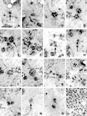
The first merogonial stage was observed in midgut epithelial cells at 24 h post-inoculation (p.i.) and was characterized by spherical binucleate meronts measuring 1·88 μm (1·6–2·3, ±0·2; n=10) in diameter (Fig. 1A). Growth of the meronts was followed by a nuclear division, resulting in quadrinucleate meronts, measuring 5·94 μm (5·3–6·6, ±0·45; n=10) in diameter (Fig. 1B). These meronts have diffused nuclei and an additional division resulted in an octanucleate form with a diameter of 7·09 μm (6·6–7·5, ±0·34; n=10). This stage eventually divided into tetra- and binucleate daughter diplokarya (Fig. 1C–E). Division of meronts was by binary fission of tetranuleate forms. After 48 h p.i., stages with 1, 2 or 4 diplokarya were the most common in the midgut, and nuclei were large and irregular and occupied about half of the area of the meronts (Fig. 1F–H). Giemsa stained the cytoplasm of meronts moderately blue and the nuclei purple. We were unable to identify sporonts, sporoblasts and primary spores with light microscopy in the midgut tissues.

Fig. 1. Giemsa-stained light micrograph of merogonic and sporogonic stages of Nosema sp. in Spodoptera litura. Scale bar on A applies also to B–P. (A) Early diplokaryotic meront. (B,C) Diplokaryotic meronts with 2 and 4 nuclei. (D) Dividing octanucleate meront undergoing cytoplasmic division. (E) Meront with 6 nuclei. (F) Binucleate meront. (G, H) Dividing meronts. (I) Octanucleate meront with horseshoe-like diplokaryotic nuclei. (J) Meront with 12 horseshoe-like diplokaryotic nuclei. (K) Binucleated sporont. (L) Tetranucleate sporont. (M) Early sporoblast. (N) Sporoblast (arrow). (O) Mature spores. (P) Spores after treatment with 0.1N HCl showing diplokarya.
Second merogony and sporogony cycle
After 72 h p.i., large multinucleate meronts with small compact horseshoe-like nuclei of uniform size and shape were observed in the fat body tissues (Fig. 1I, J). These forms developed by repeated karyokinesis without immediate cytokinesis, resulting in the production of 8 and 12 nucleated forms. They subsequently gave rise to binucleated daughter meronts which developed into sporonts. In addition, bi- and tetranucleate meronts similar to that of primary merogony cycle were also observed. This probably represents a second phase of the merogony since we did not observe any infection in the fat body tissues at 48 h p.i.. After 96 h p.i., binucleate sporonts measuring 4·84 μm (4·05–5·84, ±0·54; n=10) in diameter, and with deeply stained compact nuclei that occupied a central position in the cells, were observed (Fig. 1K). Further nuclear and cytoplasmic divisions resulted in tetranucleated sporonts of size 5·59 μm (4·64–6·52, ±0·70, n=10) (Fig. 1L). Subsequently, the tetranucleate sporonts divided into 2 sporoblasts measuring 5·4 μm (4·52–6·82, ±0·45, n=10) in length and 2·07 μm (1·84–2·34, ±0·25, n=10) in width, characterized by the presence of 2 small intensely stained and centrally placed nuclei (Fig. 1M). After 144 h p.i., binucleate sporonts, sporoblasts and immature spores were simultaneously observed in the fat body tissues. The cytoplasm of the sporoblasts stained lightly with Giemsa.
Mature spores
At 168 h p.i., sporoblasts were observed in the fat body. The binucleate condition was apparent in immature spores (Fig. 1N), while in the mature environmentally resistant (‘environmental’) spores nuclei were not distinguishable (Fig. 1O). However, on hydrolysis with 1 M HCl at 60 °C for 1 min, the binucleate state became clear (Fig. 1P). In fresh smears of infected tissues, different stages of spore development were observed. Immature spores were relatively large, with a conspicuous vacuole at one end. After 10 days p.i., environmental spores were observed in large numbers in the cytoplasm of fat body cells. They were highly refractive, ovocylindrical in shape, measuring 3·91 μm (3·8–4·0, ±0·0123, n=10) in length and 1·91 μm (1·8–2·0, ±0·0143, n=10) in width. Mature spores completely replaced the fat body tissue at 15 days p.i.. The polar filament of the spore was extruded upon rehydration of air-dried spores (Kellen and Lindegren, 1969; Watanabe, 1976) and the mean length of the extruded polar filaments was found to be approximately 90 μm.
Sequencing and phylogenetic analyses of 16S SSU-rRNA gene
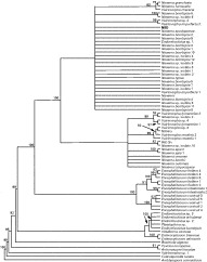
The 1231 bp 16S SSU rRNA gene of NSD was sequenced using the primers 18f and 1537r. The sequences of 72 non-redundant microsporidian isolates were downloaded from the GenBank and aligned along with the sequence of NSD and the best-fit model of evolution was identified using MODELTEST, which selected the ‘TIM+G’ model as the best-fit model of nucleotide substitution based on Akaike Information Criterion (AIC) (Akaike, 1974). However, because this model was not available in the Phylip package, we used the nearest alternative GTR+G model (AIC=33501·762) for phylogenetic analyses. The 50% consensus ML tree using gamma correction resulted in a single large cluster of Nosema/Vairimorpha clades (Fig. 2). The SSU rRNA gene sequence of NSD strongly suggested that this species is closely related to the typical Nosema subgroup within the Nosema/Vairimorpha clade as identified by Baker et al. (1994). The phylogenetic analyses using the sequence data of 72 microsporidia (Table 1) indicated that NSD is closely related to N. bombycis and N. spodopterae. The sequence of NSD differed by 18 bases from N. bombycis (GenBank Accession no. L39111). Altogether there were 2 insertions, 5 deletions, 8 transitions and 3 transversions. NSD also differed by 11 bases from N. spodopterae (AY211392), including 2 insertions, 3 deletions and 6 transitions.

Fig. 2. The 50% majority consensus maximum likelihood tree of 73 non-redundant microsporidian 16S SSU rRNA gene sequences. Amblyospora connecticus was included as an outgroup. Numbers on the nodes indicate the number of times a particular branch was recorded per 100 bootstrap replications following 1000 replicates. The new Nosema sp. isolate (NSD) is shown in bold.
Structure analyses of 16S SSU rRNA
Secondary structure prediction tool, mfold was used to obtain the folding patterns of the 16SSU-rRNA sequences. The default parameters were used for obtaining the most feasible structure at a minimum free energy (ΔG value of −260±4). The result indicated that the secondary structures of N. bombycis (L39111) and N. spodopterae (AY211392) were similar to each other whereas the structure of the NSD differed from those of both N. bombycis and N. spodopterae (Fig. 3). Such a difference is obvious because of a 3 nucleotide deletion in the new isolate at the position 918.

Fig. 3. Comparison of the 16S SSU rRNA structure of (A) Nosema sp. from Spodoptera litura (NSD) (B) Nosema bombycis (L39111) and (C) Nosema spodopterae. Only a portion of the structure (875–950) corresponding to the deleted region in NSD is shown in the figure. For all the species, the most conserved structure with similar minimum free energy (ΔG=−260±4 Kcal/mol) was used.
Host range
Cross-transmission tests to insects of 5 different insect orders (Table 2) showed that NSD was infective to all the 9 lepidopteran species tested namely, Bombyx mori (L.), Chilo infuscatellus Sn, Earias vitella (F.), Spodoptera exigua (H.), Helicoverpa armigera (H.), Leucinodes orbonalis Gn., Pericallia ricini (Fab.), Plutella xylostella (L.), and Tricoplusia ni (Hb.), although lower percentages of some species were infected. B. mori, S. litura, and P. ricini showed 100% infection. However, other insects of Orthoptera, Coleoptera, Hemiptera, and Diptera did not develop any infection.
Pathogenicity
The inoculation of NSD spores to third-instar larvae at 3 different concentrations of 2·4×102, 2·4×103, and 2·4×104 spores/larva resulted in larval mortality rates of 13·33%, 46·66%, and 73·33% respectively. The LD50 was 3885 spores/larva and the 95% limit lies between 2558·51–6110·06 spores/larva.
DISCUSSION
The microsporidium isolated from S. litura from the Dindigul, Tamil Nadu State, India is an obligatory intracellular parasite, multiplying in host cells in the form of small binucleate meronts and sporonts. Heavy infection resulted in change of larval colour to pink, frequently accompanied by swelling caused by hypertrophy of infected fat body cells. Occasionally, the body of the insect appeared deformed. The spores were found in the faecal material of the infected larvae, which probably provide the primary means of transmission to the healthy individuals due to contamination of the food source. Though cannibalistic behaviour is uncommon in S. litura, the parasitized larvae feed on the dead diseased larvae in the laboratory, suggesting that transmission of disease could also be due to cannibalism. Although experiments were not conducted to establish the transovarial transmission, presence of spores in ovarian tissues suggested that the parasite is probably incorporated into the eggs as most of the lepidopteran Nosema, for example N. pyraustae (Andreadis, 1987), N. heliothidis (Brooks, 1968) N. bombycis (Hatakeyama and Hayasaka, 2002) and also Vairimorpha pathogenic to lepidopterans (Haque, Canning and Wright, 1999; Nageswara Rao et al. 2004). The new microsporidian isolate appears to be apansporoblastic since the spores were in direct contact with the host cell cytoplasm possessing no sporophorous vesicle. All developmental stages were observed to be diplokaryotic. NSD shares all the salient features of the genus Nosema namely, apansporoblastic, and diplokaryotic developmental stages.
Sprague and Vernick (1971) characterized Nosema as having sporonts that divide into 2 sporoblasts and distinguished Nosema from the genera Glugea and Encephalitozoon by their nuclear arrangement. In the present study, the sporonts were binucleate and developed into 2 sporoblasts. Our observations show the NSD to be very similar in its development to N. bombycis, as described by Ishihara (1969), and therefore we placed it in the genus Nosema. The frequent occurrence of multinucleate meronts with 12 nuclei, however, easily distinguished NSD from N. bombycis.
A few reports have described the microsporidia from S. litura. Watanabe (1976) reported a Nosema sp. (NSW) from S. litura with development of binucleated meronts very similar to NSD. But the monosporous nature (sporont developed into single sporoblast) and uninucleate meronts of NSW distinctly differentiate it from NSD. Hsu, Hsu and Yen (1991, 1992), isolated a microsporidian species from S. litura in Taiwan and named it as Nosema spodopterae since they observed dimorphism of sporoblasts in their transmission electron microscopic study. The authors reported that one type of sporoblast possessed a plasmalemma inside of plasma membrane, and the other appeared as an amoeba-like shape without a plasmalemma within the spores. In general, the plasma membrane and plasmalemma are two names of the same structure. The internal layer of the spore wall is always referred to as a plasma membrane. The sporoblast derives its plasma membrane from the membranes of the polaroplast during the process of eversion through the polar filament during germination. No microsporidia have an amoeba-like stage in the spores. The sporoblast is the stage most difficult to fix properly for electron microscopy and electron microscopy fixation often destroys the shape of the sporoblast (Larsson, personal communication) leading to a star-like form in fixed tissues (Sokalova and Lange, 2002). Hence the two kinds of sporoblasts reported by Hsu et al. (1992) in N. spodopterae are probably an artifact arising due to fixation. Though the size of NSD sporoblasts ranges from 4·52 to 6·52 μm, the shape of the sporoblasts did not vary. The fresh spore size of NSD (3·91±0·012×1·91±0·014 μm) was similar to that of N. spodopterae (4·0±0·26×1·9±0·12 μm). Li and Wenn (1987) described a microsporidium, Nosema liturae, obtained from S. litura collected from China. The fresh spore size of N. liturae (4·34±0·27×1·99±0·14 μm) exceeds the size of NSD (3·91±0·012×1·91±0·014 μm). Bombyx mori is refractive to N. liturae, while the NSD is highly pathogenic to the same host species. The multinucleated meronts with up to 12 compact horseshoe-like nuclei reported in NSD were not reported in N. spodopterae and N. liturae.
Since many attempts to amplify large subunit rDNA did not succeed across all microsporidian species (Vossbrinck et al. 1993; Kent et al. 1996), 16S SSU rDNA has become the standard sequence for molecular characterization of microsporidia (Weiss and Vossbrinck, 1999; Vossbrinck and Debrunner-Vossbrinck, 2005). Here we aligned the SSU rRNA gene sequence of NSD with those of other known microsporidia that infect lepidopteran as well as non-lepidopteran hosts. The results lead us to confirm that NSD is the member of true Nosema namely, N. bombycis complex and belongs to the genus NosemaNageli, 1857.
Comparison of the 16S rRNA gene sequence of NSD with the corresponding sequence from several Nosema species showed that the parasite is closer to N. bombycis. We used N. bombycis ‘L39111’ as a reference strain in the present study since it is the most studied isolate for phylogenetic analyses (Baker et al. 1995; Canning et al. 2002; Choi et al. 2002; Vossbrinck and Debrunner-Vossbrinck, 2005). In NSD, the absence of 3 nucleotides at position 918 as compared to L39111 and other N. bombycis suggested a possible difference between these two isolates. The published sequence of N. spodopterae is, in fact, closer to that of L39111 than to NSD, suggesting that the new isolate is possibly a new species of Nosema from S. litura. Consistently, the secondary structure of N. bombycis and N. spodopterae 16S SSU rRNA was identical (Huang et al. 2004; Tsai, Huang and Wang, 2005) whereas the structure of NSD was different. The significance of these structural differences needs to be experimentally verified, which is out of purview of the present manuscript. These observations compel us to consider that the microsporidian isolate NSD as a new species, confirmation of which awaits further ultrastructural characterization.
The pathogenicity of NSD appears to be proportional to the inoculation dosage. The LD50 for larvae inoculated in third-instar was calculated to be 3·88×103 spores/larva at 20 days p.i. and is comparable with the published reports of other Nosema isolates from S. litura. Tsai and Wang (2001) obtained an LD50 of 1·13×104 spores/larva after 21 days when S. litura third-instar larvae were treated with N. spodopterae spores. The LD50 of Taiwan isolate in third-instar S. litura larvae was lower, 1·78×103 spores/larvae at 30 days p.i. (Tsai et al. 2003).
Host specificity is an important consideration for pathogens that are used for biological control. Microsporidia usually do not infect species in other insect orders when larvae are fed spores, but interordinal infections have been reported in laboratory infectivity studies (Fantham and Porter, 1958; Undeen and Maddox, 1973). All the lepidopteran hosts tested were susceptible to NSD indicating that this species appear to have a wider host range under laboratory conditions. Previous host specificity studies under lab conditions (Undeen and Maddox, 1973; Solter, Maddox and McManus, 1997; Solter and Maddox, 1998) suggest that terrestrial microsporidia have a broader laboratory host range within the taxonomic order of the natural host, but atypical microsporidia/host interactions suggested that the pathogens often do not reproduce optimally in non-target hosts, and data from surveys of microsporidian pathogens of various native and introduced insect hosts from field collections suggest that the ecological host ranges of entomopathogenic microsporidia are relatively narrow (Campbell and Podgwaite, 1971; Andreadis et al. 1983; Siegal et al. 1988; Jeffords et al. 1989). In the present study, lower percentages of infection rate observed in some lepidopteran species like P. xylostella and C. infuscatellus also reflected that these may be non-target hosts to NSD under field conditions. Hence, the use of NSD as a biological control agent against S. litura should be investigated.
Financial support to J.N. by the Department of Biotechnology, New Delhi under the project ‘Molecular Ecology and Systematics (Grant no. BT/012/01/NBDB/2004)’ is greatly acknowledged. We thank Dr Leellen F. Solter, University of Illinois, Urbana, USA, for her valuable help in life-cycle studies.