Introduction
Controlling gastrointestinal nematode infections in sheep still relies heavily on the use of synthetic anthelmintics. However, their indiscriminate use has led to the selection of resistant nematode populations (Calvete et al. Reference Calvete, González, Ferrer, Ramos, Lacasta, Delgado and Uriarte2020). Reducing the use of commercial antiparasitic drugs by incorporating medicinal plants into parasite control strategies is a widespread practice worldwide. Tannins are secondary plant metabolites that play an important role in plant defence mechanisms. They are widely distributed in nature, particularly in grasses, woody species, and herbaceous plants (Min et al. Reference Min, Barry, Attwood and McNabb2003). According to their chemical structure, tannins are classified into hydrolysable tannins (HT) and condensed tannins (CT) (Bravo, Reference Bravo1998). HTs are low-molecular-weight compounds that are easily degraded by rumen microorganisms and absorbed in the intestine (Olivas-Aguirre et al. Reference Olivas-Aguirre, Wall-Medrano, González-Aguilar, López-Díaz, Álvarez-Parrilla, Rosa and Ramos-Jimenez2015; Ortiz-Pineda et al. Reference Ortiz-Pineda, Bulla-Castañeda, Garcia-Corredor and Pulido-Medellín2020). This property makes them potentially toxic to ruminants, as they are absorbed as pyrogallol, a compound with hepato- and nephrotoxic effects (Min et al. Reference Min, Barry, Attwood and McNabb2003). CTs, or proanthocyanidins, are high-molecular-weight polymers capable of forming bonds with amino acids, polysaccharides, alkaloids, minerals, and proteins, which can have beneficial effects on animal nutrition (Hagerman, Reference Hagerman, Robbins, Weerasuriya, Wilson and Mcarthur1992; Reed, Reference Reed1995; Frutos et al. Reference Frutos, Hervas, Giraldez, Fernandez and Mantecon2000; Schofield et al. Reference Schofield, Mbugua and Pell2001).
Published literature indicates that CT-rich forages enhance protein absorption and reduce gastrointestinal nematode infections. Moreover, some studies have shown that parasitised sheep increase their preference for tannin-rich plants (Lisonbee et al. Reference Lisonbee, Villalba, Provenza and Hall2009; Juhnke et al. Reference Juhnke, Miller, Hall, Provenza and Villalba2012). Despite their beneficial effects on ruminant health, tannins can exert toxic or antinutritional effects due to their adverse effects by negatively influencing feed intake and animal performance (Mueller-Harvey, Reference Mueller‐Harvey2006). The magnitude of these effects depends on the tannin type, concentration per kilogram of dry matter (DM), molecular weight, and galactocyanin content (Armand Ugon Rostán & Orihuela Peruchena, Reference Armand Ugon Rostán and Orihuela Peruchena2018). Low (<2%) and moderate (3–6% of ingested DM) concentrations of tannins have beneficial effect on ruminants, whereas levels exceeding 7% can be detrimental (Barry & McNabb, Reference Barry and McNabb1999; Hoste et al. Reference Hoste, Jacksn, Athanasiadou, Thamsborg and Hoskin2006). The negative effects are mainly attributed to the high astringency of these compounds, which bind to salivary proteins and the oral mucosa, thereby reducing feed palatability and digestibility through the inhibition of proteolytic enzymes. Consequently, this leads to a reduction in daily weight gain (Escaray et al. Reference Escaray, Pesqueira, Pieckenstain, Carrasco and Ruiz2007; Lasa et al. Reference Lasa, Mantecón and Gómez2010).
Tannin extracts from red quebracho (Schinopsis balansae) and chestnut (Castanea sativa) trees are currently used in animal nutrition as functional feed ingredients. Quebracho heartwood is a major source of CT, whereas chestnut wood provides HT (Girard & Bee, Reference Girard and Bee2020; Caprarulo et al. Reference Caprarulo, Giromini and Rossi2021). In the last decade, increasing research has focused on the beneficial effects of combined chestnut-quebracho tannins on animal health. To date, tannin extracts have been shown to exert antimicrobial activity, improve intestinal health, and production parameters have been reported in poultry (Redondo et al. Reference Redondo, Redondo, Bruzzone, Diaz-Carrasco, Cabral, Garces and Fernandez-Miyakawa2022). In pigs, both in vitro assays and in vivo trials have demonstrated antioxidant capacity and antimicrobial effects against enterotoxigenic E. coli (Reggi et al. Reference Reggi, Giromini, Dell’Anno, Baldi, Rebucci and Rossi2020), as well as positive effects on growth performance, faecal and blood metabolites (Caprarulo et al. Reference Caprarulo, Hejna, Giromini, Liu, Dell’Anno, Sotira and Rossi2020), and modulation of the gut microbiota (Miragoli et al. Reference Miragoli, Patrone, Prandini, Sigolo, Dell’Anno, Rossi, Barbato, Senizza, Morelli and Callegari2021). In ruminants, supplementation with quebracho–chestnut tannin extract has been reported to influence the ruminal microbial community and fatty acid profile in dairy ewes (Buccioni et al. Reference Buccioni, Pauselli, Viti, Minieri, Pallara, Roscini, Rapaccini, Marinucci, Lupi, Conte and Mele2015). Furthermore, several studies have evaluated its impact on production performance, nitrogen partitioning, rumen microbiota and fermentation, and on reducing neonatal diarrhoea in preweaning calves (Díaz Carrasco et al. Reference Díaz Carrasco, Cabral, Redondo, Pin Viso, Colombatto, Farber and Fernández Miyakawa2017; Dell’Anno et al. Reference Dell’Anno, Frazzini, Ferri, Tuberti, Bonaldo, Botti, Grossi, Sgoifo Rossi and Rossi2024; Wang et al. Reference Wang, Li, Ren, Shen, Chen, Cui, Cao, Li, Xu, Sun, Ma, Gao and Li2024). The aim of this study was to investigate the potential anthelmintic effects of a chestnut–quebracho tannin blend against Haemonchus contortus through in vitro assays – egg hatching assay (EHA) and larval migration inhibition assay (LMIA) – as well as an in vivo trial. Additionally, scanning electron microscopy (SEM) was used to evaluate the damage caused by the extract on eggs and larvae recovered from the in vitro tests, and on adult worms obtained at necropsy.
Material and methods
Tannin extract
A commercial tannin extract consisting of 72% CT (from Schinopsis lorentzii, quebracho) and 28% HT (from Castanea sativa, chestnut) was used.
In vitro assay
Preparation of the extracts
For obtaining the extract, 5 g of the product were weighed and mixed with 100 mL of water preheated to 60°C. The mixture was kept on a magnetic stirrer until the tannin particles were completely dissolved.
Eggs and infective larvae of H. contortus
Eggs and infective larvae (L3) of H. contortus were obtained using two 3-month-old lambs (Dorper lamb F2). The animals were experimentally infected with 5000 L3 of a pure, drug-sensitive strain of H. contortus. Twenty-one days postinoculation, faecal samples were collected, and eggs were recovered following the egg recovery protocol described by the Animal Health Laboratory of Embrapa Pecuária Sudeste, São Carlos-São Paulo, Brazil, with some modifications (De Souza Chagas, personal communication).
L3 was obtained using the Corticelli-Lai technique (Corticelli & Lai, Reference Corticelli and Lai1963). Briefly, faeces from infected sheep were collected 21 days post-inoculation (PI) and placed in Petri dishes at room temperature for 10 days to allow larval development. The resulting L3 was then recovered and stored at 4°C until use.
Egg hatching assay
The EHA was carried out using an assay described by Torres-Acosta et al. (Reference JDJ, Chan-Pérez, López-Arellano, Rosado-Aguilar, Soberanes-Céspedes, Neri-Orantes and Encalada-Mena2015). Approximately 100 eggs of H. contortus were added to each well of a 24-well plate with the extracts from each plant at concentrations of 50, 25, 12.5, 6.25, 3.125, and 1.56 mg/mL, water (negative control), and benzimidazole 2 mg/mL (positive control). All the procedures were performed in triplicate. After incubation of the plate for 24 h at 37°C and 75% relative humidity, the number of eggs and larvae per well were counted under an inverted microscope. The rate of egg hatching inhibition was calculated according to the following formula: %= 100 × A/(A + B), where A is the total number of eggs and B is the total number of larvae contained in each well.
Larval migration inhibition assay
The LMIA was carried out using a modification of the method described by Demeler et al. (Reference Demeler, Küttler and Von Samson-Himmelstjerna2010). Approximately 100 L3 of H. contortus were added to each well of a 24-well plate containing plant extracts at concentrations of 50, 25, 12.5, 6.25, 3.125, and 1.56 mg/mL, water (negative control), and levamisole phosphate 22.3% (positive control). All the procedures were performed in triplicate. After 24 h of incubation of the plate at 37°C, the larvae were transferred to another 24-well plate with a 25 μm filter for 24 h. Then, the larvae retained in the filter were recovered, and the percentage of larval migration inhibition was calculated using the following formula: % = 100 × A/(A + B), where A represents the total number of migrated larvae and B represents the total number of non-migrated larvae per well.
In vivo experimental design
Eighteen 6-month-old lambs from the province of Corrientes, Argentina, were used to evaluate the anthelmintic activity of a quebracho-chestnut tannin mixture administered at 0.3% and 3% of dry matter intake (DMI). Before the start of the experiment, all animals were dewormed with a single oral dose of Naftalofos® and monitored by faecal egg counts (FEC) to confirm the absence of gastrointestinal nematode infection. The animals were allocated according to body weight into three homogeneous groups (mean 25 kg) as follows: Group 1 (g1): control (no tannin supplementation), Group 2 (g2): tannins at 0.3% of DMI (1.5 g/animal/day), and Group 3 (g3): tannins at 3% of DMI (15 g/animal/day).
All animals were experimentally infected on day 0 with a single dose of 5000 L3 of the H. contortus sensitive strain (CEDIVE strain). From day –7 to day 3 PI, g2 and g3 were supplemented with tannin extract. The extract was administered in the feed trough, spread over the alfalfa pellet ration, and moistened to ensure proper adhesion to the pellets and minimize feed losses. From day 16 to day 35 PI, faecal samples were collected from all animals three times per week to perform FEC and coprocultures. On day 35 PI, all lambs were slaughtered for adult worm recovery and further analyses.
Faecal samples
The FEC were performed using a modified McMaster technique adapted for cattle and sheep (Roberts & O’Sullivan, Reference Roberts and O’Sullivan1950). Faecal samples were collected directly from the rectum of each lamb on days 16, 18, 21, 23, 25, 29, 31, 33, and 35 PI.
The recovery of L3 of Haemonchus contortus was performed according to the method described by Corticelli and Lai (Corticelli & Lai, Reference Corticelli and Lai1963). On each sampling day, three pooled coproculture (one for each experimental group) were prepared using 30 g of faeces from each group.
Isolation of H. contortus adults
On day 35 PI all animals were slaughtered, and their abomasa were removed. The abomasa contents were collected and washed thoroughly with running water to recover the adult worms. The mucosa and its folds were carefully rinsed to detach any adhering parasites, and the washings were combined and diluted with water to a final volume of 4 L. The suspension was homogenised by gentle stirring, and several aliquots were taken until a final volume of 1 L was obtained for parasite quantification.
Statistical analysis
The dose-response data were fitted to a four-parameter log-logistic model using the drc package (Ritz et al. Reference Ritz, Baty, Streibig and Gerhard2015) in R (R Core Team, 2024). The 50%, 90%, and 99% effective concentrations (EC50, EC90, and EC99) were estimated with the ED() function (type = “relative”) of the drc package, which also provided the 95% confidence interval for EC50.
FEC were analysed using a mixed negative binomial model, with treatment and time as fixed effects, and time and animal as random effects. For the L3 counts, only time was included as a random effect. Adult worm counts in the abomasum were analysed using a generalised least squares model. All post hoc comparisons between groups were performed using the emmeans package with Tukey adjustment for multiple comparisons.
Scanning electronic microscopy of H. contortus
Eggs and L3 obtained from the in vitro assay exposed to 50 mg/mL of tannins, as well as adult worms recovered from necropsies of animals treated with 0.3% and 3% quebracho-chestnut tannin extract, were fixed in 2% glutaraldehyde. After successive washes, the parasites were dehydrated in ascending concentrations of absolute alcohol, dried by critical point drying with Emitech K850 and coated with gold-palladium with SPI-Module brand sputter coater. Samples were examined to evaluate surface morphology using Fei Quanta 200 scanning microscope operated at 20.00 kV.
Results
In vitro assay
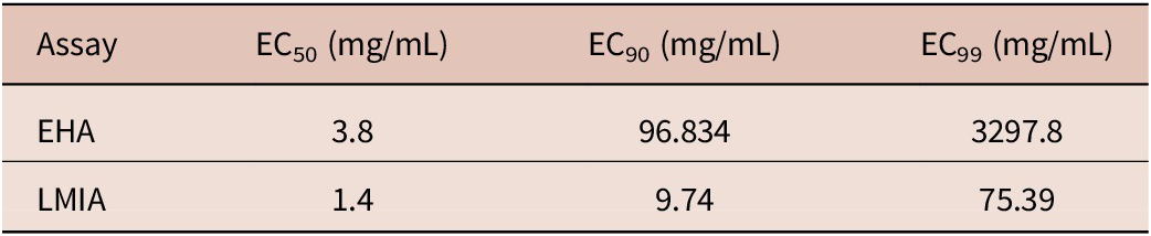
Dose-response analysis demonstrated that tannins possess potent in vitro anthelmintic activity in both assays (Table 1). The EC50 values were low, particularly in the LMIA (1.4 mg/mL), indicating high potency. The EC90 and EC99 values, which characterise the upper range of the response, were also relatively low. This confirms that high levels of inhibition were achieved at low doses.
Effective concentrations (EC50, EC90, EC99) from EHA and LMIA of Haemonchus contortus exposed to chestnut-quebracho tannin extract

The 95% confidence interval for EC₅₀ was 1.6396–6.0 mg/mL for egg hatching inhibition and 1.1–1.8 mg/mL for larval migration inhibition.
SEM of eggs and L3 of Haemonchus contortus
Fig. 1 shows the normal structure and the changes observed in H. contortus eggs and L3 after in vitro exposure to 50 mg/mL of quebracho–chestnut tannin extract. In the nonexposed control group, no structural alterations were detected. Eggs presented an intact shell and normal embryogenesis (Fig. 1A), and L3 appeared coiled with a normal intact cuticle (Fig. 1B). In contrast, eggs exposed to the quebracho–chestnut tannin extract showed deformation of the eggshell with possible rupture, and impaired embryogenesis, while exposed L3 exhibited cuticle damage and rupture (Fig. 1C and D, respectively).
SEM micrographs of egg and infective larvae of Haemonchus contortus recovered from the control group (without tannins) and exposed to 50 mg/mL of tannins. (A) Control egg. (B) Control infective larvae. (C) Egg showing loss of the air chamber and compaction of blastomere cells. (D) Infective larvae with loss of cuticle continuity.

In vivo trial
Faecal samples
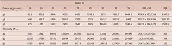
Table 2 shows the results of the faecal egg counts and the recovery of L3 from coprocultures. Infected sheep began to excrete eggs through faecal matter from day 16 PI. However, no significant differences (p > 0.05) were observed between treatments for each day evaluated.
Faecal egg counts and recovered L3 of Haemonchus contortus from lambs receiving the three treatments

Regarding the recovery of L3, despite slight variations in mean values between treatments, there is no statistical evidence that treatments g2 and g3 had a significant effect (p > 0.05) on larval count compared to g1.
Worm burden of Haemonchus contortus
Table 3 shows the H. contortus worm burden recovered from the abomasa of lambs in the three treatments. No significant differences were found between the g1 and g2 (p = 0.7950), g1 and g3 (p = 0.5935) or g2 and g3 (p = 0.9517). Additionally, worm counts were not significantly correlated (p > 0.05) with FEC at the last sampling time. However, a positive trend was observed across all groups, with the strongest correlation in the control group (r = 0.53) and lower correlations in animals treated with high (r = 0.33) and low (r = 0.16) concentrations of tannins.
Worm burden of Haemonchus contortus in lambs from the three treatments

SEM of adult of Haemonchus contortus
Fig. 2 shows normal structure of recovered adults from the control group. In the anterior region, the oral cavity presents no alterations, the cuticular striations remain continuous, and the cervical papilla appears without damage. The median region shows preserved natural folds and striations remain continuous. In the posterior region, the vulvar flap in the female is unmodified, and the male exhibits a bursa copulatrix without structural changes.
Normal structure of adult Haemonchus contortus in the control group (without tannins). (A) Anterior region. (B) Longitudinal lines of the cuticle. (C) Posterior region of a male (spicules and copulatory bursa). (D) Posterior region of a female (vulvar flap).

Fig. 3 shows the morphological damage observed in adults exposed to 0.3% DMI quebracho–chestnut tannin extract associated with the cuticle. Loss of normal structure and incision were observed in the cuticle until its rupture in the anterior and middle regions of the parasite, and tannin deposits were observed on the cuticle.
Damaged structure of adult Haemonchus contortus exposed to 0.3% DMI quebracho–chestnut tannin extract. (A) Cuticle disruption in the cephalic region. (B) Irregularities in the longitudinal striations of the cuticle. (C) Tannin adhesion to the cuticle. (D) Loss of cuticle integrity in the middle region.

Fig. 4 shows the morphological damage observed in adult worms exposed to 3% DMI quebracho–chestnut tannin extract. The parasites exhibited severe alterations affecting the cuticle, oral cavity, and reproductive organs. Tannin deposits were observed in the oral cavity (over the lancet), at the excretory pore, and around the vulvar flap in some adult worms. In addition, the cuticle lost its normal appearance, showing longitudinal and transverse folds and thicker ridges.
Damaged structure of adult Haemonchus contortus exposed to 3% DMI quebracho–chestnut tannin extract. (A) Tannin deposition in the oral cavity and rupture of the oral cavity. (B) Tannin deposition in the excretory pore. (C) Thickening and irregularities of the cuticle. (D) Tannin deposition around the vulvar flap.

Discussion
Haemonchosis is considered one of the most common parasitic diseases in sheep, causing substantial economic losses in production worldwide. In recent decades, control of this disease has relied almost exclusively on the use of synthetic anthelmintics, which has contributed to the emergence of resistant nematode populations. In this context, phytomedicine has gained increasing global attention as an alternative tool for controlling parasitic diseases, particularly through the use of tannins.
The results of the present study demonstrated the potent anthelmintic activity in vitro of quebracho-chestnut tannin extract against H. contortus. However, this effect could not be replicated in vivo, highlighting the well-known gap that often exists between controlled laboratory conditions and the complexity of the host-parasite interaction. The efficacy observed in vitro was consistent with the structural damage observed by SEM. The changes observed proved the occurrence of strong interactions between the tannin extracts and the external structures of the egg and larvae. The changes observed in the eggs could be attributed to the formation of tannin-protein complexes on the egg coat, altering permeability, modifying oxygen exchange, and inhibiting the release of enzymes that degrade the egg coat (Cortes-Morales et al. Reference Cortes-Morales, Olmedo-Juárez, Trejo-Tapia, González-Cortazar, Domínguez-Mendoza, Mendoza-de Gives and Zamilpa2019). Regarding L3, changes in their cuticle were also observed. Montero (Reference Montero2017) stated that the mechanism of action of tannins is to prevent them from unsheathing, which directly interferes with infectivity of the larvae, thus preventing the continuation of their development. However, this last statement could not be demonstrated under in vivo conditions in the present study, as no statistically significant differences in parasitic load were detected among the three experimental groups.
Although no in vivo effect of tannins were detected in the present experiment (whether on egg counts, larvae recovery, or adult burdens), several authors have confirmed the efficacy of tannin extracts in vivo trial. Martínez-Ortíz-de-Montellano et al. Reference Martínez-Ortíz-de-Montellano2010 stated that the consumption of 60 g/kg of body weight of balanced tannin reduced egg excretion and female fertility without affecting the nematode load. In turn, Iqbal et al. (Reference Iqbal, Sarwar, Jabbar, Ahmed, Nisa, Sajid and Yaseen2007) reported that supplying 20–30 g/kg DM of tannins in the diet over a longer period (60–120 days) significantly reduced egg counts starting on day 60. Similarly, Minho et al. (Reference Minho, Bueno, Louvandini, Jackson, Gennari and Abdalla2008) also reported a marked reduction in abomasal parasite burden following the administration of 1.6 g/kg live weight of Acacia molissima CT for two consecutive days, with a repeated dose on day 30. Likewise, Pathak et al. (Reference Pathak, Dutta, Banerjee, Goswami and Sharma2016) observed a significant decrease in H. contortus burden in lambs inoculated with 2000 L3 after receiving 1.5% tannin in the diet for 90 days. According to the damage observed using SEM, marked cuticular alterations were observed, along with tannin deposits in the oral cavity and vulvar region. These findings suggest a possible inhibition of feeding and impaired reproductive function, which could lead to a reduction in egg excretion. This damage was consistent with previous reports, which describe tannin deposition followed by tissue degradation around the oral cavity, anus, vulva, and copulatory bursa, along with loss of integrity, cuticular thickening, formation of ridges and folds, and altered permeability (Martínez-Ortíz-de-Montellano et al. Reference Martínez-Ortíz-de-Montellano, Arroyo-López, Fourquaux, Torres-Acosta, Sandoval-Castro and Hoste2013; Tresia et al. Reference Tresia, Evvyernie and Tiuria2016; Barone et al. Reference Barone, Zajac, Manzi-Smith, Howell, Reed, Krueger and Petersson2018 and Del Carmen Acevedo-Ramírez et al. Reference Del Carmen Acevedo-Ramírez, Hallal-Calleros, Flores-Pérez, Alba-Hurtado, Mendoza-Garfías, Del Campo and Barajas2019).
The different efficacy obtained in this work between the in vitro assay and the in vivo trial raises methodological aspects that should be addressed in future studies. In the in vitro assays, the range of concentrations tested was sufficient to estimate EC50 values with precision; however, it was not adequate to reliably calculate higher effective concentrations (EC90 and EC99), as these estimates required extrapolation beyond the experimental range. Future in vitro studies should therefore explore a broader range of lower and intermediate concentrations to better characterize the full dose–response relationship. On the other hand, in the in vivo trial, lambs were evaluated after receiving two different concentrations of quebracho-chestnut tannin for 10 days, accompanied by a single experimental infection of 5000 L3. This experimental design, which differs from the gradual and continuous exposure to infective larvae under natural grazing conditions, could not demonstrate the nematocidal effect that tannins would have if the infection rate were lower. Also, the different efficacy obtained in both trials could be attributed to the interaction factor between the host and parasite substances in an in vivo trial (Torres-Fajardo et al. Reference Torres-Fajardo, González-Pech, Sandoval-Castro and Torres-Acosta2020). According to Borges et al. (Reference Borges, de Araújo, Carollo, Carollo, Lifschitz, Conde and de Almeida Borges2020), the bioavailability of an active compound is affected by metabolic transformation and elimination processes, and in turn, its efficacy will depend on the parasite’s developmental stage and degree of susceptibility. The reduced sample size used in the in vivo trial was justified under the principles of the 3Rs and supported by robust statistical analyses; however, increasing the number of animals in future trials would allow a more accurate assessment of variability and treatment effects under conditions closer to natural infection dynamics.
In conclusion, the pronounced dose-response relationship observed in the in vitro assays suggests strong biological activity of tannins, which could be a useful tool in an integrated control programme, serving as an alternative to chemical antiparasitic, especially in sheep-farming areas where anthelmintic resistance is widespread. However, future studies should include a broader in vitro concentration range to allow for a more robust estimation of higher effective concentrations and increase the number of animals in future in vivo trials to more clearly establish the dose-effect-host relationship.
Acknowledgements
We gratefully acknowledge the farm staff for their valuable assistance with animal handling throughout the study.
Financial support
This study was funded by the diagnostic area service of the parasitology laboratory, Institute of Veterinary Pathobiology (IPVET), Dual Dependency Unit, National Institute of Agricultural Technology (INTA), and National Council for Scientific and Technical Research (CONICET).
Competing interests
The authors declare that they have no conflict of interest.
Ethical standard
The authors affirm that all procedures conducted in this study were conducted in accordance with the ethical standards established by the relevant national and institutional guidelines for the care and use of laboratory animals.