INTRODUCTION
Lice are medically and economically important blood-feeding ectoparasites. The lice that infest humans are the head louse, Pediculus capitis, the body (clothes) louse, Pediculus humanus, and the crab or pubic louse, Pthirus pubis. The Pediculus have infected humans for thousands of years (Mumcuoglu et al. Reference Mumcuoglu, Zias, Tarshis, Lavi and Stiebel2003; Leo and Barker, Reference Leo and Barker2005) causing localized skin irritations. Furthermore, the body louse is a vector of 3 pathogenic bacteria that may cause louse-borne epidemic typhus, relapsing fever and trench fever (Maurin and Raoult, Reference Maurin and Raoult1996; Schaub, Reference Schaub and Mehlhorn2001; Fournier et al. Reference Fournier, Ndihokubwayo, Guidran, Kelly and Raoult2002). In addition, body lice can transmit Yersinia pestis in the laboratory (Houhamdi et al. Reference Houhamdi, Lepidi, Drancourt and Raoult2006).
Head lice are a persistent problem in most parts of the world (Gratz, Reference Gratz1997) with the United States of America alone spending approximately US$100 million per annum on pediculicides to control head lice (Jones and English, Reference Jones and English2003). In Australia, head lice are a continuing problem in preschool and elementary (primary) school aged children. The most recent report from Victoria, Australia, stated that 16·6% of 1838 primary school children had head lice (13·3%) or dead/hatched eggs (3·3%) (Counahan et al. Reference Counahan, Andrews, Buttner, Byrnes and Speare2004). There are several reasons for the high prevalence of head lice, including poor compliance when using pediculicides, resistance to the current actives in the pediculicide (e.g. permethin, malathion, pyrethrins) and an inability to effectively kill all of the eggs in the hair. Indeed, at present no 100% effective ovicide is available for any species of louse.
Egg-hatch is a critical process in the life-cycle of insects. Larvae and nymphs of insects emerge from their eggs using structures including hatching spines, egg bursters and egg teeth (reviewed by Young et al. Reference Young, Mancuso and Bowles1999). Indeed, previous studies of Pt. pubis and P. humanus egg-hatch described a ‘latch’ and spikes on the ellipsoid structures that are associated with the vitelline membrane of the eggs (Berman and Firstenberg, Reference Berman and Firstenberg1979; Berman et al. Reference Berman, Carter and Brodkin1980). It was thought that the ellipsoid structure with spikes and the ‘latch’ ruptured the vitelline membrane and thus allowed the nymph to escape from the egg through the operculum. Egg-hatch in Pt. pubis and P. humanus was therefore thought to be by the mechanical action of the latch and spike and by physical movement of the nymph in the egg. However, evidence is now mounting for the importance of a biochemical component to egg-hatch in insects. For example, in the grasshopper, Melanoplus differentialis, a chitinase enzyme facilitates egg-hatch by digesting a specific region of the egg known as the white cuticle (Slifer, Reference Slifer1937). More recently, a potential role for serine and/or metalloproteases in egg-hatch was described in Lucilia cuprina, the sheep blowfly (Young et al. Reference Young, Mancuso and Bowles1999, Reference Young, Mancuso, Meeusen and Bowles2000). In the present study, we identify, partially characterize and inhibit the activity of proteases expressed during egg-hatch in the body louse, P. humanus. Based on these findings, we propose that metalloproteases play a key role in this very important process.
MATERIALS AND METHODS
Source of insects
Body lice were from the Barker isolate of the Orlando strain of body (clothes) lice, P. humanus, at the University of Queensland. Dr Barker has maintained this lice colony in his laboratory for over 10 years. The Barker isolate was founded with lice from the isolate of Dr Mumcuoglu from the Hebrew University, Jerusalem. The Orlando strain was originally founded from body lice from a small, but unspecified number of people in Washington DC and Orlando, Florida, USA, around 1942 (Culpepper, Reference Culpepper1944). The Orlando strain was maintained at the United States Department of Agriculture, Bureau of Entomology and Plant Quarantine, Orlando, Florida from 1942–1945 on human volunteers (Culpepper, Reference Culpepper1944, Reference Culpepper1946). The strain was adapted to feed on the blood of rabbits in 1945 (Culpepper, Reference Culpepper1948) and has been maintained in this manner ever since. Lice were kept in an incubator at 32±1°C, 50% relative humidity with a photo-period of 3 h: 21 h (light: dark).
Collection of egg-shell-washings (ESW)
Gravid adult female P. humanus were incubated with pieces of human hair at 32°C, 50% relative humidity for 12–16 h. Washings from the freshly laid eggs termed egg-shell-washings (ESW) and other solutions were then collected as follows. Sample 1, from unhatched eggs placed in 20 μl of distilled water and incubated at 32°C for 30 min. Sample 2, from human hair (following egg removal) added to 20 μl of distilled water and incubated at 32°C for 30 min. Sample 3, from human hair washed in 1% sodium hypochlorite for 1 min, followed by 5×1 min washes in distilled water before being incubated as for sample 2. Sample 4, from unhatched eggs washed with 1% sodium hypochlorite and distilled water prior to incubation as previously described for sample 1. Sample 5, was collected from hatched eggs; 24 h prior to hatch, eggs were washed with 1% sodium hypochlorite, rinsed and allowed to hatch at 32°C. The empty egg-shells were collected 0–2 h post-hatch and incubated as for samples 1–4. From samples 1–5 the 20 μl of fluid was recovered, freeze-dried and stored at −70°C.
One-dimensional gelatin SDS-PAGE analysis
The method for gelatin SDS-PAGE was adapted from Heussen and Dowdle (Reference Heussen and Dowdle1980) and described by Young et al. (Reference Young, Meeusen and Bowles1996) with the following modification. Gels were incubated for 3 h at 37°C in 0·1 m Tris/HCl containing 1 mm calcium chloride, pH 8·0.
Two-dimensional gelatin SDS-PAGE analysis of egg-shell-washings (ESW)
Hatched egg shells were collected in 200 μl of distilled water as described for sample 5. For analysis, ESW were resuspended in rehydration buffer (8 m urea, 2% 3-[(3-cholamidopropyl) dimethylammonio]-1-propanesulfonate, 2% immobilized pH gradient buffer 3–10; GE Healthcare) and used to rehydrate 7 cm Immobiline Dry strips, pH 3–10 (GE Healthcare) overnight. Strips were transferred to the Multiphor II (GE Healthcare) apparatus, electrophoresed in the first dimension at 200 V for 1 min, increasing to 3500 V over the next 90 min followed by 65 min at 3500 V, equilibrated (6 m urea, 30% glycerol, 50 mm Tris, pH 8·8 and 2% SDS) and then run on a 10% SDS-PAGE gel containing 0·1% gelatin for the second dimension as for one-dimensional gelatin SDS-PAGE analysis.
Characterization of proteases in the egg-shell-washings (ESW)
For mechanistic characterization, the protease inhibitors 10 mm ethylenediamine tetraacetic acid (EDTA), 5 mm phenylmethylsulfonyl fluoride (PMSF), 10 mm 1,10-phenanthroline, 10 μm E-64 or 5 μm pepstatin were added to the wash and incubation buffers of the one-dimensional gelatin SDS-PAGE. In addition, following separation of the ESW on a two-dimensional gelatin SDS-PAGE gel, the gel was incubated in the presence of 10 mm 1,10-phenanthroline as described above.
In vitro egg-hatch assay
Protease inhibitors in solvent or the equivalent solvent control were added for 10 min to P. humanus eggs that were within 24 h of hatching. The eggs (15–20 per replicate) were then removed from the solutions, blotted dry with tissue paper, placed in an incubator at 32°C and left to hatch. The proportion of hatched eggs in the treated groups was compared to the proportion of successfully hatched eggs in the solvent control eggs and a percentage inhibition calculated. Percentage hatch inhibition was defined as the percentage reduction in egg hatching produced by a particular treatment when compared to the untreated control.
Statistical analysis
The egg-hatch assays were performed at least twice, in triplicate. Chi-squared analysis was used on the data from the egg-hatch inhibitor studies to determine the level of significance.
RESULTS
Identification of proteases present during egg-hatch
Gelatin SDS-PAGE was used to analyse the protease activity in ESW from louse eggs both before and after hatch, and from human hair samples. We attempted to determine protein levels in each of the different ESW samples; however, this was unsuccessful due to the very low protein levels present. Therefore, for comparative purposes, samples have been described in terms of the number of louse eggs from which the washings were obtained.
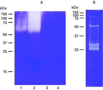
Protease activity was detected in washings from unhatched eggs within 12 h of hatching (sample 1) in the higher molecular weight region of the gel, above 50 kDa (Fig. 1A, lane 1). A similar pattern of protease activity was detected in the washings from human hair samples following the removal of the eggs (sample 2) (Fig. 1A, lane 2). Treatment of the hair with 1% sodium hypochlorite prior to collecting the washings (sample 3) completely eliminated all protease activity (Fig. 1A, lane 3). Hypochlorite treatment also eliminated all protease activity from unhatched eggs (sample 4) (Fig. 1A, lane 4). To remove extraneous protease activity, hypochlorite was used to treat unhatched eggs prior to the collection of ESW for all subsequent protease analyses.
(A) One-dimensional gelatin SDS-PAGE gel of egg-shell-washings (ESW) from 50 unwashed eggs of Pediculus humanus (sample 1, lane 1), unwashed human hair (sample 2, lane 2), washed human hair following treatment with 1% sodium hypochlorite (sample 3, lane 3) and washings from 50 unhatched eggs following treatment with 1% sodium hypochlorite (sample 4, lane 4). (B) One-dimensional gelatin SDS-PAGE gel with ESW from 66 freshly hatched P. humanus egg-shells treated with 1% sodium hypochlorite prior to egg-hatch. The positions of pre-stained Mr standards (kDa) are shown to the left of panels A and B.

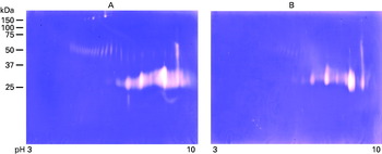
ESW collected from freshly hatched eggs revealed several distinct proteases around 25–30 kDa and 50 kDa. There were also a few fainter bands above 75 kDa (sample 5) (Fig. 1B). Using two-dimensional gelatin SDS-PAGE the proteases in the 25–30 kDa molecular weight range resolved to at least 7 proteases with isoelectric points in the neutral to alkaline pH range, whereas the band of protease activity around 50 kDa resolved to at least 11 distinct protease regions with iso-electric points in the acidic to neutral pH region (Fig. 2A). The regular banding pattern of the proteases in the 25–30 kDa and 50 kDa regions suggests that these proteases may be isoenzymes. At least 5 proteases with molecular weights above 75 kDa were also observed.
Two-dimensional gelatin SDS-PAGE gels of ESW from 2125 freshly hatched egg-shells of Pediculus humanus. Samples were separated by their isoelectric point in the first dimension (pH range 3–10) and by size in the second dimension. The positions of pre-stained Mr standards (kDa) are shown to the left. (A) No protease inhibitors were present during protease development. (B) The gel was incubated in the presence of 10 mm 1,10-phenanthroline during protease development. Note the marked reduction in protease activity following exposure to 1,10-phenanthroline.

To identify the ESW proteases, proteins of an equivalent size and isoelectric point to the major proteases (4 in total) were selected from non-gelatin containing gels and sent to the Australian Proteome Analysis facility for 2D Liquid Chromatography Electrospray Ionization tandem Mass Spectrometry (2D LC ESI MS/MS) analysis. The LC/MS/MS data were searched using the program Mascot against the NCBI non-redundant database and a number of the peptide spectra were de novo sequenced for further database searching; however, no unambiguous protein identifications could be made (data not shown).
Characterization of the proteases in egg-shell-washings (ESW)
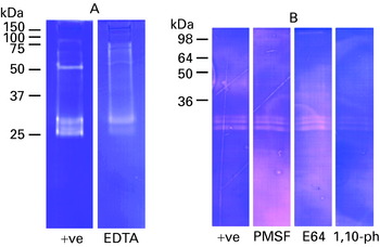
The proteases present in the louse ESW were further characterized by their mechanistic class. Incubation with the metal-chelating agents EDTA and 1,10-phenanthroline, to inhibit metalloproteases, resulted in a reduction in protease activity compared with the untreated controls (Fig. 3A and 3B, respectively). In contrast, there was no apparent reduction in protease activity when the ESW were incubated with the serine/cysteine protease inhibitor PMSF (Fig. 3B), the cysteine protease inhibitor E-64 (Fig. 3B) or the aspartic protease inhibitor pepstatin (data not shown), although lower amounts of ESW were used for the characterization with these inhibitors. The inhibition observed with 1,10-phenanthroline was further investigated following a second dimension separation of the ESW. The results confirmed the inhibitory effect of this metalloprotease inhibitor on the activity of the louse egg proteases (Fig. 2B) with a general reduction in protease activity in this 25–30 kDa region that contained the most abundant proteases and a clear reduction in the region around 50 kDa and above 75 kDa.
One-dimensional gelatin SDS-PAGE mechanistic investigation of proteases contained within ESW obtained from 66 louse eggs per lane (A) or 20 louse eggs per lane (B). Each of the gel lanes was treated with a different inhibitor during protease development. (A) No treatment control and 10 mm EDTA. (B) No treatment control, 5 mm PMSF, 10 μm E-64 and 10 mm 1,10-phenanthroline. The positions of pre-stained Mr standards (kDa) are shown to the left of panels A and B.

The effect of protease inhibitors on the hatching of P. humanus eggs
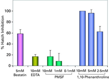
The effects of protease inhibitor exposure on louse eggs were determined using the in vitro egg-hatch assay. Of the 4 protease inhibitors tested, 2, bestatin and 1,10-phenanthroline (both of which are metalloprotease inhibitors), produced significant inhibition of louse egg-hatch in vitro (Fig. 4). The metal-chelating protease inhibitor, 1,10-phenanthroline, was the most effective inhibitor of louse egg-hatch. This response was shown to be dose dependent, with complete inhibition observed at 10 mm which declined to approximately 60% at 2·5 mm (P<0·05). The aminopeptidase inhibitor, bestatin, also resulted in significant inhibition of louse egg-hatch at 5 mm (P<0·05). In contrast, neither the metalloprotease inhibitor, EDTA (10 mm), or the serine/cysteine protease inhibitor PMSF, significantly inhibited louse egg-hatch.
The percentage egg-hatch inhibition for Pediculus humanus eggs. Louse eggs were exposed to protease inhibitors for a period of 10 min before being removed, blotted dry and placed in an incubator at 32°C until the eggs hatched. The percentage hatch inhibition was then determined compared to the control eggs that were exposed to the solvent only. Significant (P<0·05) inhibition of louse egg hatching was observed when the louse eggs were exposed to bestatin and 1,10-phenanthroline.

DISCUSSION
This paper describes, for the first time, the presence of proteases in ESW from freshly hatched eggs of the Orlando strain of the body (clothes) louse, P. humanus. At least 23 distinct regions of protease activity were detected over a range of molecular weights and isoelectric points. Mechanistic class studies with gelatin SDS-PAGE indicated that the ESW contained numerous metalloproteases, based on their inhibition with 1,10-phenanthroline and EDTA. The identification of louse proteases around the time of egg-hatch implies a role(s) for proteases in the egg-hatch process of these parasites. Indeed the finding that the metalloprotease inhibitors, 1,10-phenanthroline and bestatin, were highly effective at inhibiting louse egg-hatch, further supports this possibility.
In invertebrates, hatching enzymes belong to a large family of zinc-dependent metallo-enzymes referred to as matrixins or matrix metalloproteinases (MMP) (Geier and Zwilling, Reference Geier and Zwilling1998; Katagiri et al. Reference Katagiri, Maeda, Yamashika, Mita, Sargent and Yasumasu1997). For example, in the free-living nematode, Caenorhabditis elegans, a hch-1 metalloprotease is secreted into the perivitelline fluid before hatching (Hishida et al. Reference Hishida, Ishihara, Kondo and Katsura1996) and a zinc-dependent leucine aminopeptidase has been identified in the egg homogenates of the soybean cyst nematode, Heteroderaglycines glycines (Tefft and Bone, Reference Tefft and Bone1985). In contrast, there are few reports of proteases associated with insect egg-hatch and where these molecules have been implicated, they have been associated with serine/cysteine proteases as in the sheep blowfly, Lucilia cuprina (Young et al. Reference Young, Mancuso, Meeusen and Bowles2000), and in the silkworm Bombyx mori (Maki and Yamashita, Reference Maki and Yamashita2001).
Analysis by two-dimensional gelatin SDS-PAGE highlighted the proteolytic complexity of the ESW. At this point it is not possible to conclude which proteases in the ESW may be associated with louse egg-hatch; however, further studies using a range of different substrates may assist in identifying the proteases involved. The finding that 1,10-phenanthroline could inhibit a number of the proteases detected by gelatin SDS-PAGE and also completely inhibited louse egg-hatch, in a dose-dependent manner, supports a role for metalloproteases in louse egg-hatch. More specifically, the inhibition of louse egg-hatch by bestatin implies that egg-hatch may be facilitated, at least in part, by metalloproteases of the aminopeptidase subclass. Bestatin has also been shown to disrupt the hatching of the parasites Schistosoma mansoni and Haemonchus contortus, presumably through its ability to inhibit leucine aminopeptidase activity (LAP) present in the hatching fluid (Rogers and Brooks, Reference Rogers and Brooks1977; Xu and Dresden, Reference Xu and Dresden1986). Taken together, these results indicate that further studies on the role of aminopeptidases in louse egg-hatch are warranted.
It is unusual that although both 1,10-phenanthroline and EDTA were able to significantly inhibit the activity of the proteases present in the ESW, only 1,10-phenathroline inhibited egg-hatch. This pattern of inhibition was also observed when sheep blowfly ESW and eggs were exposed to metalloprotease inhibitors (Young et al. Reference Young, Mancuso, Meeusen and Bowles2000). 1,10-phenanthroline has also been reported to have a greater inhibitory effect on some aminopeptidases than EDTA, which might explain the observed difference in the inhibitory effect of these two inhibitors on louse egg hatch (Laeremans et al. Reference Laeremans, Demaegdt, De Backer, Le, Kersemans, Michotte, Vauquelin and Vanderheyden2005). These chelating inhibitors have differing affinities for certain divalent ions, with 1,10-phenanthroline having significantly greater affinity for zinc than calcium compared to EDTA, which would affect the rate at which these compounds inhibit specific proteases. Further work with the isolated proteases responsible for acting on the egg-shell proteins would enable additional characterization that may explain the observed difference in action between these two metalloprotease inhibitors. Alternatively, it may be that EDTA is unable to penetrate the louse egg, as this compound was dissolved in water not in an aqueous solvent. The natural waxy barriers or osmotic potentials of the egg may inhibit the penetration of water-soluble molecules into the egg, whereas alcohol may have altered the permeability of the aeropyles on the operculum to facilitate entry of alcohol-soluble compounds like 1,10-phenanthroline and bestatin. Presently, there are no published data on factors that affect the permeability of louse eggs that may lead to a rational basis for selecting potential inhibitors.
It must also be considered that these inhibitors are adversely affecting proteins other than metalloproteases during egg-hatch. Indeed, 1,10-phenanthroline could be acting by removing metal ions from one or more proteins that are important in embryonic development or egg-hatch. However, the non-chelating metalloprotease inhibitor, bestatin was also able to inhibit egg-hatch, supporting the possibility that metalloproteases/aminopeptidases are important in this process. It is also interesting to note that the highly toxic protease inhibitor PMSF, did not significantly inhibit egg-hatch, even at the relatively high concentration of 10 mm, suggesting that the inhibition is not merely due to the toxicity of the treatments to the developing embryo.
These are the first studies on the potential role of proteases in egg-hatch in species from the Order Phthiraptera (lice) which comprises 4994 species ((Durden and Musser (Reference Durden and Musser1994) for the Anoplura and (Price et al. Reference Price, Hellenthal and Palma2003) for the Amblycera, Ischnocera, Rhynchophthirina)). While it is possible that egg-hatch in lice may also involve some mechanical disruption of the vitelline membrane, the identification of proteases in the ESW from P. humanus and the subsequent inhibition of louse egg-hatch with metalloprotease inhibitors, indicates a role for these enzymes in louse egg-hatch and provides a foundation for evaluating the use of metalloprotease inhibitors as therapeutic agents against these parasites.
We thank Ms Maryam Ashafi for her expert and enthusiastic technical assistance in rearing louse eggs and for collecting egg-shell-washings. The research was supported by Hatchtech Pty Ltd. V. B. is a director of Hatchtech.