Introduction
Nervous system transmitters take part in a number of key processes during cleavage division (Shmukler & Buznikov, Reference Shmukler and Buznikov1998) such as triggering of the cell cycle (Buznikov, Reference Buznikov1990), formation of primary cellular interactions, completion of cleavage furrow formation (Shmukler et al., 1998) and direct blastomere interactions (Shmukler, Reference Shmukler1981, Reference Shmukler1992, Reference Shmukler1993). Evidence is presented that serotonin (5-HT) or 5-HT-like substances take part in all the above-mentioned processes in sea urchin early embryos and that corresponding receptors (or their functional analogues) are localized either intracellularly (Buznikov & Shmukler, Reference Buznikov and Shmukler1978; Shmukler et al., Reference Shmukler, Grigoriev, Buznikov and Turpaev1986) or on the surface of blastomeres membrane (Shmukler, Reference Shmukler1992, Reference Shmukler1993; Shmukler & Tosti, Reference Shmukler and Tosti2002).
In particular, 5-HT-antagonists may evoke functional isolation of blastomeres of intact embryos, by influencing intracellular receptors (Buznikov & Shmukler, Reference Buznikov and Shmukler1978, Reference Buznikov and Shmukler1981), that is at least partially due to blockage of ‘post-division adhesion’ (Vacquier & Mazia, Reference Vacquier and Mazia1968). At the same time in the ‘micromere model’ (Shmukler et al., Reference Shmukler, Chailakhian, Smolianinov, Bliokh, Karpovich, Gusareva, Naidenko, Khashaev and Medvedeva1981; Shmukler, Reference Shmukler1981), in which the process of adhesion is excluded by experimental conditions, serotonergics specifically influenced a cleavage pattern of sea urchin half-embryos, imitating or eliminating the interblastomere signal, probably via corresponding receptors localized at the surface membrane of blastomeres (Shmukler, Reference Shmukler1992, Reference Shmukler1993) However the absence of well-developed serotonergics pharmacology did not allow us to determine the type of the 5-HT-receptor of interest.
In our following study, the effect of 5-HT3-receptor ligands on Ca2+-levels in sea urchin Lytechinus pictus embryos was found (Shmukler et al., 1998). Moreover, poorly penetrating the cell membrane, the 5-HТ3-agonist 5-HTQ was shown to evoke specific inward currents selectively in Paracentrotus lividus blastomeres during the first and the second cleavage furrow formation. This finding was in contrast to the 5-HT2-agonist DOI, which evoked currents over whole cell cycle and the 5-HТ1-agonist 8-OH-DPAT, which did not generate any currents (Shmukler & Tosti, Reference Shmukler and Tosti2002). These findings provide evidence of the presence of 5-HT-receptive structures on the surface membrane of blastomeres and of their certain pharmacological specificity.
The ‘protosynapse’ concept stated by us previously (Shmukler, Reference Shmukler1992, Reference Shmukler1993; Buznikov et al., Reference Buznikov and Shmukler1996; Shmukler & Buznikov, Reference Shmukler and Buznikov1998) suggests that the rise of internal asymmetry of blastomeres might be due to an increase of 5-HT or 5-HT-like substance levels in the interblastomere compartment and not dependent on the spatial distribution of corresponding receptors over the blastomere surface. In this paper we have provided evidence on the distribution and the possible role of such receptors on the surface membrane of blastomeres.
Materials and Methods
Gametes
Female gametes of sea urchin Paracentrotus lividus (Bay of Naples, Mediterranean) were obtained by 0.05 ml 0.5 M KCl injection into the coelom. Spermatozoa were collected from male gonads and a drop of sperm was diluted and added to an oocyte suspension immediately before the artificial fertilization.
Fertilization and removal of vitelline envelope and hyaline layer were performed as described earlier by Yazaki et al. (Reference Yazaki, Tosti and Dale1995). Embryos were transferred to normal seawater before electrophysiological recording or blastomere isolation.
Electrophysiology
Nude embryos were voltage clamped in the whole-cell configuration as previously described (Yazaki et al., Reference Yazaki, Tosti and Dale1995). Briefly, patch pipettes of 3–5 MOhm (MΩ) resistance and 1–2 μm tip diameter, were filled with an intracellular-like solution (ICS) containing 200 mM K2SO4, 20 mM NaCl, 200 mM sucrose, 10 mM EGTA and 10 mM HEPES, with pH adjusted to 7.5. After GigaΩ-seals were obtained, the pipette voltage was set to the desired value and the patch ruptured by applying negative pressure to gain access to the cytosol. Observation of a stable negative resting potential signalled access to a cytosol. Only cells with a holding current below 200 pA were used and ion currents were reported after subtraction of a holding current.
Currents were recorded using the List EP7 amplifier, the Digidata 1322A digitizer and the Axoscope 9.0 software (pClamp 9) for the acquisition. The data were analysed using Clampfit software 9.0. Sporadic outward currents, evoked by ligand applications, were averaged as zero to strict the evaluation of the results.
Measurements were started at the first signs of the first cleavage division furrow formation.
Administration of ligands
2-Methyl-serotonin (Sigma-RBI), 5-HTQ (trimethylserotonin iodide, Sigma-RBI), N-methylquipazine dimaleate salt (Sigma) and nicotine (Sigma-RBI) were administered into a 2 ml experimental chamber to a final concentration of 100 μm, using a micropipette. Substances were added to the most distant part of the chamber from the patch micropipette to prevent breakage of the patch and in order to exclude an excessive concentration of ligand and/or dimethylsulpho-xide (DMSO, Sigma) used as a solvent for stock solutions of neurochemicals. The final concentration of DMSO in the experimental bath did not exceed 0.5%.
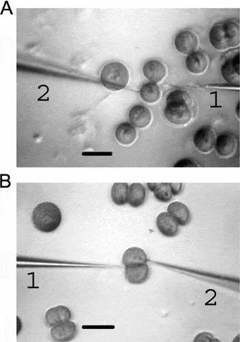
Microapplication of ligands
The tip of an application micropipette, filled with the ligand solution at a final concentration of 1 mM and connected to a micro-injector Picospritzer, was placed either ~10 μm from the first cleavage division furrow or from cell membrane out of contact area (Fig. 1). The location of the application micropipette tip close to the surface of embryo was assessed by determining the acutance of its image to patch one. Microapplication was performed by a single pulse of 10 pl volume. A variety of 5-HТ3-receptor agonists [5-HTQ, quipazine (2-(1-piperazinyl)quinoline maleate), SR 57277A (4-amino-1-(6-chloro-2-piridyl)-piperidine hydrochloride) and chlorphenyl-biguanide] and agonists of n-acetylcholine receptors [epibatidine and methylcarbamylcholine chloride (Sigma)] were used in microapplication experiments; DMSO was used as the solvent.
Arrangement of patch-clamp and application micropipettes. (a) Application into contact area before adhesion, (b) after adhesion 1 – application micropipette; 2 – patch-clamp micropipette. Scale bar: 100 μm.

Blastomeres isolation
An embryo suspension was transferred to normal seawater immediately before isolation. Isolation of blastomeres was carried out during the first cleavage division before (B1-half-embryos) or after ‘post-division adhesion’ (A1-half-embryos) using a Γ-shape glass needle with the tip diameter of ~5 μm, as described earlier (Shmukler et al., Reference Shmukler, Chailakhian, Smolianinov, Bliokh, Karpovich, Gusareva, Naidenko, Khashaev and Medvedeva1981; Shmukler, Reference Shmukler1993), in Petri dishes in presence of the ligand. Control blastomeres were isolated similarly in absence of ligands. After a 10 min incubation in the ligand solution, isolated blastomeres were transferred to Petri dishes with normal seawater using Pasteur pipettes with minimal amount of liquid. The micromere formation moment was checked in experimental and control half-embryos. The influence of quipazine, imipramine and two chemical analogues that differed in cell membrane permeability, 5-HT3-antagonists 3-tropanylindole-3-carboxilate hydrochloride (tropisetron) and methiodide, were studied.
Statistical analysis
Data were subjected to statistical analysis using Microsoft Excel software. Significance of the difference between data from various series was evaluated using Student's t-test.
Results
Membrane currents at administration of ligands
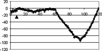
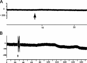
The specific effect of 5-HT3-agonist 5-HTQ, shown earlier, was assessed experimentally with the addition of other ligands into an experimental chamber. 2-Methyl-serotonin (100 μm) evoked inward currents (−335 ± 55 pA, n = 7), similar to the effect found with 5-HTQ (Fig. 2). N-Methylquipazine (dimaleate salt, 100 μm) did not evoke inward currents when added before first cleavage furrow formation, but during cleavage this compound had a pronounced effect (−114.6 ± 23.2 pA, n = 7). At the same time, nicotine (100 μm) had no significant effect (−52 ± 19 pA, n = 16), inward currents were observed only in six experiments.
Inward current evoked by the addition of 2-methyl-serotonin (100 μm) into an experimental chamber during P. lividus first cleavage division. Arrows show the moment of the pulse. Abscissa – time (s), ordinate – current (pA).

Membrane currents at local application of ligands
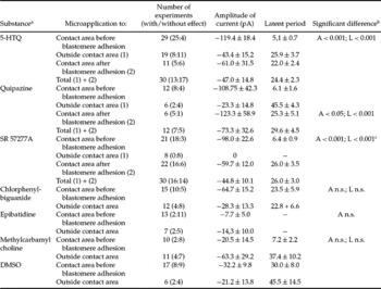
Results of the local application of neuropharmacological substances on sea urchin embryos during the first cleavage division are summarized in Table 1.
Effects of microapplication of chemicals on P. lividus blastomeres during first cleavage division

A – significance of amplitude difference, L – significance of latent period difference.
n.s. – statistically no significant difference.
aIn all cases ligand concentration in the micropipette was 1 mM.
bSignificant differences are shown between the effects of application into the interblastomere cleft and into an area outside the contact area.
cSignificant differences for latent periods are estimated with respect to experiments with microapplication into the cleavage furrow after adhesion, as this was the most appropriate comparison.
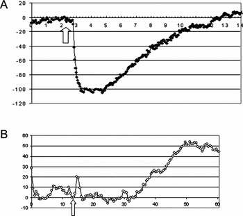
Inward current evoked by microapplication of 5-HTQ into the interblastomere cleft before ‘post-division adhesion’ (−103 pA, latent period about 0.5 s) is shown in Figure 3a and by microapplication on the free surface of the blastomere is shown in Figure 3b.
Membrane currents at microapplication of 5-HТQ (single pulse) on the free surface of a P. lividus blastomere during first cleavage division. Arrows show the moment of the pulse (a) to the contact area of P. lividus embryos during first cleavage division, abscissa – time (s), ordinate – current (pA); (b) on free surface of a P. lividus embryo during first cleavage division, abscissa – time (s), ordinate – current (pA).

As seen in Table 1, the current amplitude that was produced following 5-HTQ application into an interblastomere cleft immediately before adhesion differed significantly from that produced either from application on the free surface or from the application on the free surface and on the cleft following adhesion (in both cases р < 0.01). Furthermore, these differences were statistically insignificant when compared with application to the cleft before and after adhesion. More important is that the latent periods of currents evoked by 5-HTQ application into the contact area were significantly shorter than in all other variants (outer surface, cleft after adhesion and their sum) (in all cases p < 0.001). The latent period could clearly be determined for cases in which the current rose after application. The duration of the latent periods following microapplication of 5-HTQ into the cleft after adhesion and on the free surface did not differ significantly. In three control experiments, in which microapplication of 5-HTQ onto the free surface did not evoke pronounced inward currents, the addition of the same ligand into the experimental chamber (to a 100 μm final concentration) evoked pronounced inward currents (Fig. 4). This finding confirmed the sensitivity of these embryos to 5-HT3-agonists. The results of microapplications performed both at the cleft and at the free surface, in an arbitrary order on the same embryo (n = 7), confirmed the above-mentioned statistical data.
Effects following microapplication of 5-HTQ and its addition to the experimental chamber during P. lividus first cleavage division. (a) Microapplication onto the free surface of blastomeres. (b) Addition of 5-HTQ to an experimental chamber to a final concentration of 100 μm. Abscissa – time (s), ordinate – current (pA).

Other 5-HT3-agonists quipazine and SR 57277A also evoked pronounced inward currents when microapplicated into an interblastomere cleft before adhesion (see Table 1). For these two agonists there was a clear difference between the effects of microapplication into a cleft and onto a free surface. There was no significant difference, however, between current amplitudes induced by microapplication into a cleft before and after adhesion, although a difference in latent periods was striking. The only 5-HT3-agonist that did not show any significant difference in the effects of microapplication into the cleft and onto free surface of blastomeres was chlorphenyl-biguanide (Table 1). Furthermore, no significant current were evoked by n-acetylcholine-receptor agonists epibatidine and methylcarbamylcholine.
Application of DMSO into the contact area of embryo evoked sporadic currents of different directivity and low amplitude. Nevertheless, it must be noted that the latent period of such currents was close to that observed when ligands were applied onto the free surface of blastomeres (Table 1).
Action of serotonergics on isolated blastomeres
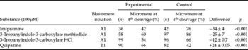
Results of experiments on the action of serotonergics on the cleavage pattern of P. lividus half-embryos are summarized in Table 2.
Effects of serotonergics on the pattern of cleavage of P. lividus half-embryos

A1, isolation of blastomeres after post-division adhesion during first cleavage division.
B1, isolation of blastomeres before post-division adhesion during first cleavage division.
Imipramine, which was used as a reference preparation in previous studies, significantly decreases the fraction of A1-half-embryos that form micromeres at the fourth cleavage division, when compared with controls (without antagonist). The 5-HT3-receptor antagonist 3-tropanylindole-3-carboxylate methiodide had a similar effect, but its tertiary analogue 3-tropanylindole-3-carboxylate hydrochloride shows less effectiveness, although it was statistically significant.
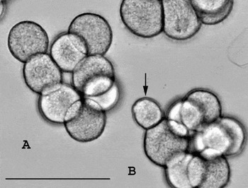
The modification of the conditions of blastomere isolation allowed us to obtain stable fractions of B1-half embryos that do not form micromeres at the fourth cleavage division (Fig. 5). Thus, along with the outcomes of the antagonists, it became possible to study the effect of a serotonin agonist that, as was shown earlier (Shmukler, Reference Shmukler1981), imitates an interblastomere signal and increases the fraction of half-embryos that form micromeres at fourth cleavage division. Indeed, the 5-HT3-receptor agonist quipazine significantly increased the fraction of B1-half embryos that formed micromeres at the fourth cleavage division as compared with control B1-half embryos (without agonist).
Cleavage patterns of P. lividus half-embryos at fourth cleavage division. (a) Equal cleavage, (b) unequal cleavage. Arrow shows the micromere. Scale bar: 100 μm.

Discussion
Previously we have shown the induction of specific inward currents evoked by the 5-HT3-receptor agonist 5-HTQ that were timed to the formation of furrows of the first and the second cleavage divisions, this was in contrast to previously found effects of 5-HT1- and 5-HT2-receptor agonists (Shmukler & Tosti, Reference Shmukler and Tosti2002). According to these data it emerged that: (i) embryonic receptive structures that function during cleavage furrow formation are similar to 5-HT3-receptors found in adults; and (ii) such receptive structures are organized in a temporal way.
In this present work, the effectiveness of 5-HT3-receptor ligands is confirmed by experiments with quipazine, SR 57277A and 2-methyl-serotonin, whereas n-acetylcholine-receptor agonists and DMSO did not evoke considerable currents. Very low membrane permeability to 5-HTQ provides evidence in favour of surface membrane localization of the corresponding receptors, but not of intracellular ones that are involved, in particular, in processes of embryonic cell-cycle triggering (Buznikov, 1990).
In the present work spatial organization of 5-HT3-receptor-linked processes was shown (see also Shmukler et al., 1998), together with their temporal organization. The evidence for the localization of receptive structures, similar to those of 5-HT3-receptors, in the contact area of a 2-cell embryo is supported by the increase in current amplitude evoked by the application of 5-HT3-receptor agonists (5-HTQ, SR 57277A and quipazine) into the cleft rather than onto the free surface. However, the difference in the latent periods under various experimental conditions (application into the cleft before adhesion, after adhesion and onto the free surface) seems unequivocal. Latent periods following application into the cleft before adhesion are clearly shorter than in other experimental conditions, even when current amplitude did not differ significantly, or example after the application of SR 57277A and quipazine into the cleft before and after adhesion (see Table 1). In the case of chlorophenyl-biguanide, there was no difference between the lengths of the latent period following application into a cleft or onto a free surface, this might be explained speculatively by the presence of different 5-HT3-receptor-like structures in embryos and adults.
It must be considered that the variability of latent periods that were found in this study may be due to: (i) difficulty in standardizing the position of the pipette tip with respect to the angle of the blastomere surface; and (ii) the time taken for the embryo to reform the hyaline layer, which could influence the velocity of ligand diffusion to the receptors. Nevertheless, four- to five-fold differences in latent period duration following application into the contact area or onto the free surface of the 2-cell embryo provides convincing evidence in favour of localization of corresponding structures in the contact area of blastomeres.
The increase in current following the application of substances onto the free surface of blastomeres can be explained the flow of part of the ligand into the contact area. Normally in these cases latent periods are prolonged, because of longer distance from the tip of the micropipette, and current amplitude is decreased, due to ligand dilution at the further distance. That the sensitivity of embryos to 5-HT3-agonists is linked to the contact area is supported indirectly by the effect found from the addition of agonists into the chamber, in cases in which application on the free surface evoked no currents. Furthermore, a difference in 5-HT3 sensitivity was confirmed by consecutive applications onto the free surface and into the contact area.
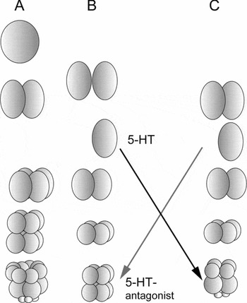
The physiological activity of 5-HT3-receptor ligands is not restricted to the ability to evoke membrane currents in early sea urchin blastomeres. A modification of the method of blastomere isolation allowed us to obtain the fraction of half-embryos that do not form micromeres at the fourth cleavage division, i.e. to prevent the interblastomere signal in the first cleavage division in accordance with the so-called ‘micromere model’ (Fig. 6; Shmukler et al., Reference Shmukler, Chailakhian, Smolianinov, Bliokh, Karpovich, Gusareva, Naidenko, Khashaev and Medvedeva1981; Shmukler Reference Shmukler1981, Reference Shmukler1993) and show the effect of serotonin agonist. Administration of quipazine to blastomeres that were isolated before adhesion in the first cleavage division, led to an increase in the fraction of half-embryos that form micromeres at the fourth cleavage division. This administration imitated the interblastomere signal that restricts totipotency of blastomeres and directed their development to the path of half of a whole embryo, but not whole half-size embryos. Conversely, the administration of 5-HT3-receptor antagonists led to a decrease in the fraction of half-embryos that formed micromeres at the fourth cleavage division, i.e. eliminated the interblastomere signal. These effects are in agreement with the effects of serotonergics demonstrated previously (Shmukler Reference Shmukler1981, Reference Shmukler1993), including a more pronounced effect with poorly penetrating cell membrane protein quaternary 3-tropanylindole-3-carboxylate methiodide as compared with its tertiary analogue. It is probable that the effects of specially synthesized serotonin antagonists, such as methiodides of inmecarb and preparation KYuR-14, described earlier (Shmukler, Reference Shmukler1992), resulted from their interaction with 5-HT3-receptor-like structures. Similarly, it is possible that both demonstrated effects of 5-HT3-receptor ligands (membrane currents and the influence on the cleavage pattern) are due to their interaction with receptive structures at the contact area during cleavage division. This hypothesis might explain how the non-localized administration of ligands is able to imitate a localized interblastomere signal. In such a way, the signal can change the state of cytocortex locally and, respectively, predetermine the position of further mitotic spindles and the moment of micromere formation.
The ‘micromere model’. (a) Normal development of whole embryo. (b) Development of half-embryos, isolated before ‘post-division-adhesion’. (c) Development of half-embryos, isolated after ‘post-division adhesion’.

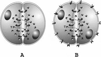
All these observations are in good agreement with the ‘protosynapse’ concept (Shmukler, Reference Shmukler1992, Shmukler & Buznikov, Reference Shmukler and Buznikov1998) on the double-sided symmetrical structure of blastomere interaction. This model (Fig. 7) suggests that each blastomere is both the source and the target of the interblastomere signal (serotonin or serotonin-like substance) and simultaneously presents a barrier for leakage of a signal substance from an interblastomere cleft to an outer medium. Such a concept is not dependent on receptor distribution on the surface of blastomeres, but the results of our present work allow us to propose this based on the temporal restriction of corresponding receptors to the moment of cleavage and their spatial restriction to the contact area.
Models of a ‘protosynapse’ (a) with receptor localization in the interblastomere contact area; (b) with uniform distribution of receptors over the blastomere surface. R – receptor, Т – transmitter.

Acknowledgements
This work is supported partially by an RFBR grant 05-04-48293 to Y.S. We thank Giuseppe Gargiulo for computer graphics and the Marine Resources for Research Service of the Stazione Zoologica for animals and gametes supply.