
Introduction
Several worldwide studies have shown that dogs and cats play an important role in the transmission of zoonotic parasites (Torgerson and Macpherson, Reference Torgerson and Macpherson2011; Otranto et al., Reference Otranto, Dantas-Torres, Mihalca, Traub, Lappin and Baneth2017). Domestic animals can act as definitive hosts for various helminth and protozoan parasites with zoonotic potential, e.g. Toxocara canis, Toxocara cati, Taenia spp., Echinococcus spp., Dipylidium caninum, Ancylostoma spp., Giardia spp., Toxoplasma gondii, Cryptosporidium spp. Particularly, toxocariasis in humans is a systemic larval parasitosis which is usually asymptomatic but can sometimes present respiratory symptoms, eosinophilia, fever, hepatomegaly, splenomegaly, hypergammaglobulinemia, lymphadenopathy, the involvement of the central nervous system, myocardium and skin, and may even lead to death (Rostami et al., Reference Rostami, Ma, Wang, Koehler, Hofmann, Chang, Macpherson and Gasser2019). Clinically, it can present as visceral larva migrans, ocular larva migrans, neurological toxocariasis and covert toxocariasis, being very common mainly in children (Fisher, Reference Fisher2003; Archelli and Kozubsky, Reference Archelli and Kozubsky2008). Sometimes toxocariasis can be associated with complications, such as allergic and/or neurological disorders, possibly including cognitive or developmental delays in children (Rostami et al., Reference Rostami, Ma, Wang, Koehler, Hofmann, Chang, Macpherson and Gasser2019). The literature published from Argentina in recent years reveals that the prevalence of toxocariasis infections in children is between 19.5% and 38.3% (Archelli et al., Reference Archelli, Santillan, Fonrouge, Céspedes, Burgos and Radman2014; Ciarmela et al., Reference Ciarmela, Pezzani and Minvielle2016).
In the canine host, heavy infections with T. canis via the transplacental route in neonates and puppies, may result in pneumonia and acute death owing to enteritis and gastrointestinal blockage as early as 10 days of age. Heavy burdens with T. canis in pups may produce ill thrift, stunting, abdominal discomfort (pups adopt a straddle-legged posture and a pot-bellied appearance), anorexia, diarrhoea and vomiting (adult worms may be expelled). Occasional gastrointestinal obstruction and death may also occur (Dantas-Torres et al., Reference Dantas-Torres, Ketzis, Mihalca, Baneth, Otranto, Tort, Watanabe, Linh, Inpankaew, Jimenez Castro, Borrás, Arumugam, Penzhorn, Ybañez, Irwin and Traub2020). In the feline host, clinical signs depend on the burden of infection, low burden T. cati infections may be subclinical in kittens but those infected with T. cati, especially by the trans-mammary route, may present with cachexia, pot-bellied appearance, respiratory disorders, diarrhoea, vomiting and other signs as early as 3 weeks of age. Heavy infections may cause intestinal blockage or intussusceptions in kittens, which are potentially fatal (Dantas-Torres et al., Reference Dantas-Torres, Ketzis, Mihalca, Baneth, Otranto, Tort, Watanabe, Linh, Inpankaew, Jimenez Castro, Borrás, Arumugam, Penzhorn, Ybañez, Irwin and Traub2020).
Diagnosis of intestinal zoonotic parasites is based on the microscopic detection of cyst/oocysts/eggs in the feces (Dantas-Torres et al., Reference Dantas-Torres, Ketzis, Mihalca, Baneth, Otranto, Tort, Watanabe, Linh, Inpankaew, Jimenez Castro, Borrás, Arumugam, Penzhorn, Ybañez, Irwin and Traub2020). The eggs of T. canis and T. cati can be distinguished morphologically from each other in the definitive host (Warren, Reference Warren1969; Uga et al., Reference Uga, Matsuo, Kimura, Rai, Koshino and Igarashi2000). However, it is known that the latter has low sensitivity, associated to low parasitic burden, intermittent egg output and the requirement of trained and experienced human resources for accurate identification of parasitic structures (Deng et al., Reference Deng, Zhong, Kamolnetr, Limpanont and Lv2019). Nonetheless, the sensitivity may be improved through the use of new copro-microscopic techniques with higher sensitivity, such as Mini-FLOTAC and FLOTAC, as described by Maurelli et al. (Reference Maurelli, Rinaldi, Alfano, Pepe, Coles and Cringoli2014). Techniques based on the detection of antigens in feces have also been used and have shown a wide range of sensitivity and specificity (Deplazes et al., Reference Deplazes, Gottstein and Stingelin1990; Sen et al., Reference Sen, Yildirim, Bişkin, Düzlü and Inci2011; Christy et al., Reference Christy, Hencke, Escueta-De Cadiz, Nazib, Von Thien, Yagita, Ligaba, Haque, Nozaki, Tannich, Herbein and Petri2012; Shimelis and Tadesse, Reference Shimelis and Tadesse2014; Jara et al., Reference Jara, Rodriguez, Altamirano, Herrera, Verastegui, Gímenez-Lirola, Gilman and Gavidia2019). Due to the different sensitivity and specificity presented by both microscopic and antigen detection techniques and with the advent of molecular techniques based on the detection of nucleic acids, certain aspects of sensitivity and specificity were improved (Jacobs et al., Reference Jacobs, Zhu, Gasser and Chilton1997; Li et al., Reference Li, Lin, Chen, Sani, Song and Zhu2007; Pinelli et al., Reference Pinelli, Roelfsema, Brandes and Kortbeek2013; Knapp et al., Reference Knapp, Umhang, Poulle and Millon2016); however, supplies and equipment are usually expensive and the procedures require human resources with specific training which are not widely available in endemic areas, complicating its implementation. Nonetheless, techniques based on isothermal amplification of nucleic acids are promising for the transfer of molecular diagnostics to the field given their lower complexity, lower cost and the lack of specialized equipment for its implementation. Particularly, the loop-mediated isothermal amplification (LAMP) developed by Notomi et al. (Reference Notomi, Okayama, Masubuchi, Yonekawa, Watanabe, Amino and Hase2000) uses two or three pairs of primers that recognize different regions flanking a central DNA fragment thus providing higher specificity for this sequence since the primers generate looped structures that serve as a template to start a new polymerization cycle. The Bacillus stearothermophilus DNA polymerase I (Bst) used in the technique causes DNA strand displacement and therefore it does not require denaturing of the double-strand, thus the technique can be carried out in a thermostatic bath without the need of a thermocycler. The LAMP reaction has been proposed as an attractive diagnostic method since it meets all the criteria proposed by the World Health Organization (WHO) for an ‘ideal’ diagnostic test: affordable, sensitive, specific, user-friendly, rapid, equipment-free and delivered to those who need it (Mabey et al., Reference Mabey, Peeling, Ustianowski and Perkins2004). A LAMP technique for the separate detection of T. canis and T. cati has been previously developed (Macuhova et al., Reference Macuhova, Kumagai, Akao and Ohta2010), although it is not currently routinely used because two LAMP reactions are needed for each environmental sample collected (either feces or sand/soil). Although the separate detection of Toxocara spp. maybe useful for application in the definitive host, for epidemiological and environmental studies, simultaneous detection of T. canis and T. cati in the feces from definitive hosts could be more useful for wide-scale implementation. Previous studies have shown the presence of T. cati in dog feces with a prevalence ranging from 7.3 to 34.5% (Fahrion et al., Reference Fahrion, Schnyder, Wichert and Deplazes2011; Nagy et al., Reference Nagy, Ziadinov, Schweiger, Schnyder and Deplazes2011; Vienažindienė et al., Reference Vienažindienė, Joekel, Schaper, Deplazes and Šarkūnas2018; Maurelli et al., Reference Maurelli, Santaniello, Fioretti, Cringoli, Rinaldi and Menna2019). The presence of T. cati in dogs could be mainly attributed to coprophagia of cat feces (Vienažindienė et al., Reference Vienažindienė, Joekel, Schaper, Deplazes and Šarkūnas2018).
Due to its simplicity and high sensitivity and specificity, LAMP assays have already been implemented for the detection of protozoan (Ikadai et al., Reference Ikadai, Tanaka, Shibahara, Matsuu, Uechi, Itoh, Oshiro, Kudo, Igarashi and Oyamada2004; Poon et al., Reference Poon, Wong, Ma, Chan, Chow, Abeyewickreme, Tangpukdee, Yuen, Guan, Looareesuwan and Peiris2006; Han et al., Reference Han, Watanabe, Sattabongkot, Khuntirat, Sirichaisinthop, Iriko, Jin, Takeo and Tsuboi2007; Karanis et al., Reference Karanis, Thekisoe, Kiouptsi, Ongerth, Igarashi and Inoue2007; Njiru et al., Reference Njiru, Mikosza, Armstrong, Enyaru, Ndung'u and Thompson2008; Plutzer and Karanis, Reference Plutzer and Karanis2009; Singh et al., Reference Singh, Mirdha, Ahuja and Singh2013; De Ruiter et al., Reference De Ruiter, Van Der Veer, Leeflang, Deborggraeve, Lucas and Adams2014; Karani et al., Reference Karani, Sotiriadou, Plutzer and Karanis2014; Gallas-Lindemann et al., Reference Gallas-Lindemann, Sotiriadou, Plutzer, Noack, Mahmoudi and Karanis2016; Besuschio et al., Reference Besuschio, Llano Murcia, Benatar, Monnerat, Cruz Mata, Picado de Puig, Curto, de los, Kubota, Wehrendt, Pavia, Mori, Puerta, Ndung'u and Schijman2017; Durand et al., Reference Durand, La Carbona, Geffard, Possenti, Dubey and Lalle2019; Nzelu and Kato, Reference Nzelu, Kato and Peters2019), as well as helminth parasites, and are even available in the market for epidemiological surveys (Kumagai, et al., Reference Kumagai, Furushima-Shimogawara, Ohmae, Wang, Lu, Chen, Wen and Ohta2010; Deng et al., Reference Deng, Zhong, Kamolnetr, Limpanont and Lv2019). LAMP has also been implemented for the diagnosis of pathogens associated with food-borne diseases, such as Salmonella typhi, Campylobacter jejuni, Helicobacter pylori, Listeria monocytogenes, Leptospira interrogans, Bucella sp, Escherichia coli, and for water control (Hara-Kudo et al., Reference Hara-Kudo, Konishi, Ohtsuka, Hiramatsu, Tanaka, Konuma and Takatori2012; Abdullah et al., Reference Abdullah, Saffie, Sjasri, Husin, Abdul-Rahman, Ismail, Aziah and Mohamed2014; Pham et al., Reference Pham, Trinh, Khamrin, Ukarapol, Kongsricharoern, Yamazaki, Komine-Aizawa, Okitsu, Maneekarn, Hayakawa and Ushijima2015; Trangoni et al., Reference Trangoni, Gioffré, Cerón Cucchi, Caimi, Ruybal, Zumárraga and Cravero2015; Wang et al., Reference Wang, Wang, Ma, Li, Luo, Liu, Hu, Jin, Liu, Ye and McPhee2015, Reference Wang, Zheng, Cai, Liu, Liao, Li, Zhang and Lin2019; Bakhtiari et al., Reference Bakhtiari, Alvandi, Pajavand, Navabi, Najafi and Abiri2016; Löffler et al., Reference Löffler, Leiva, Scialfa, Redondo, Florin-Christensen, Martínez, Romero and Brihuega2016; Azizi et al., Reference Azizi, Zaferani, Cheong and Abbaspourrad2019). Nonetheless, this technique has some disadvantages, including the use of expensive commercial kits (CKs) for DNA extraction as well as reagents for the detection and visualization of the amplified product. Another complication is the false positive reactions generated by contamination of the sample with unwanted amplification products. These disadvantages are probably the reason why this technique is not presently routinely and widely used.
In this study, a LAMP assay that was developed for the simultaneous detection of T. canis and T. cati, causal agents for human toxocariasis is described. An alternative in-house method (IHM) for DNA extraction and basic reagents, widely available in most laboratories was used. This low-cost strategy aims at making this technique feasible and easier to implement in laboratories from endemic areas with scarce resources.
Materials and methods
Parasite material
All fecal samples collected from dogs and cats of San Juan Province, Argentina, were stored at −20°C for 1 month. T. canis and T. cati adult worms were obtained from spontaneous elimination from their respective definitive hosts and were preserved in 70% ethanol.
Optical microscopy
Feces were processed using two different flotation techniques, Sheather method (1.25 specific gravity) (Sheather, Reference Sheather1923) and Willis method (1.20 specific gravity) (Willis, Reference Willis1921) as well as a sedimentation technique, Telemann method (Telemann, Reference Telemann1908). The techniques chosen for this study are standard concentration techniques that increase the chances of detecting intestinal parasitic structures, including helminth parasites such as Toxocara spp. Each sample was microscopically examined at 100× and 400× magnifications.
DNA extraction
Genomic DNA (gDNA) extraction from cestode parasites was performed using the DNeasy Blood & Tissue Kit® (QIAGEN), following the manufacturer's instructions. The extraction of gDNA from nematode parasites was performed according to Repetto et al. (Reference Repetto, Soto, Cazorla, Tayeldin, Cuello, Lasala, Tekiel and González Cappa2013), and DNA from E. coli (abundant in canine and feline feces) was extracted using the phenol–chloroform method (Sambrook and Russell, Reference Sambrook and Russell2001). In each case, DNA concentrations were determined using a Nanodrop® Nd 1000 (Thermo Fisher Scientific, Waltham, Massachusetts, United States) and DNA integrity was assessed by electrophoresis in 1% agarose gel stained with GelRed® (Biotium®, San Francisco, California, United States) and UV visualization. The DNA extraction and purification from feces (fDNA) was performed using both a CK (ADN Puriprep SUELO-kit® from Inbio-Highway, Tandil, Buenos Aires, Argentina) and an IHM adapted from Macuhova et al. (Reference Macuhova, Kumagai, Akao and Ohta2010). Briefly, each canine/feline stool sample was homogenized and filtered through 250 and 63 μm sieves, respectively. The filtered material was homogenized in 50% sodium hypochlorite diluted from commercially sold aqueous solution with 5% effective chlorine concentration. The material was then thoroughly mixed and incubated at room temperature for 10 min, followed by centrifugation at 800 rpm for 3 min. The supernatant containing the eggs was recovered in a separate tube. The eggs were washed with distilled water, followed by a centrifugation step (3000 rpm for 5 min), and finally, the supernatant was discarded. This procedure was repeated three times. DNA was extracted by incubating samples with 50 mm NaOH in a water bath at 95°C for 30 min while mixing thoroughly every 15 min. This solution was used as a template for the LAMP assay. The DNA obtained by CK was conserved at −20°C, while the DNA obtained by IHM was cooled and used as a template for the LAMP reaction.
LAMP assay
Primer design
The selection of the target gene for the design of the LAMP assay for the simultaneous detection of T. canis and T. cati was based on the general rules indicated by Notomi et al. (Reference Notomi, Okayama, Masubuchi, Yonekawa, Watanabe, Amino and Hase2000), as well as the Primer V5 design software guide (Eiken (2018), Eiken Chemical Co., Ltd.). The selected target for primer design was a 261 bp region of the mitochondrial gene cox-1 (Genbank accession number AM411108.1, nucleotides: 6046–7623 and AM411622.1, nucleotides: 6055–7632). This region was selected due to the high identity between the two Toxocara species (T. canis and T. cati) and the presence of several mismatches with respect to other parasites that might coexist in the same sample. This strategy is fully described in previous studies (Avila, Reference Avila2019; Avila et al., Reference Avila, Mozzoni, Trangoni, Cravero, Pérez, Valenzuela, Gertiser, Butti, Kamenetzky, Jensen and Rosenzvit2020).
Master mix
The LAMP reaction was performed in a 12.5 μL final reaction mixture containing: 20 mm Tris (pH 8.8), 50 mm KCl, 8 mm MgSO4, 10 mm (NH4)2SO4, 8 mm betaine, 1.4 mm dNTPs and 4 U Bst 2.0 polymerase (New England Biolabs). The primer concentration was 20 pmol for each FIP and BIP primer, 2.5 pmol for each F3 and B3 primer, and 5 pmol for each LB and LF primer. In all cases, 1 μL of DNA was used as a template. All reactions were performed on ice. The amplification conditions were evaluated using a temperature gradient (since 52°C–68°C), different incubation times (15–120 min) and different concentrations of malachite green dye (0.004–0.4% w/v). The use of malachite green dye and the IHM were evaluated with the aim of obtaining a low-cost strategy for the detection of Toxocara spp.
Analytical sensitivity
The analytical sensitivity was evaluated in two conditions: with ten-fold serial dilutions of gDNA from T. canis and T. cati (ranging from 10−12 to 10−15 g μL−1), while the sensitivity of the LAMP reaction was evaluated with serial dilutions of fDNA (ranging from 100 to 10−6) extracted from feces using CK and IHM. In each case, DNA obtained was diluted in ultrapure water to obtain serial dilutions. The feces used were positive for Toxocara spp. eggs under microscopic observation.
Specificity evaluation
The specificity of the LAMP reaction was evaluated by using gDNA from Canis lupus familiaris, Felis catus, E. coli, since these DNAs are abundant in canine and feline feces and also gDNA from other helminth parasites usually present in feces such as: Toxascaris leonina, Ancylostoma caninum, Echinococcus granulosus sensu stricto and Taenia hydatigena. Ten picograms of gDNA were used in all cases.
Environmental samples from endemic areas
A total of 38 environmental fecal samples were collected from San Juan Province, Argentina, and were analyzed by optical microscopy. Samples were considered positive when Toxocara spp. eggs were observed by at least one of the methods used (Telemann concentration technique, as well as Willis and Sheather flotation techniques). All samples were analyzed by the LAMP assay using triplicate DNA samples obtained by both CK and IHM. The final cost (USD $) of reagents per reaction for each method was estimated using the cost of each necessary reagent for both the LAMP reaction and the method for DNA extraction. This cost was obtained from each of the commercial providers in Argentina.
Results
Primer design
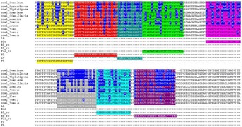
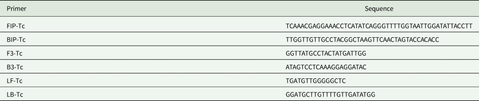
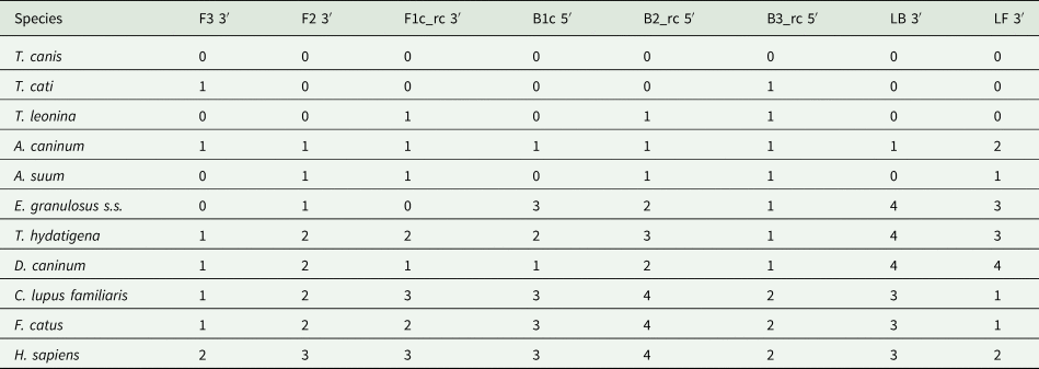
The T. canis cox-1 gene was selected as a target for the primer design. The region of the gene was chosen according to the number of mismatches between sequences (Fig. 1). The primer sets (Table 1) were selected according to the number of mismatches in the 3′ and 5′ ends, where each of the primers aligns (Table 2).
Design of LAMP primers: Multiple alignment of target sequences for the design of LAMP primers for Toxocara canis and Toxocara cati simultaneous detection, using sequences of orthologous genes of T. canis_dCOX1 (AM411108.1:6046–7623) T. cati_dCOX1 (AM411622.1:6055–7632), Echinococcus granulosus sensu stricto G1_dCOX1 (AF297617.1:6760-8367), T. leonina (NC_023504.1:1–1578), A. caninum (NC_012309.1:1–1578), Ascaris suum (NC_001327.1:8771–10347), Dipylidium caninum (AB732959.1:9743–11485), Taenia hydatigena (GQ228819.1:6831–8450), Felis catus (U20753.1), Canis lupus familiaris (U96639.2), and Homo sapiens sapiens (J01415.2). Mismatches are highlighted in blue.

Sequence data of primer set designed for simultaneous detection of a fragment of mitochondrial cox1 genes from Toxocara canis and Toxocara cati species

Determination of the number of mismatches between LAMP primers and orthologous gene sequences

Mismatches in the 3′ end from Forward Outer Primer (F3), F2 region from Forward Inner Primer (FIP), reverse complement sequence from F1c region from Forward Inner Primer (FIP), Backward Loop Primer (LB), Forward Loop Primer (LF) and mismatches in the 5′ end from B1c region from Backward Inner Primer (BIP), reverse complement sequence from B2 region from Backward Inner Primer (BIP), reverse complement sequence from Backward Outer Primer (B3).
Amplification conditions
Optimal conditions for DNA amplification were 60 min at 58°C with a final incubation at 80°C for polymerase inactivation (Supplementary Table S1). The optimal malachite green dye final concentration was 0.016% w/v (Supplementary Table S2).
Sensitivity
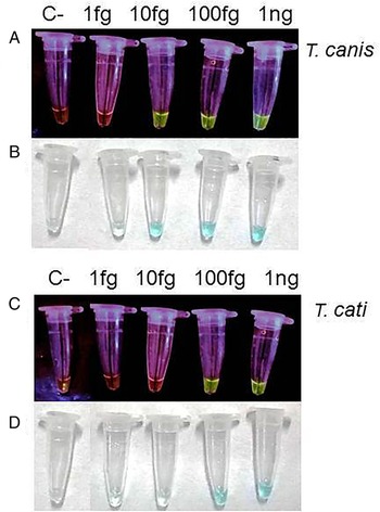
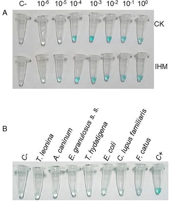
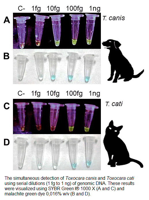
The LAMP reaction was able to detect 10 fg of gDNA from T. canis and 100 fg of gDNA from T. cati (Fig. 2). In each case, the results were obtained by direct visualization of green fluorescence in the reaction tube, using 1 μL of SYBR Green I®, and 0.016% w/v malachite green dye final concentration. The LAMP reaction showed positive results with fDNA prepared from canine feces positive for T. canis eggs by using CK (diluted up to 1/10 000 or 10−4) and IHM (diluted up to 1/100 000 or 10−5) (Fig. 3A).
Analytical sensitivity of the LAMP assay. The simultaneous detection of T. canis and T. cati was evaluated by using serial dilutions (1 fg to 1 ng) of genomic DNA extracted from adult worm parasites of each species. These results were visualized using SYBR Green I® 1000 × (A and C) and malachite green dye 0.016% w/v (B and D). C−: water control.

(A) Sensitivity of the LAMP reaction with serial dilutions of DNA (100 to 10−6) extracted from feces by using a commercial kit (CK) and an in-house method (IHM). The feces used were positive for T. canis eggs by optical microscopy. C−: water control. (B) Specificity of the LAMP assay: The specificity of LAMP for simultaneous T. canis and T. cati detection was evaluated using 10 pg of genomic DNA from C. lupus familiaris, Escherichia coli, F. catus, and the following parasites: D. caninum, T. hydatigena, Toxascaris leonine, and A. caninum. C−: water control, C+: 1 pg of genomic DNA from T. canis.

Specificity evaluation
The specificity of the LAMP reaction was tested with 1 ng of DNA from C. lupus familiaris, F. catus and E. coli. In addition, the specificity was evaluated with gDNA from parasite species commonly found in canine and feline feces (E. granulosus s. s., T. leonina, A. caninum, T. hydatigena) (Fig. 3B). No amplification products were observed with these gDNAs while the positive control (gDNA from T. canis) showed the expected amplification signal.
LAMP analysis of environmental samples from endemic areas
The LAMP reaction was applied to 30 canine fecal samples and 8 feline fecal samples in order to determine the usefulness of the technique for the simultaneous detection of T. canis–T. cati from feces collected from the environment in endemic areas in Argentina. Out of the 38 samples, 13 (34%) were positive for T. canis–T. cati eggs (Table 3) when using optical microscopy methods. On the other hand, when using the LAMP assay, a total of 28 samples (74%) had detectable DNA of T. canis–T. cati, regardless of the extraction method used (CK of IHM). Moreover, the 13 samples detected as positive by microscopy were also detected as positive by the LAMP assay. LAMP determination using CK for DNA extraction and SYBR Green I dye for visualization resulted in a final cost of reagents per reaction of approximately USD$ 6.12. On the other hand, use of the IHM method for extraction and malachite green dye for visualization, the final cost of reagents was reduced to approximately USD$ 1.16 per reaction (Table 3).
Detection of T. canis and T. cati in environmental samples by optical microscopy and LAMP assay

Each sample (N = 38) was considered positive if Toxocara spp. eggs were observed by at least one of the methods (8–10). LAMP assay was performed using DNA obtained from CK and IHM. All results were confirmed in three independent assays. The final cost of reagents per reaction (USD$) was estimated according to quotations from each commercial provider.
Discussion
Pathogen's diagnoses through techniques that detect nucleic acids are increasingly used. The analytical sensitivity and specificity values place them among the best options for diagnosis. However, their high economic value and complexity make them hard to implement in areas where laboratory equipment is basic and budget for reagents is low. Although the detection of T. canis and T. cati eggs by optical microscopy is the most widely used technique, due to its high specificity and low cost, its sensitivity can vary according to the parasite burden, egg shedding dynamics or sample size; the sensitivity also depends on the experience of the technician that performs the analysis. Although different PCR's, with high sensitivity and specificity have been developed specifically for these parasites (Jacobs et al., Reference Jacobs, Zhu, Gasser and Chilton1997; Li et al., Reference Li, Lin, Chen, Sani, Song and Zhu2007; Pinelli et al., Reference Pinelli, Roelfsema, Brandes and Kortbeek2013; Knapp et al., Reference Knapp, Umhang, Poulle and Millon2016), they are not routinely and widely implemented due to their high cost and the need of sophisticated equipment which are not widely available in endemic areas. Nonetheless, with the advent of isothermal amplification techniques, such as the LAMP, which uses the same principle as a regular PCR but does not need specialized equipment, various researchers are studying the possibility of using it as a routine technique, even for point-of-care testing (Durant et al., Reference Durant, Irenge, Fogt-Wyrwas, Dumont, Doucet, Mignon, Losson and Gala2012; Njiru, Reference Njiru2012; Wong et al., Reference Wong, Othman, Lau, Son and Chee2017; Deng et al., Reference Deng, Zhong, Kamolnetr, Limpanont and Lv2019; Nzelu and Kato, Reference Nzelu, Kato and Peters2019).
Specific LAMP primer sets for separate T. canis and T. cati detection have been previously designed using a partial sequence of the internal transcriber spacer 2 rRNA region (ITS-2), with an analytical sensitivity of 100 fg of gDNA (Macuhova et al., Reference Macuhova, Kumagai, Akao and Ohta2010) and two to three eggs in 30 g of feces (Khoshakhlagh et al., Reference Khoshakhlagh, Spotin, Mahami-Oskouei, Shahbazi and Ozlati2017). On the other hand, the LAMP assay developed herein was able to simultaneously detect T. canis and T. cati in a single reaction with analytical sensitivity of 10 fg for T. canis gDNA, which is more sensitive than previously reported values for both LAMP (Macuhova et al., Reference Macuhova, Kumagai, Akao and Ohta2010) and PCR assays (Jacobs et al., Reference Jacobs, Zhu, Gasser and Chilton1997; Pinelli et al., Reference Pinelli, Roelfsema, Brandes and Kortbeek2013; Knapp et al., Reference Knapp, Umhang, Poulle and Millon2016). The analytical sensitivity for T. cati detection was lower (100 fg) and could be related to the primer design, since they had two mismatches in the 3′ regions of the F3 and B3 primers (Table 1), this was previously observed for Echinococcus spp. (Avila et al., Reference Avila, Mozzoni, Trangoni, Cravero, Pérez, Valenzuela, Gertiser, Butti, Kamenetzky, Jensen and Rosenzvit2020). These mismatches could influence initial LAMP reaction steps, due to an incorrect match with the target sequence (Avila, Reference Avila2019). The LAMP reaction developed is able to detect low concentrations of DNA from Toxocara spp in feces. Nevertheless, further studies are needed to evaluate the detection limit with known concentrations of Toxocara spp eggs.
The LAMP assay proved to be more sensitive than optical microscopy for the identification of Toxocara spp., as previously reported (Khoshakhlagh et al., Reference Khoshakhlagh, Spotin, Mahami-Oskouei, Shahbazi and Ozlati2017; Avila et al., Reference Avila, Mozzoni, Trangoni, Cravero, Pérez, Valenzuela, Gertiser, Butti, Kamenetzky, Jensen and Rosenzvit2020). Furthermore, in this study, the use of the IHM for DNA extraction adapted from (Macuhova et al., Reference Macuhova, Kumagai, Akao and Ohta2010), was as efficient as CK, confirming that it could be used in laboratories that do not have funds to purchase a CK, thus reducing costs. However, for the method to be effective, it is necessary to use the entire sample in order to increase sensitivity. Moreover, the interpretation of results was performed with the commonly used malachite green dye which is usually available in parasitology labs since it is routinely used for the Kato–Katz thick smear method for helminths (Katz, et al., Reference Katz, Chaves and Pellegrino1972). This dye has been used before in other LAMP reactions (Nzelu et al., Reference Nzelu, Gomez, Cáceres, Sakurai, Martini-Robles, Uezato, Mimori, Katakura, Hashiguchi and Kato2014, Reference Nzelu, Cáceres, Guerrero-Quincho, Tineo-Villafuerte, Rodriquez-Delfin, Mimori, Uezato, Katakura, Gomez, Guevara, Hashiguchi and Kato2016; Lucchi et al., Reference Lucchi, Ljolje, Silva-Flannery and Udhayakumar2016; Chahar et al., Reference Chahar, Anvikar, Dixit and Valecha2018; Kudyba et al., Reference Kudyba, Louzada, Ljolje, Kudyba, Muralidharan, Oliveira-Ferreira and Lucchi2019; Serra-Casas et al., Reference Serra-Casas, Guetens, Chiheb, Gamboa and Rosanas-Urgell2019) and it proved to be useful for the current study as well. The use of this dye also reduces the possibility of amplicon contamination, since the tube opening after incubation is no longer necessary. The use of the IHM for DNA extraction and malachite green dye for visualization of the results guarantee their use in almost any laboratory since they reduce the cost of reagents for the reaction by 81%. The IHM along with the use of malachite green was applied in 38 environmental samples and the high sensitivity of the LAMP assay against conventional helminth egg concentration techniques was observed. The LAMP assay could be compared with copro-microscopic techniques with high sensitivity such as Mini-FLOTAC and FLOTAC (Maurelli et al., Reference Maurelli, Rinaldi, Alfano, Pepe, Coles and Cringoli2014).
Herein, a sensitive, specific and economic LAMP assay was developed in order to simultaneously detect T. canis and T. cati in fecal material from definitive hosts. This method may be implemented in low complexity laboratories in endemic areas for epidemiological purposes. Wider field studies need to be conducted in order to corroborate its performance at a larger scale. In the future, this methodology could be tested in other matrixes such as food, water and sand, among others, in order to identify environmentally contaminated areas and prevent infection in humans, especially children.
Conclusions
The results of this study showed that the LAMP technique can provide a specific and sensitive method for the simultaneous detection of T. canis and T. cati, etiological agents of toxocariasis in humans. Detection in a single assay, using an IHM method for DNA extraction, and malachite green dye for visualization of results, make up an easy and low-cost option for diagnosis, providing a new tool to improve toxocariasis diagnosis in facilities from endemic areas lacking sophisticated equipment.
Supplementary material
The supplementary material for this article can be found at https://doi.org/10.1017/S0031182021000342
Financial support
This work was supported by Fundación Mundo Sano, Consejo Nacional de Investigaciones Científicas y Tecnológicas (CONICET), Ministerio de Salud de la Provincia de San Juan, and Universidad Católica de Cuyo.
Conflict of interest
The authors declare none.
Ethical standards
Not applicable.