Introduction
Body image disturbance occurs as a central symptom in anorexia nervosa (AN) and bulimia nervosa (BN) (Cash & Deagle, Reference Cash and Deagle1997) and is also manifested in DSM-IV-TR diagnostic criteria for these disorders (APA, 2000). Beyond an overestimation of one's own body size (Farrell et al. Reference Farrell, Lee and Shafran2005; Vocks et al. Reference Vocks, Legenbauer, Rüddel and Troje2007a), body image disturbance is characterized by body dissatisfaction and exaggerated weight and shape concerns as well as by body-related avoidance and checking behaviour (Cash, Reference Cash2004; Thompson, Reference Thompson2004). Accordingly, females with eating disorders display stronger negative emotional and cognitive reactions when looking at their bodies compared with healthy controls (Tuschen-Caffier et al. Reference Tuschen-Caffier, Vögele, Bracht and Hilbert2003; Vocks et al. Reference Vocks, Legenbauer, Wächter, Wucherer and Kosfelder2007b).
Recently, the neuronal base of body image disturbance in eating disorders has been examined using functional magnetic resonance imaging (fMRI). Seeger et al. (Reference Seeger, Braus, Ruf, Goldberger and Schmidt2002) found that adolescents with AN display stronger activations in the amygdala, the fusiform gyrus and the brainstem region relative to healthy controls when looking at distorted photographs of their own body, indicating an activation of the brain's fear network. However, in a subsequent study using the same paradigm in a larger sample, these results were not sustained. Instead, participants with AN displayed a higher activation in the inferior parietal lobule including the anterior part of the intraparietal sulcus (Wagner et al. Reference Wagner, Ruf, Braus and Schmidt2003), areas that are specifically involved in visuospatial processing and attentional processes (Coull & Nobre, Reference Coull and Nobre1999; Clower et al. Reference Clower, West, Lynch and Strick2001). In a mixed sample consisting of participants with AN and BN, Uher et al. (Reference Uher, Murphy, Friederich, Dalgleish, Brammer, Giampietro, Phillips, Andrew, Ng, Williams, Campbell and Treasure2005) found lower activations to looking at line drawings of female bodies in the inferior parietal lobule and in the lateral fusiform gyrus, covering the extrastriate body area (EBA), a region that is specialized in the visual perception of human bodies (Downing et al. Reference Downing, Jiang, Shuman and Kanwisher2001; Peelen & Downing, Reference Peelen and Downing2007). Sachdev et al. (Reference Sachdev, Mondraty, Wen and Gulliford2008) found that when looking at undistorted pictures of another female, patients with AN and healthy controls showed activations in the inferior and middle frontal gyri, in the superior parietal lobule and in the thalamus. Eating-disordered participants displayed additional stronger activations in the medial frontal gyrus. However, when viewing self-images, patients with AN displayed no significant activations, probably pointing to a decreased activity of the attentional system, which might contribute to body image distortion. Using a similar paradigm, Vocks et al. (Reference Vocks, Busch, Grönemeyer, Schulte, Herpertz and Suchan2010a) found that when viewing their own body, participants with AN and BN showed a lower activity in the inferior parietal lobule compared with healthy females, possibly reflecting decreased attention due to body-related avoidance behaviour (Coull & Nobre, Reference Coull and Nobre1999). In response to looking at another female's body, a higher activity in the amygdala was observed in participants with AN relative to those with BN and healthy controls, which might be seen as a neural correlate of negative emotional activation, probably following unfavourable social comparison processes (Troop et al. Reference Troop, Allan, Treasure and Katzman2003; Friederich et al. Reference Friederich, Uher, Brooks, Giampietro, Brammer, Williams, Herzog, Treasure and Campbell2007).
Since there is growing evidence that body image disturbances are not only a symptom of AN and BN, but also contribute to the maintenance and relapse process of eating disorders (Fairburn et al. Reference Fairburn, Stice, Cooper, Doll, Norman and O'Connor2003b; Jacobi et al. Reference Jacobi, Hayward, de Zwaan, Kraemer and Agras2004; Keel et al. Reference Keel, Dorer, Franko, Jackson and Herzog2005), interventions that directly aim to improve body image disturbances have been applied in the treatment of eating disorders (e.g. Cash & Hrabosky, Reference Cash, Hrabosky and Thompson2004). Various studies provide hints that cognitive behavioural body image therapy is effective in reducing body image disturbance in patients with eating disorders (Rushford & Ostermeyer, Reference Rushford and Ostermeyer1997; Vocks et al. Reference Vocks, Legenbauer, Troje and Schulte2006). Furthermore, previous research has demonstrated that affective and cognitive reactions elicited by looking at one's own body can be reduced from pre- to post-body image therapy (Vocks et al. Reference Vocks, Wächter, Wucherer and Kosfelder2008). Based on these findings, the question can be raised of whether altered body image-processing brain circuits can be changed by cognitive behavioural interventions.
In a growing number of fMRI studies, the outcome of cognitive behavioural therapy is assessed using symptom provocation paradigms, mimicking diagnosis-specific psychopathological states before and after treatment (Linden, Reference Linden2006). Taken together, these studies indicate that cognitive behavioural therapy seems to influence neuronal correlates of the specific symptoms of various forms of anxiety disorders, mainly located in limbic and paralimbic structures (Furmark et al. Reference Furmark, Tillfors, Marteinsdottir, Fischer, Pissiota, Langstrom and Fredrikson2002; Paquette et al. Reference Paquette, Lévesque, Mensour, Leroux, Beaudoin, Bourgouin and Beauregard2003; Straube et al. Reference Straube, Glauer, Dilger, Mentzel and Miltner2006; Schienle et al. Reference Schienle, Schäfer, Hermann, Rohrmann and Vaitl2007). Given that previous research indicated that females with eating disorders display abnormal brain responses to looking at their own body and another female's body (Seeger et al. Reference Seeger, Braus, Ruf, Goldberger and Schmidt2002; Wagner et al. Reference Wagner, Ruf, Braus and Schmidt2003; Uher et al. Reference Uher, Murphy, Friederich, Dalgleish, Brammer, Giampietro, Phillips, Andrew, Ng, Williams, Campbell and Treasure2005; Sachdev et al. Reference Sachdev, Mondraty, Wen and Gulliford2008; Vocks et al. Reference Vocks, Busch, Grönemeyer, Schulte, Herpertz and Suchan2010a), the question can be raised of whether these brain responses can be modified by interventions directly aiming at an improvement of body image disturbance (Cash & Hrabosky, Reference Cash, Hrabosky and Thompson2004). A previous paper, which included a subsample from the present study consisting of five patients with AN, provided first hints on the treatment-induced changeability of the activity of the EBA (Vocks et al. Reference Vocks, Busch, Schulte, Grönemeyer, Herpertz and Suchan2010b). The aim of the current study – which is the first randomized controlled intervention study using fMRI in eating disorders research – is to test the effects of a cognitive behavioural body image therapy as compared with a waiting list control condition on neuronal correlates of viewing one's own and another female's body.
Method
Participants
Patients with eating disorders were recruited from the waiting list of the Centre for Psychotherapy (Ruhr-University, Bochum) and through newspaper announcements. Due to the similarities between the various forms of eating disorders, especially concerning body image problems (Fairburn et al. Reference Fairburn, Cooper and Shafran2003a), and because of the comparability of treatments for body image disturbances in various forms of eating disorders (Vocks et al. Reference Vocks, Wächter, Wucherer and Kosfelder2008), patients with AN, BN and an eating disorder not otherwise specified were examined together. Inclusion criteria were female sex and aged between 18 and 50 years. Additionally, only right-handed females were included in the study. Exclusion criteria were pregnancy, personality disorders, claustrophobia, suicidal tendencies and deliberate self-harm, as well as piercings, earrings or other pieces of metal in any part of the body. Diagnoses were assessed by experienced clinical psychologists using the Structured Clinical Interview for DSM-IV (First et al. Reference First, Spitzer, Gibbon and Williams1996; Wittchen et al. Reference Wittchen, Zaudig and Fydrich1997). The design of the study was explained to each participant and written consent was obtained. The study protocol was approved by the local ethics committee.
Stimuli
For the present study, one set of photographs of each participant's own body and one with photographs of an unknown female's body were used. The unknown female was 28 years old and had a body mass index (BMI) of 19 kg/m2. The photographs were all taken under standardized conditions in the same room while participants wore a uniform pink bikini in their size. Photographs from each participant were taken without the head from 16 different perspectives, i.e. front, back, right, left and the 12 between perspectives. The photographs of one's own body and of the other female's body were presented using a block design without repetition of the same block, starting with the block of pictures of one's own body. Before the presentation of each block, participants were shown a slide indicating whether they would next see the set of photographs of their own or of the other female's body. Each of the sets of stimuli was presented three times during the scanning session. The photographs were shown in a standardized order and each picture was shown for 3 s, i.e. each block lasted for 48 s altogether. Between each set of photographs, a fixation cross was presented for 48 s (see Fig. 1). The participants were instructed to look at the pictures and not to close their eyes during the scanning session.
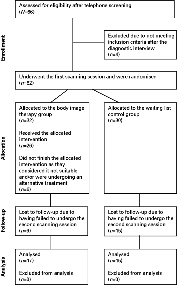
CONSORT flow chart displaying the progress of all participants through the trial (www.consort-statement.org).

Image acquisition
Image acquisition was performed on a 1.5 T Symphony scanner (Siemens, Germany). In each condition, a total of 440 T2*-weighted whole brain volumes (omitting parts of the cerebellum) were acquired in ascending order. Each volume consisted of 25 slices of 3 mm with an interslice gap of 1 mm. The repetition time was 80 ms per slice with a flip angle of 90°. Additional high-resolution T1-weighted images with a voxel size of 1×1×1 mm, a repetition time of 2110 ms, an echo time of 3.93 ms and a flip angle of 15° were acquired in order to facilitate localization and co-registration of the functional data.
Questionnaire measures
In order to assess various aspects of body image and eating disturbance, several self-report questionnaires were answered by the participants at pre- and post-test. From the Eating Disorder Examination Questionnaire (Fairburn & Cooper, Reference Fairburn, Cooper, Fairburn and Wilson1993; Hilbert et al. Reference Hilbert, Tuschen-Caffier, Karwautz, Niederhofer and Munsch2007), the subscales restraint, eating concern, shape concern and weight concern were used. Additionally, the subscales drive for thinness, bulimia and body dissatisfaction from the Eating Disorder Inventory (Garner, Reference Garner1991; Paul & Thiel, Reference Paul and Thiel2005) were assessed. Furthermore, negative cognitions elicited when looking at one's own body were measured using the Thoughts Checklist (Cooper & Fairburn, Reference Cooper and Fairburn1992; Vocks et al. Reference Vocks, Legenbauer, Wächter, Wucherer and Kosfelder2007b).
Body image therapy
Body image therapy was conducted in a group setting at the Centre for Psychotherapy (Ruhr-University, Bochum). It consisted of 10 weekly sessions, each lasting approximately 90 min. Altogether, 3–6 participants were treated in each group. Two female clinical psychologists, trained in cognitive behavioural therapy and experienced in eating disorder treatment, led the groups. The body image therapy was manualized (Vocks & Legenbauer, Reference Vocks and Legenbauer2005), with fixed contents for each session. The various components of a disturbed body image were introduced in the first two sessions and a model of the development and maintenance of body image disturbance was derived. Dysfunctional body-related thoughts were identified and modified in the following two sessions. In the next three sessions, a mirror and video feedback were used for body exposure tasks. Further exposure tasks were introduced in the following session and were arranged in various real-life situations in order to reduce body avoidance and body-checking behaviour. The final two sessions encompassed enhancement of the frequency of positive body-related activities and the incorporation of techniques for relapse prevention. After each session, homework was assigned in order to further elaborate the contents of each session. Several studies have established the efficacy of this therapy programme (Vocks et al. Reference Vocks, Legenbauer, Troje and Schulte2006, Reference Vocks, Wächter, Wucherer and Kosfelder2008).
Procedure
After participants had been informed about the study and agreed to participate, the Structured Clinical Interview for DSM-IV was conducted in order to assess the diagnoses. Afterwards, participants filled in the questionnaire battery containing the Eating Disorder Examination Questionnaire and the Eating Disorder Inventory. On a separate day, photographs of each participant were taken and participants were weighed on a standardized weighing scale, followed by the first scanning session, in which they were presented with the sets of photographs of their own body and of the other female's body. Presentation of these photographs was repeated after the scanning session on a laptop computer to enable assessment of cognitive responses to looking at one's own body using the Thoughts Checklist. In the next step, participants were randomly allocated to the body image therapy group (BITG) or the waiting list control group (WLCG) by throwing a die. Afterwards, body image therapy was conducted or the waiting time began, respectively. After the final session or at the end of the waiting time, respectively, participants filled in the aforementioned questionnaire battery once again. Following this, on a separate day, photographs of the participants were taken a second time. They were presented to the participants in the second scanning session, using the same scanning paradigm as before treatment. Subsequently, the photographs were presented once again on a computer screen in order to assess cognitive reactions of the participants. Additionally, participants were weighed again. Finally, participants of both groups talked to a clinical psychologist about their experiences during their participation in the study and further therapeutic steps were planned.
Statistical analysis
Statistical analysis for the questionnaire data was conducted using the SPSS v. 16 software package (SPSS Inc., USA). In order to compare the BITG and the WLCG before the treatment, a two sample t test including Levene's test of homogeneity of variance was calculated. In order to test for group differences in pre–post changes, a two-way analysis of variance (ANOVA) with the between-subjects factor group (BITG versus WLCG) and the within-subjects factor time (pre versus post) was conducted. Additional paired t tests were applied to specify the pre–post differences within each of the two groups. For these analyses, the significance level was fixed at p<0.05. For significant t tests, effect sizes according to Cohen (Reference Cohen1977) were calculated.
Brain imaging data were analysed using SPM8. First, slice time correction was conducted, followed by motion correction. Each of the scans was realigned to the first scan of the session. Images were normalized to the standard brain of the Montreal Neurological Institute as provided by SPM8. Images were then smoothed using a Gaussian kernel of 8 mm. A general linear model was applied to these data. In the first-level analysis, contrasts of one's own and the other female's body were calculated with the fixation cross used as implicit baseline. Contrast images were fed into a second-level analysis using a three-way ANOVA with the between-subjects factor group and the two within-subjects factors time and stimulus. An additional two-way ANOVA with the factors group and time was calculated separately for the two stimulus conditions of viewing one's own and the other female's body. Furthermore, paired t tests were applied to compare pre–post differences within each of the two groups, separately for the two stimulus conditions. Results were thresholded at p<0.001 (uncorrected). Resulting coordinates were transformed into the space defined by Talairach & Tournoux (Reference Talairach and Tournoux1988), using the algorithm suggested by Brett (http://imaging.mrc-cbu.cam.ac.uk/downloads/MNI2tal/mni2tal.m).
Moreover, percentages of signal changes in the left middle temporal gyrus covering the EBA (coordinates: x=−54, y=−60, z=−6) were extracted for the two conditions of looking at one's own and the other female's body using the Marsbar software package (marsbar.sourceforge.net/). Signal changes were analysed using a three-way ANOVA and a two-way ANOVA for each condition and were followed up by paired t tests for each condition and group. Furthermore, difference scores in percentage of signal change in the EBA from pre- to post-treatment were correlated with the pre–post differences from the body image questionnaires, applying the Pearson coefficient.
Results
Sample characteristics
As indicated in detail in the CONSORT flow chart displaying the progress of all participants through the trial (www.consort-statement.org), from N=66 potential participants who were tested for eligibility, n=32 females completed their participation in this study (see Fig. 2). Of these, n=17 were randomly assigned to a BITG (AN: n=5; BN: n=8; eating disorder not otherwise specified: n=4) and n=15 to a WLCG (AN: n=1; BN: n=5; eating disorder not otherwise specified: n=9).
Stimulus material with examples of the two sets of the 16 standardized pictures of one's own and an unknown female's body photographed from the front, back, right and left and 12 between-perspectives.

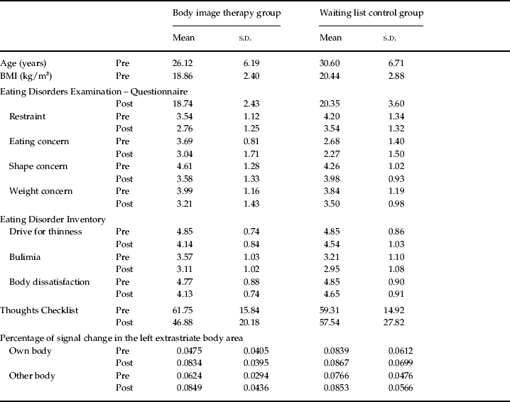
The BITG and WLCG did not significantly differ in age [t(30)=−1.97, p=0.059] and BMI [t(30)=−1.50, p=0.143; Table 1]. Furthermore, no significant differences were found in the pre-measures of the scales restraint [t(30)=−1.43, p=0.162], shape concern [t(30)=0.61, p=0.549] and weight concern [t(30)=0.10, p=0.925]. However, the BITG showed significantly higher scores on the eating concern subscale compared with the WLCG [t(22.03)=2.26, p=0.034]. In addition, no group differences were found on the scales drive for thinness [t(30)=0.07, p=0.945], bulimia [t(30)=0.38, p=0.705] and body dissatisfaction [t(30)=−0.66, p=0.517] or the Thoughts Checklist [t(28)=0.26, p=0.796].
Means (s.d.) of age, body mass index (BMI), questionnaire measures and percentage of signal change in the left extrastriate body area at pre- and post-test in the two groups

Changes in BMI and in self-reported body image and eating disturbance
In the two-way ANOVA for the BMI, the group×time interaction was not significant [F(1, 27)=0.11, p=0.915] and post-hoc t tests revealed no significant changes in the BITG [t(15)=0.94, p=0.362] or in the WLCG [t(15)=0.26, p=0.796].
For the Eating Disorder Examination Questionnaire, the group×time interaction reached significance for the subscale shape concern [F(1, 28)=4.26, p=0.048], but was not significant for the subscales restraint [F(1, 28)=0.10, p=0.751], eating concern [F(1, 28)=0.29, p=0.596] and weight concern [F(1, 28)=1.07, p=0.311]. Post-hoc t tests demonstrate a reduction from pre- to post-test in the BITG on the subscales restraint [t(15)=2.82, p=0.013; d=0.69], shape concern [t(15)=3.97, p=0.001; d=0.80] and weight concern [t(15)=2.51, p=0.024; d=0.67], but not for the scale eating concern [t(15)=1.57, p=0.136]. Furthermore, in the WLCG, a significant reduction was found from pre- to post-test on the scales restraint [t(13)=2.77, p=0.016; d=0.49] and eating concern [t(13)=3.02, p=0.010; d=0.29], but stable values were observed for the subscales shape concern [t(13)=1.11, p=0.289] and weight concern [t(15)=1.22, p=0.245].
For the Eating Disorder Inventory, the ANOVA indicated that the interaction group×time failed to reach statistical significance for the subscales drive for thinness [F(1, 28)=2.43, p=0.131], bulimia [F(1, 28)=0.43, p=0.516] and body dissatisfaction [F(1, 28)=2.67, p=0.114]. Post-hoc t tests showed that, in the BITG, values for drive for thinness [t(15)=4.55, p<0.001; d=0.96] and body dissatisfaction [t(15)=3.36, p=0.004; d=0.73] significantly decreased from pre- to post-test. In the WLCG, no significant change was detected for the subscales drive for thinness [t(13)=1.52, p=0.152] and body dissatisfaction [t(13)=1.07, p=0.305]. For the subscale bulimia, no significant change was found from pre to post in the BITG [t(15)=1.93, p=0.073] or in the WLCG [t(15)=1.77, p=0.100].
For the Thoughts Checklist, the ANOVA indicated that the group×time interaction failed to reach statistical significance [F(1, 27)=2.61, p=0.118]. However, the post-hoc t tests revealed that, in the BITG, negative body-related thoughts were significantly reduced from pre- to post-treatment [t(15)=3.11, p=0.007; d=0.94], whereas in the WLCG, no significant change was detected [t(12)=0.26, p=0.800]. Means and standard deviations are provided in Table 1.
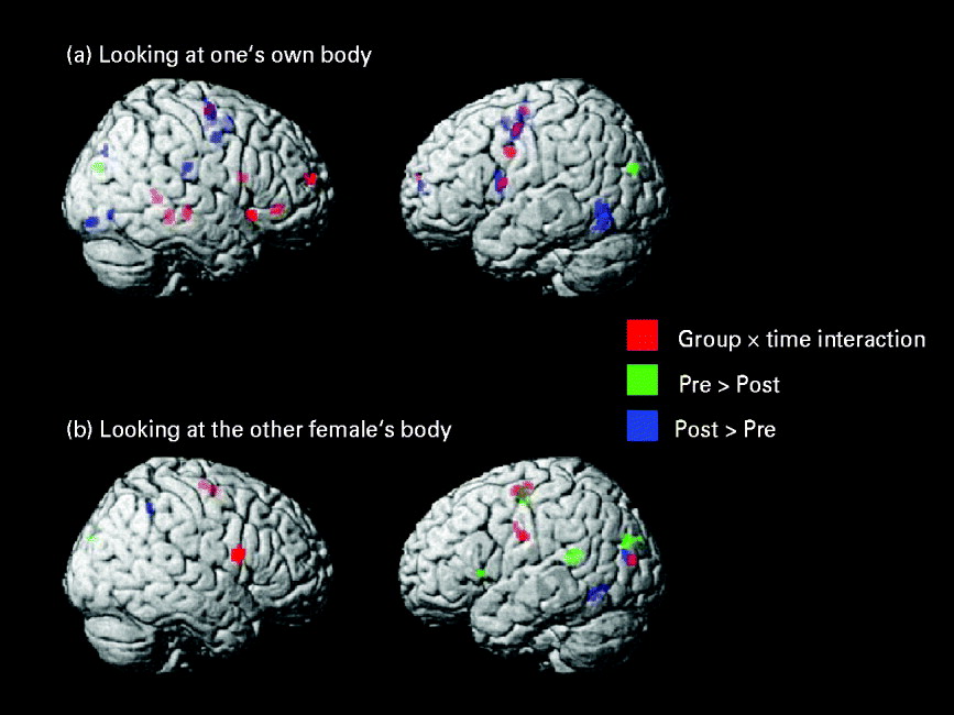
Group×time interaction from the two-way analysis of variance and follow-up t tests indicating changes from pre- to post-body image therapy for the condition of looking at one's own body (a) and looking at the other female's body (b) projected on a rendered brain.

Changes of neuronal correlates to looking at one's own and the other female's body
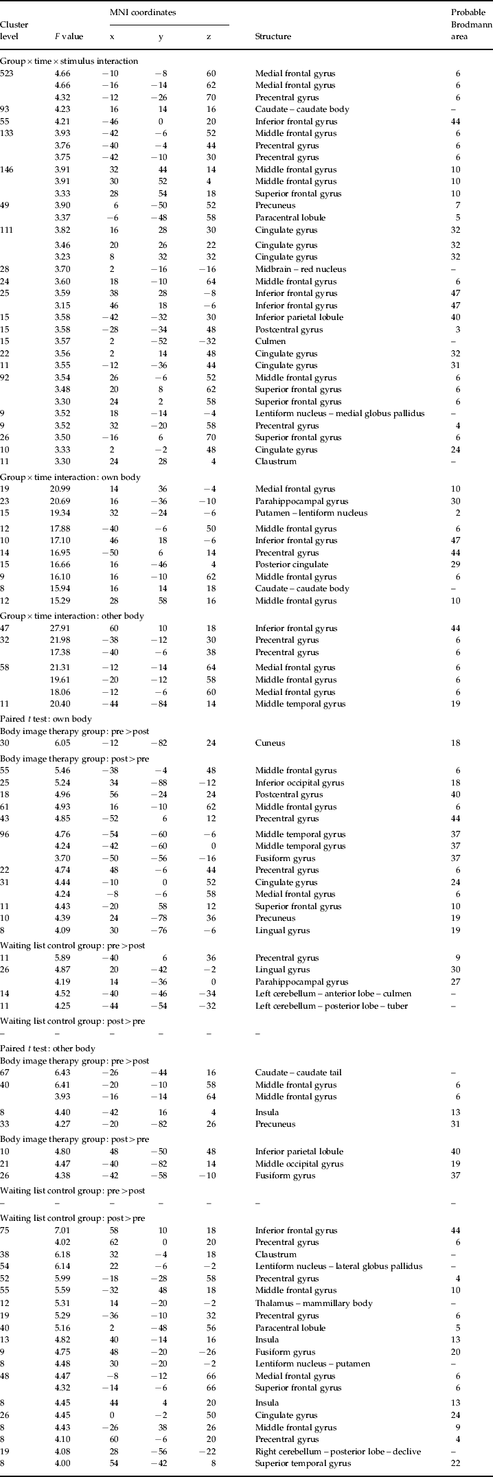
The group×time×stimulus interaction effects of the three-way ANOVA and the group×time interaction effects of the two-way ANOVA for the two conditions of looking at one's own and the other female's body as well as results of the paired t tests comparing activations at pre- and post-test separately for the two conditions and groups are displayed in Table 2 and Fig. 3.
Interaction effects of the three-way analysis of variance (ANOVA) with the factors group (body image therapy group versus waiting list control group), time (pre versus post) and stimulus (own body versus other body) in brain activations and of the two-way ANOVA with the factors group and time for the two stimulus conditions as well as paired t tests comparing brain activations at pre- and post-test for the two stimulus conditions and groups

MNI, Montreal Neurological Institute.
An additional ANOVA using the extracted signal changes from the left EBA as dependent variable indicated no significant group×time×stimulus interaction [F(1, 30)=1.17, p=0.288; Table 1]. However, for the condition of looking at one's own body, a significant group×time interaction [F(1, 30)=6.38, p=0.017] was found, which was due to a significant increase in EBA activity from pre- to post-treatment in the BITG [t(16)=−5.33, p<0.001; d=−0.89] and unchanged values in the WLCG [t(14)=−0.24, p=0.814]. In contrast, for the condition of looking at the other female's body, no significant group×time interaction [F(1, 30)=1.20, p=0.283] was found. While the t test comparing pre- and post-values reached statistical significance for the BITG [t(16)=−2.41, p=0.029; d=−0.77], it was not significant for the WLCG [t(14)=−1.04, p=0.316].
Moreover, a significant correlation of the difference in percentage of signal change in the left EBA from pre- to post-body image therapy in the condition of viewing one's own body with the pre–post difference of the scale body dissatisfaction (r=0.525, p=0.037) was found, whereas the correlations with the difference scores of the other body-image-related questionnaire scales drive for thinness (r=0.233, p=0.385), weight concern (r=0.269, p=0.314), shape concern (p=0.439, p=0.089) and the Thoughts Checklist (r=0.316, p=0.234) were not significant.
Discussion
The purpose of the present randomized controlled trial was to assess alterations in the neuronal bases of visual processing of one's own and another female's body through cognitive behavioural body image therapy. One general finding was that in the condition of looking at one's own body, nearly all of the changes in the various brain regions from pre- to post-treatment in the BITG were activation increases. However, in the WLCG exclusively, activation decreases from pre- to post-waiting time were observed. This overall finding might be regarded in the context of a previous study (Sachdev et al. Reference Sachdev, Mondraty, Wen and Gulliford2008), which demonstrated that when presented with their own body, patients with AN showed no significant activations. This was suggested to be an indication of a reduced activation of the attentional system regarding the processing of one's own body. In line with these findings, Vocks et al. (Reference Vocks, Busch, Grönemeyer, Schulte, Herpertz and Suchan2010a) found that subjects with AN did not display higher activations in any brain region compared with healthy controls when looking at the pictures of their own body. Instead, they displayed weaker activations in regions belonging to the attentional system, i.e. the inferior and superior parietal lobule (Coull & Nobre, Reference Coull and Nobre1999; Clower et al. Reference Clower, West, Lynch and Strick2001). It has been suggested that the reduced perceptual processing of persons with AN may provide the basis for perceptual disturbance in this disorder, possibly associated with the distorted view that persons with eating disorders have of their bodies (Sachdev et al. Reference Sachdev, Mondraty, Wen and Gulliford2008). Consequently, the findings of the present study, pointing to activation increases in general brain activity after treatment when looking at oneself, might be a result of a more intense processing of one's own body, probably due to the repeated body exposure tasks set within the sessions and as homework assignments, which are one main component of the cognitive behavioural body image therapy (Vocks et al. Reference Vocks, Wächter, Wucherer and Kosfelder2008).
Further support for a treatment-induced intensified body-related neuronal processing of one's own body was provided by the activation increases from pre- to post-body image therapy in the left middle temporal gyrus covering the EBA in the whole-brain analysis, confirming the results of our previous separate analysis, which included a subsample of five patients with AN (Vocks et al. Reference Vocks, Busch, Grönemeyer, Schulte, Herpertz and Suchan2010a). This finding was further confirmed by additional analyses using the extracted signal changes from the EBA, in which a significant group×time interaction was found, resulting from a significant activation increase from pre to post in the BITG and unchanged values in the WLCG. This finding of increased EBA activity after treatment might be related to previous research on body image disturbance in eating disorders, providing indications of a low functionality (Uher et al. Reference Uher, Murphy, Friederich, Dalgleish, Brammer, Giampietro, Phillips, Andrew, Ng, Williams, Campbell and Treasure2005) and focused reduction of grey matter density in this brain area (Suchan et al. Reference Suchan, Busch, Schulte, Grönemeyer, Herpertz and Vocks2010). As the EBA is specialized in the visual processing of the human body (Peelen & Downing, Reference Peelen and Downing2007), the observed under-activation in AN and BN was assumed to underlie body image disturbance (Uher et al. Reference Uher, Murphy, Friederich, Dalgleish, Brammer, Giampietro, Phillips, Andrew, Ng, Williams, Campbell and Treasure2005; Suchan et al. Reference Suchan, Busch, Schulte, Grönemeyer, Herpertz and Vocks2010). The increase in neuronal activity in the EBA after the body image therapy found in the present study is likely to reflect a stronger visual processing of the body in this highly selective brain area. The potential relevance of the EBA in treatment-induced body image improvement was emphasized by the correlation of changes in its activity from pre- to post-treatment with improvement of self-reported body dissatisfaction.
Moreover, group×time×stimulus interaction effects were observed in the inferior, middle, medial and superior frontal gyri, which were partly followed up by a significant two-way interaction effect in these regions in the condition of looking at one's own body, resulting from an enhanced activity after body image therapy, but no changes after the waiting period. Accordingly, the bilateral, but right-dominated middle and inferior frontal gyri have been shown to be involved in self-recognition (see meta-analysis by Platek et al. Reference Platek, Wathne, Tierney and Thomson2008). In line with this, previous research has demonstrated that, in general, the middle frontal gyrus is more strongly activated by self-related tasks such as self-reflection (Heatherton et al. Reference Heatherton, Wyland, Macrae, Demos, Denny and Kelley2006; Modinos et al. Reference Modinos, Ormel and Aleman2009), and also by viewing pictures of oneself compared with pictures of others (Hodzic et al. Reference Hodzic, Muckli, Singer and Stirn2009). Based on these findings, it might be speculated that in addition to the aforementioned increase in attention towards one's own body, the body image therapy might have led to a more pronounced sense of ownership and internal representation of one's own body.
In general, it can be speculated that after treatment, brain activations of the participants with eating disorders come closer to what is observed in healthy individuals. However, we did not find an enhanced activation in the inferior and superior parietal lobule when looking at one's own body and no reduced activation in the amygdala when viewing the other female's body, which would have been expected based on the results of our previous study comparing eating disordered and healthy individuals using the same stimulus material as in the present study (Vocks et al. Reference Vocks, Busch, Grönemeyer, Schulte, Herpertz and Suchan2010a). This unexpected finding might be explained by the fact that mechanisms of the development and maintenance of a disorder might not necessarily be identical to those involved in recovery (Grawe, Reference Grawe2006).
Although the body image intervention had a positive outcome on the body image questionnaire data with moderate to high effect sizes, which were broadly comparable with the effects found in previous research using the same treatment in different samples (e.g. Vocks & Legenbauer, Reference Vocks and Legenbauer2005; Vocks et al. Reference Vocks, Wächter, Wucherer and Kosfelder2008), the body image therapy obviously did not significantly affect the questionnaire data covering the eating pathology. This finding indicates that although body image and eating disorder symptoms are closely linked in general (e.g. Ricciardelli et al. Reference Ricciardelli, Tate and Williams1997), the improvement of body image disturbance did not generalize to the eating pathology at the end of the intervention. Consequently, treatment components directly addressing dysfunctional eating patterns such as food restriction and binge eating/purging behaviour are necessary.
As a limitation of the present study, the rather small sample size should be emphasized. The resulting low statistical power might be responsible for the absence of significant interaction effects for some of the variables despite the significant pre–post-changes occurring in the BITG, but not in the WLCG. Additionally, it has to be considered that the attrition from the study was rather high, thus limiting the generalizability of the results. The high drop-out rate might not only be regarded in the context of the high rates of premature termination of treatments in general in eating disorders (e.g. Treat et al. Reference Treat, Gaskill, McCabe, Ghinassi, Luczak and Marcus2005), but can also be suspected to result from the high effort required from the participants of the present study.
Furthermore, it is not clear from the design of the present study whether the observed changes in brain activity result from the intervention as a whole or whether they can be explained by mirror and video exposure alone, which might meet the specific requirements of the scanning task. As patients are repeatedly shown their bodies during body exposure, this intervention might have influenced the neuronal processing of viewing one's own body during the scanning sessions, independently of a more general behavioural improvement. Therefore, in future research, component analyses should be conducted in order to test for the specific contribution of each therapeutic ingredient.
The question can be raised of whether differences in neuronal correlates of viewing one's own body are due to BMI changes from pre to post, resulting in differences in the stimulus material. However, as BMI did not differ significantly from pre- to post-test in either of the groups, it seems improbable that a different BMI at the two time points might have considerably influenced the neuronal responses.
To conclude, the present randomized controlled trial indicates that cognitive behavioural body image therapy can change the impaired processing of one's own body in eating disorders in the form of an enhancement in general brain activity, including focused activation increases in the EBA and in frontal cortical areas, which are responsible for the visual processing of the human body and self-awareness, respectively. A more in-depth insight into the phenomenon of body image disturbance in eating disorders, which is not yet fully understood, might help to foster focused intervention strategies targeting specific symptoms more precisely in order to improve the generally still unsatisfactory treatment results in these severe mental disorders.
Acknowledgements
We thank Marie Hennecke, Kathrin Hubbert, Karin Liebenrodt, Serban Mateiescu, Claudia Moswald, Lena Schloßmacher, Beate Steinfeld, Annika Vogt and Sabrina Zuleger for helpful assistance during collection and management of the data.
Declaration of Interest
None.