Introduction
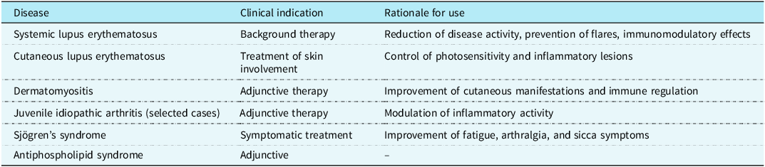
Hydroxychloroquine has been used for decades in the treatment of various rheumatic diseases, such as rheumatoid arthritis, juvenile idiopathic arthritis, systemic lupus erythematosus, and cutaneous-articular lupus. Both the Brazilian Society of Rheumatology and the European League Against Rheumatism also recommend its use in patients with Sjögren’s syndrome and antiphospholipid syndrome. Its efficacy and safety are well established at the recommended dose of 5 mg/kg/day. Reference Klumb, Silva and Lanna1–11 Table 1 summarises the main indications for hydroxychloroquine use in paediatric rheumatic diseases.
Main clinical indications for hydroxychloroquine use in paediatric rheumatic diseases according to the Brazilian Society of Rheumatology (SBR) and the European League Against Rheumatism (EULAR)

Table 1 Long description
The table presents clinical indications and rationales for hydroxychloroquine use in pediatric rheumatic diseases, including systemic lupus erythematosus, cutaneous lupus erythematosus, dermatomyositis, juvenile idiopathic arthritis, Sjogren's syndrome, and antiphospholipid syndrome. It has six rows and three columns. The columns are labeled Disease, Clinical Indication, and Rationale for Use. Each row lists a specific disease, its clinical indication for hydroxychloroquine use, and the rationale behind its use. For example, systemic lupus erythematosus is used for background therapy to reduce disease activity and prevent flares, while cutaneous lupus erythematosus is used to control photosensitivity and inflammatory lesions.
In patients with lupus, hydroxychloroquine has well-recognised benefits, as it improves disease activity and reduces the risk of recurrence, while also improving manifestations such as joint symptoms and skin lesions. Reference van Vollenhoven, Mosca and Bertsias10,11 Neuropsychiatric adverse effects have been described as rare and associated with doses above the recommended range. 11,Reference Hamm and Rosenthal12 Ocular toxicity and QT interval prolongation are potential adverse effects, making careful screening essential before and during treatment.
In 2020, during the COVID-19 pandemic, several studies evaluated hydroxychloroquine use in this context, highlighting its association with cardiac abnormalities, including QT interval prolongation, which can lead to potentially fatal arrhythmias. However, these patients were often also receiving medications such as azithromycin and presented additional factors, including the inflammatory process itself, which may have contributed to these outcomes. Reference Tuncer, Karaci, Boga, Durmaz and Guven13,Reference Samuel, Friedman and Sharma14
Monitoring the cardiac effects of hydroxychloroquine is therefore of huge importance. Electrocardiography emerges as a vital tool in this setting, allowing early detection of abnormalities that may help prevent severe complications. Nevertheless, studies evaluating the association between hydroxychloroquine and QT prolongation in paediatric patients with immune-mediated diseases are scarce, leaving much to be investigated.
Hydroxychloroquine sulphate may induce QT interval prolongation by increasing the duration of ventricular repolarisation. This disturbance in intracardiac electrical conduction raises the risk of polymorphic ventricular tachycardia, known as Torsades de Pointes. Its adverse effects are dose-dependent, and inappropriate use may lead to serious complications such as syncope, ventricular fibrillation, and sudden death.
Hydroxychloroquine, a pillar in the management of systemic lupus erythematosus and rheumatoid arthritis, has the potential to influence the QT interval on electrocardiography. However, there is a lack of studies addressing these effects in the paediatric population with autoimmune diseases. Reference Tuncer, Karaci, Boga, Durmaz and Guven13–Reference Fiehn, Ness and Weseloh20
Thus, the aim of this study was to evaluate paediatric patients with rheumatologic diseases receiving hydroxychloroquine. Specifically, we investigated the prevalence, mechanisms, and clinical implications of QT interval prolongation and other electrocardiographic abnormalities associated with hydroxychloroquine use in patients followed at a tertiary reference centre for paediatric cardiology and rheumatology.
Methods
This was a retrospective and prospective, cross-sectional, and analytical study including children and adolescents up to 18 years of age using hydroxychloroquine, with a total of 26 evaluations at different time points, relating electrocardiography findings to the cumulative hydroxychloroquine dose at the corresponding moment. The mean duration of hydroxychloroquine use was 2 years and 10 months. This study was carried out in the Paediatric Cardiology and Pediatric Rheumatology outpatient clinics at the Martagão Gesteira Institute of Childcare and Pediatrics – Federal University of Rio de Janeiro. This study was approved by the Ethics Committee of the Federal University of Rio de Janeiro, and the parents gave their informed consent.
Study population
Children and adolescents ≤18 years of age with rheumatologic diseases on hydroxychloroquine therapy, followed by paediatric rheumatology and cardiology services at Martagão Gesteira Institute of Childcare and Pediatrics. The retrospective review included cases from January 2020 to March 2025, while the prospective arm included patients between 2024 and 2025. The sample was selected by convenience, including all consecutively diagnosed patients within the study period.
Inclusion criteria were the following: (1) children and adolescents ≤18 years with a confirmed diagnosis of rheumatologic disease; (2) continuous use of hydroxychloroquine; (3) regular follow-up at the paediatric rheumatology and cardiology services. Exclusion criteria were the following: (1) patients with a prior diagnosis of CHD or arrhythmia, particularly long QT syndrome; (2) presence of comorbidities (e.g., chronic kidney disease, endocrine disorders) that could prolong the QT interval; (3) family history of long QT syndrome; (4) concomitant use of QT-prolonging medications; and (5) significant uncorrected electrolyte disturbances (hypokalaemia, hypomagnesaemia or hypocalcaemia).
For each patient, the variables collected were (1) demographic data; (2) date of onset of disease; (3) date of first visit to the centre; (4) date of confirmed diagnosis; (5) presence of comorbidities; (6) clinical manifestations; (7) hospitalisations; (8) electrocardiographic findings: normal electrocardiography, tachyarrhythmias, prolonged PR interval, prolonged QT interval, bundle branch block, atrioventricular block, ST-segment abnormalities, and atrial or ventricular overload; (9) date of medication initiation; and (10) outcomes: complete recovery without electrocardiography abnormalities or death.
Electrocardiograms were manually reviewed by a paediatric cardiologist. The corrected QT interval was calculated using Bazett’s formula (QT/√RR). Standard 12-lead electrocardiographies were performed at a paper speed of 25 mm/s and an amplitude of 10 mm/mV. The QT interval was measured from the earliest onset of the QRS complex to the end of the T wave as the mean of at least 4 cardiac cycles. Measurements were preferentially taken in leads II and V5/V6, with the longest value used. In cases with adjacent T and U waves, the end of the T wave was defined as the return to baseline of the descending limb not followed by a U wave or distinct from it. In bifid T waves of nearly equal amplitude, QT was measured to the final return to baseline. Endpoints of interest included a corrected QT interval increase > 60 ms between consecutive electrocardiographies, corrected QT interval ≥ 500 ms, and documented adverse outcomes.
Statistical analysis
Descriptive statistical analysis was performed, with absolute frequencies and percentages for categorical variables, as well as means and standard deviations or medians and interquartile ranges for continuous variables, depending on data distribution in the sample. Differences between quantitative variables were analysed using the Mann-Whitney test. Differences between proportions were calculated using Fisher’s exact test. A p-value < 0.05 was considered statistically significant.
Logistic regression was used to assess the influence of sex, age at diagnosis, time to diagnosis, duration of fever, clinical manifestations, laboratory abnormalities, and treatment on cardiac alterations. Corrected QT interval values ≥ 450 ms were considered abnormal. Data were transferred to Microsoft Excel 2010 (Microsoft Corp., Redmond, WA, USA) and analysed using PASW Statistics version 18.0 (SPSS Inc., Chicago, IL, USA).
Results
A total of 26 patients were included in the study, all under rheumatology follow-up. Demographic analysis showed a predominance of female patients (76.92%) compared to males (23.08%). Regarding underlying diseases, systemic lupus erythematosus was the most prevalent condition, present in 88.89% of the sample, followed by dermatomyositis, which accounted for 11.11% of cases.
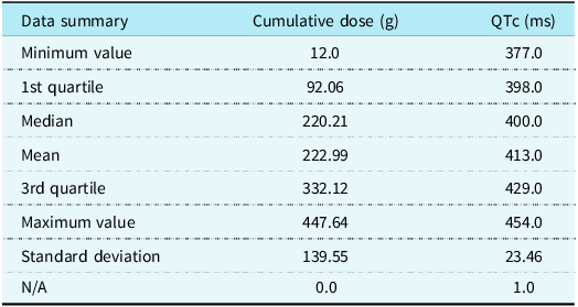
Throughout follow-up, a total of 26 evaluations were performed at different time points, aiming to relate electrocardiography findings to the cumulative hydroxychloroquine dose recorded at each period. Descriptive analysis showed that the cumulative dose ranged from 12.00 to 447.64 g, with a mean of 222.99 g and a median of 220.21 g.
With respect to the corrected QT interval, values ranged from 377.0 to 454.0 ms, with a mean of 413.0 ms and a median of 400.0 ms. One missing data point was identified for this variable, which was imputed using the mean of the remaining values to ensure consistency of subsequent analyses (Table 2). Normality of variables was assessed using the Shapiro-Wilk test, with a significance level of 5%. For the variable “cumulative dose,” the p-value was 0.3474, and for corrected QT interval it was 0.07192. Thus, the null hypothesis was not rejected in either case, indicating that the data followed an approximately normal distribution. Pearson’s correlation analysis between cumulative hydroxychloroquine dose and corrected QT interval interval yielded a correlation coefficient of –0.2396, suggesting a weak negative correlation. However, this association was not statistically significant (p = 0.3382), and therefore, no linear relationship could be established between the variables analysed (Figure 1).
Data summary of cumulative dose of hydroxychloroquine and corrected QT interval

Table 2 Long description
The table presents a data summary of cumulative dose of hydroxychloroquine and corrected QT interval. It includes minimum, 1st quartile, median, mean, 3rd quartile, maximum values, and standard deviation for both variables. The cumulative dose ranges from 12.0 to 447.64 grams, with a mean of 222.99 grams and a median of 220.21 grams. The corrected QT interval ranges from 377.0 to 454.0 milliseconds, with a mean of 413.0 milliseconds and a median of 400.0 milliseconds. The table also includes standard deviations for both variables.
NA = not available.
Correlation between corrected QT interval and cumulative dose of hydroxychloroquine (g).

Figure 1 Long description
A scatter plot with several data points representing the relationship between corrected QT interval and cumulative hydroxychloroquine dose. The x-axis represents the cumulative hydroxychloroquine dose in grams, ranging from 0 to 400 grams. The y-axis represents the corrected QT interval in milliseconds, ranging from 360 to 440 milliseconds. The data points are scattered with no clear trend, indicating no strong correlation between the two variables. All values are approximated.
Based on the normal distribution of the data, a simple linear regression was performed. The coefficients obtained were intercept = 421.98 (p < 0.0001) and slope = –0.04029 (p = 0.338). The estimated regression equation was cumulative dose = 421.98–0.04029 × QTc. The coefficient of determination (R 2) was 0.057, indicating that the model explained only 5.7% of the variability in the data.
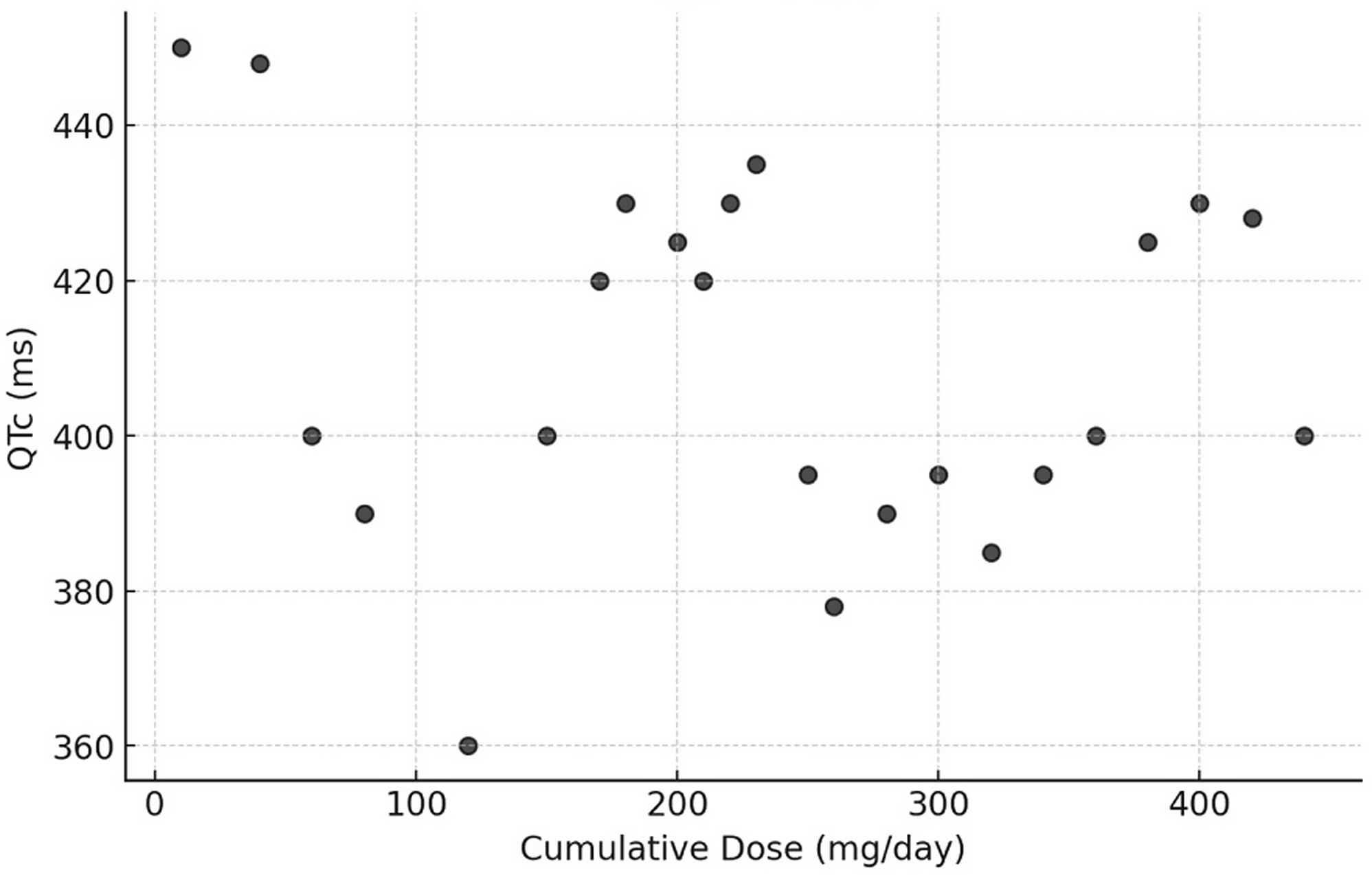
The F statistic of the model was 0.9747, with p = 0.3382, confirming that the regression was not statistically significant. A scatter plot of cumulative dose versus corrected QT interval showed a weakly negative relationship, indicating that as cumulative dose increased, corrected QT interval tended to decrease. The correlation coefficient was –0.2396245 (Figure 2).
Correlation between corrected QT interval and cumulative dose of hydroxychloroquine (mg/day).

Figure 2 Long description
A scatter plot with dozens of data points showing the relationship between corrected QT interval and cumulative dose of hydroxychloroquine. The x-axis represents the cumulative dose in milligrams per day, ranging from 0 to 400. The y-axis represents the corrected QT interval in milliseconds, ranging from 360 to 440. The data points are scattered with no clear overall trend, showing some clustering around certain dose levels. All values are approximated.
Discussion
The relationship between hydroxychloroquine use in paediatric patients with rheumatic diseases and electrocardiographic alterations is a topic of growing interest in the medical literature, as there is a scarcity of studies focusing on this population. Hydroxychloroquine has been associated with QT interval prolongation through its effects on ventricular repolarisation, primarily related to inhibition of the rapid component of the delayed rectifier potassium current (IKr), mediated by the human ether-a-go-go-related gene channel. Blockade of this current prolongs phase 3 of the ventricular action potential, which may lead to QT interval prolongation and, in susceptible individuals, increase the risk of polymorphic ventricular tachycardia. The arrhythmic risk associated with hydroxychloroquine appears to be dose-related and influenced by additional factors such as electrolyte imbalance, systemic inflammation, underlying cardiac disease, and concomitant use of other QT-prolonging medications. In paediatric populations, however, the lower prevalence of cardiovascular comorbidities, reduced exposure to polypharmacy, and strict adherence to weight-adjusted dosing may mitigate these effects. In the present cohort, no clinically significant corrected QT interval prolongation was observed, even with prolonged exposure and high cumulative doses, supporting the cardiovascular safety of hydroxychloroquine in children when appropriately prescribed and monitored. This section synthesises findings from studies in paediatric patients with a particular emphasis on QT interval changes. Reference Desmarais, Rosenbaum and Costenbader18,Reference Roden21–Reference Woosley, Heise, Gallo, Tate, Woosley and Romero23
In the study by Rey et al., Reference Rey, Berneck, Gonçalves, Silva, Skare and Silva15 the influence of chloroquine and hydroxychloroquine on the QT interval in patients with rheumatic diseases was evaluated. Among 46 users of antimalarials, the prevalence of corrected QT interval prolongation was 17.39%. All patients with altered electrocardiograms were female and chloroquine users, suggesting a possible implication of this antimalarial in the aetiology of the alteration. AlTwajery et al. Reference AlTwajery, AlMane and Al-Mayouf16 conducted a retrospective cross-sectional study in paediatric patients with systemic lupus erythematosus. Electrocardiographic abnormalities were observed in 29.3% of patients, including ST-T changes and right bundle branch block, with 90.3% of patients receiving hydroxychloroquine. This study highlighted a significant association between anti-Ro/SSA antibodies and electrocardiography abnormalities. Another study, conducted by Samuel et al., Reference Samuel, Friedman and Sharma14 focused on the incidence of arrhythmias and electrocardiographic abnormalities in symptomatic paediatric patients with SARS-CoV-2 infection, including drug-induced changes in the corrected QT interval. The study revealed that COVID-19 treatment, including hydroxychloroquine with or without azithromycin, was not associated with significant arrhythmias. Reference Samuel, Friedman and Sharma14
Tuncer et al. Reference Tuncer, Karaci, Boga, Durmaz and Guven13 reported their experience with QT interval evaluation associated with concomitant hydroxychloroquine and azithromycin use in children who tested positive for coronavirus disease 2019. The study demonstrated that neither patients receiving hydroxychloroquine monotherapy nor those receiving hydroxychloroquine plus azithromycin developed a corrected QT interval prolongation of 500 milliseconds or more during or after discontinuation of the drugs. Reference Tuncer, Karaci, Boga, Durmaz and Guven13 The study by Esmel-Vilomara et al. Reference Esmel-Vilomara, Dolader, Sabaté-Rotes, Soriano-Arandes, Gran and Rosés-Noguer17 addressed corrected QT interval interval prolongation in patients infected with SARS-CoV-2 and treated with antiviral drugs. This study observed a median corrected QT interval increase of only 15 ms between baseline electrocardiography and the recordings obtained within the first 72 hours of treatment, indicating a limited risk of arrhythmias associated with hydroxychloroquine use. Reference Esmel-Vilomara, Dolader, Sabaté-Rotes, Soriano-Arandes, Gran and Rosés-Noguer17
In the present study, no significant association was observed between cumulative hydroxychloroquine dose and corrected QT interval prolongation in paediatric patients with rheumatologic diseases. Although a negative correlation was identified, it was weak and lacked statistical significance. The literature describes hydroxychloroquine as a potentially cardiotoxic drug, particularly in adults with cardiovascular comorbidities or concomitant use of multiple medications. In this population, reports of ventricular arrhythmias and cases of torsades de pointes have underlined the need for periodic monitoring. However, our findings suggest that, in paediatrics, even in the setting of high cumulative doses, there is no evidence of a significant impact on ventricular repolarisation.
These results may reflect age-related characteristics, such as the lower prevalence of cardiovascular risk factors and reduced exposure to polypharmacy. Furthermore, the low explanatory power of the model (R 2 = 5.7%) and the limited sample size reinforce the need for caution in interpretation. Multicentre studies, with larger samples and control for confounding variables, are essential to confirm these findings. The dose employed (5 mg/kg/day) corresponds to the recommendation of the Brazilian Society of Rheumatology and the European League Against Rheumatism, Reference Klumb, Silva and Lanna1–11 reinforcing the safety of the therapeutic regimen used.
The present study evaluated the relationship between cumulative hydroxychloroquine dose and the corrected QT interval in a sample of paediatric patients. Statistical analysis indicated no significant association between increased cumulative dose and corrected QT interval prolongation, suggesting that, within the analysed parameters, hydroxychloroquine shows an appropriate cardiovascular safety profile in the studied paediatric population. Although no statistically relevant correlation was identified, the importance of electrocardiographic monitoring is emphasised, particularly in clinical situations associated with higher arrhythmic risk. The finding of no significant impact on corrected QT interval should be interpreted in light of methodological limitations, including the small sample size and the lack of control for potential confounding factors.
Conclusion
In conclusion, hydroxychloroquine, at doses used in paediatric clinical practice, showed no evidence of causing significant corrected QT interval alterations, supporting its safe use when indicated. Nevertheless, further investigations with more robust methodological designs are recommended to deepen the understanding of the drug’s cardiac effects in different clinical contexts.