Introduction
Female insect physiology changes dramatically after mating (Gillott Reference Gillott2003). The most common response is an increase in fertility (Baena Hurtado Reference Baena Hurtado2009), but our knowledge of how mating affects behavioural and physiological changes in the females is still incipient. When insects mate, females receive external and internal stimuli through copulatory courtship as well as through seminal fluid, which is composed of sperm and male accessory gland secretions (Avila et al. Reference Avila, Sirot, LaFlame, Rubistein and Wolfer2011). These secretions consist of several proteins, such as proteases, protease inhibitors, lecithins, pro-hormones, peptides, and antioxidant proteins, and their composition plays a key role in the evolutionary ecology of mating and reproduction (Perry et al. Reference Perry, Sirot and Wigby2013). The act of mating itself can also bring about changes in female behaviour, yet how these mating-driven changes may manifest independently of receiving an ejaculate is not usually studied (Peretti and Aisenberg Reference Peretti and Aisenberg2015; Rodríguez Reference Rodríguez2015).
Molecules in the male accessory glands may have a direct impact on the postcopulatory behaviour of females in a variety of organisms from arthropods to mammals (Sirot et al. Reference Sirot, LaFlamme, Sitnik, Rubinstein, Avila, Chow and Wolfner2009). Male accessory gland secretions decrease the probability of remating and function as a seminal plug or as modulators of sperm storage. They can also have antimicrobial properties and can increase the rate of ovarian development and oviposition (Lung et al. Reference Lung, Kuo and Wolfner2001; Gillott Reference Gillott2003; Avila et al. Reference Avila, Sirot, LaFlame, Rubistein and Wolfer2011). Specific peptides in the secretions, such as the sexual peptide Acp70, induce the sexual refractory period of females (Craig Reference Craig1967; Shutt et al. Reference Shutt, Stables, Aboagye, Moran and Tripet2010; Helinski et al. Reference Helinski, Deewatthanawong, Sirot, Wolfner and Harrington2012; Brent and Hull Reference Brent and Hull2014), reduce the host-seeking behaviour of females (Judson Reference Judson1967; Fernández and Klowden Reference Fernández and Klowden1995; Lee and Klowden Reference Lee and Klowden1999), can induce oviposition (Wolfner Reference Wolfner2002), and increase female fecundity (Villarreal et al. Reference Villarreal, Pitcher, Helinski, Johnson, Wolfner and Harrington2018). Other important peptides, such as ovuline (peptide 26Aa), can also induce ovarian development and are responsible for the development of eggs and egg laying (Liu and Kubli Reference Liu and Kubli2003; Fricke et al. Reference Fricke, Wigby, Hobbs and Chapman2009; Yamane and Miyatake Reference Yamane and Miyatake2010; Xu and Wang Reference Xu and Wang2011). In some insects, particularly those that produce spermatophores, prostaglandins transferred in the ejaculate increase female oviposition (Loher Reference Loher1979; Stanley-Samuelson Reference Stanley-Samuelson1994). For example, female crickets that receive prostaglandin E2 initiate oviposition and increase egg laying, whereas females mated with males that have less prostaglandin E2 in their spermatophore lay fewer eggs (Stanley-Samuelson Reference Stanley-Samuelson1994; Worthington et al. Reference Worthington, Jurenka and Kelly2015). Although we know very little about the effects of prostaglandins in other insects, the function of the male accessory gland secretions and how mating, the ejaculate, or the combination of the two modifies female insect behaviour and physiology also has yet to be fully understood.
For pest species, understanding how mating and the ejaculate can impact female reproduction can improve current control methods (Hendrichs et al. Reference Hendrichs, Robinson, Cayol and Enkerlin2002; Robinson et al. Reference Robinson, Cayol and Hendrichs2002; Lance and McInnis Reference Lance and McInnis2005; Sirot et al. Reference Sirot, Poulson, McKenna, Girnary, Woflner and Harrington2008). Specifically, in the case of tephritid fruit flies, the function of male accessory gland secretions within some species has been inferred from the results of directly injecting homogenates of these secretions into the thorax of females. For example, injected females of the Mediterranean fruit fly Ceratitis capitata (Wiedemann) (Jang Reference Jang1995; Jang et al. Reference Jang, McInnis, Kurashima and Carvalho1999), Queensland fruit fly Bactrocera tryoni (Froggatt) (Radhakrishnan and Taylor Reference Radhakrishnan and Taylor2007), and South American fruit fly Anastrepha fraterculus (Wiedemann) (Abraham et al. Reference Abraham, Caldera, Goane and Vera2012) (Diptera: Tephritidae) change their olfactory response from male pheromone to host fruits and have reduced sexual receptivity. However, for the Mexican fruit fly, Anastrepha ludens (Loew) (Diptera: Tephritidae), no inhibition in the sexual receptivity of females was found with the injection of male accessory gland secretions from sterile, fertile, or wild males (Abraham et al. Reference Abraham, Núñez-Beverido, Contreras-Navarro and Pérez-Staples2014), even though mating does reduce female receptivity in wild females (Aluja et al. Reference Aluja, Rull, Sivinski, Trujillo and Pérez-Staples2009; Meza et al. Reference Meza, Arredondo, Orozco and Pérez-Staples2014): for this species, the functions of accessory gland secretions remain unclear.
Anastrepha ludens is a multivoltine and polyphagous species. Females reach sexual maturity between 9 and 15 days of age, although some females can start mating as early as six days of age (Carey et al. Reference Carey, Liedo, Müller, Wang, Senturk and Harshman2005; Reyes-Hernández and Pérez-Staples Reference Reyes-Hernández and Pérez-Staples2017). Once sexually mature, females produce eggs throughout their life (Aluja et al. Reference Aluja, Pinero, Jacome, Diaz-Fleischer and Sivinski2000, Reference Aluja, Dıaz-Fleischer, Papaj, Lagunes and Sivinski2001). During mating, the male introduces its reproductive organ, the aedeagus, through the tip of the ovipositor into the female’s bursa copulatrix, inflates, and deflates its anal membrane while the female keeps the lateral surstylus membrane extended. Mating has been shown to reduce female response to male pheromones (Robacker et al. Reference Robacker, Ingle and Hart1985), but whether mating can enhance oviposition remains unknown. Because complex internal and external behaviours occur during mating (Briceño et al. Reference Briceño, Orozco, Luis, Hanson and Hernández2011), the mechanical stimulation of mating could impact female physiology independently of the effect of the ejaculate. This means that changes in female behaviour may be due to chemosensation through receiving male accessory gland secretions or through mechanical stimulation (Cury et al. Reference Cury, Prud’homme and Gompel2019). Here, we hypothesised that these secretions would increase ovarian development and oviposition. We predicted that mechanosensation through intromission without an ejaculate would not be enough to induce this behavioural change in females.
Material and methods
Insects
Wild A. ludens were obtained from infested sour oranges (Citrus aurantium Linnaeus) (Sapindales) collected in Tuzamapan, Veracruz, Mexico. The pupae were placed in Petri dishes inside tulle-meshed cages (30 cm × 30 cm × 30 cm) with water, sugar, and hydrolysed protein (MP Biomedicals, Irvine, California, United States of America; 3:1 ratio) at a temperature of 25 ± 2 °C. Adults were separated by sex 48 hours after they emerged. Wild males and females used for experiments were 21 days of age. Laboratory-reared A. ludens were obtained as pupae from the standard strain produced at the Moscafrut facility, Metapa de Domínguez Chiapas, Mexico.
Ejaculate transfer and oviposition
Following Miyatake et al. (Reference Miyatake, Chapman and Partridge1999) and Abraham et al. (Reference Abraham, Lara-Pérez, Rodríguez, Contreras-Navarro, Núñez-Beverido, Ovruski and Pérez-Staples2016), we generated males that could mate (i.e., insert the aedeagus) but were unable to transfer the ejaculate. In the morning (09 h 30), 25 males that were 15–17 days old were kept in the freezer (–4 °C) for five minutes. Males were then transferred to a microscope slide, and the tip of the aedeagus was cut under a stereoscopic microscope (Olympus SZX7; Olympus, Tokyo, Japan). The incision was made between the glans and the distiphallus. After the operation, the individuals were kept in a recuperation cage (30 cm × 30 cm × 30 cm) with water and food (sugar and hydrolysed protein at a ratio of 3:1) at a temperature of 25 ± 2 °C (aedeagus-cut males). As control, males handled in the same way were used, except the aedeagus was only extended using tweezers (sham males). These males were then placed in a separate cage of the same characteristics and under the same conditions. As in Abraham et al. (Reference Abraham, Lara-Pérez, Rodríguez, Contreras-Navarro, Núñez-Beverido, Ovruski and Pérez-Staples2016), aedeagus-cut males could court and mate similarly to sham males. In various pilot experiments, we checked mating likelihood, mating latency (time between access to the female and initiation of copulation), copula duration, and sperm transfer of the aedeagus-cut and sham males. Mating likelihood of the two types of males was not significantly different (Kruskal Wallis test, χ 2 = 0.039, df = 1, P = 0.844, n = 120). Also, there were no significant differences between how quickly the differently treated males mated (mating latency; one-way analysis of variance, F = 0.58, df = 1, P = 0.448, n = 153) or copula duration (F = 0.08, df = 1, P = 0.775, n = 120), indicating no discernable differences in mating behaviour or female acceptance. No sperm transfer was seen for aedeagus-cut males. Spermathecae were dissected to determine if sperm was stored in females mated with normal males (n = 15), females mated with aedeagus-cut males (n = 15), and females mated with sham males (n = 15). All females mated to sham males or normal males had sperm in their spermathecae, but only one female had sperm from mating with aedeagus-cut males. This coincides with previous studies that have used this method to prevent male ejaculation during mating (Miyatake et al. Reference Miyatake, Chapman and Partridge1999; Abraham et al. Reference Abraham, Lara-Pérez, Rodríguez, Contreras-Navarro, Núñez-Beverido, Ovruski and Pérez-Staples2016).
To observe matings, individual females were placed in plastic containers (250 mL) at 16 h 00 with either aedeagus-cut males, sham males, or normal males (n = 35 for each of the male treatments). The beginning and end of each mating interaction was observed. For pairs that mated, males were removed at the end of mating, and females were provided with an artificial host consisting of 4-cm-diameter agar sphere and with food and water (Díaz-Fleischer and Aluja Reference Díaz-Fleischer and Aluja2003). A separate group of virgin females (n = 25) was also placed individually in containers.
Twenty-four hours after mating, between 09 h 30 and 12 h 00, all eggs were collected. We chose this time frame because we wanted to see immediate postmating responses and because male accessory gland molecules have been found in various female tissues shortly after mating (Radhakrishnan et al. Reference Radhakrishnan, Nair, Raftos and Taylor2008). To facilitate egg removal, water was sprinkled onto them. Collection was carried out with tweezers, and the eggs were placed in Petri dishes (60 cm × 15 mm) with water for hydration. Eggs were then counted with an entomological pin and a manual counter under a stereoscopic microscope. The experiment was repeated five times.
Male accessory gland secretions
Males were sacrificed by placing them in a closed container with a few drops of ethyl acetate, and then they were immediately dissected under a stereoscopic microscope (Olympus SZX7). Following methodology by Jang (Reference Jang1995), Radhakrishnan and Taylor (Reference Radhakrishnan and Taylor2007), and Abraham et al. (Reference Abraham, Caldera, Goane and Vera2012, Reference Abraham, Núñez-Beverido, Contreras-Navarro and Pérez-Staples2014), the accessory glands of 20 virgin males were extracted with tweezers (Swiss forceps #5) and washed in a drop of saline solution (sodium chloride 0.9%). Subsequently, the 20 glands were placed in 100 µL of saline solution in an Eppendorf tube with a 1.5-mL conical tip and triturated with a plastic micro-homogeniser. The glands were then centrifuged at a speed of 12 000 rpm for one minute at 4 °C. Finally, the supernatant for use in injections was separated, and the pellet discarded.
In a natural mating, it is unlikely that males transfer the entire contents of their glands (Radhakrishnan and Taylor Reference Radhakrishnan and Taylor2007). Therefore, a male equivalent was defined as the quantity of liquid obtained from breaking up the accessory glands of one male in 1 µL saline solution (Jang Reference Jang1995). The concentration for the injection was standardised at 0.2 male equivalents, that is, the 20 male accessory glands (one gland from each of the 20 males) homogenised in 100 µL saline solution (dose based on Radhakrishnan and Taylor Reference Radhakrishnan and Taylor2007 and Abraham et al. Reference Abraham, Caldera, Goane and Vera2012, Reference Abraham, Núñez-Beverido, Contreras-Navarro and Pérez-Staples2014). In order to avoid protein degradation, injections were carried out immediately after homogenisation inside an air-conditioned room.
Injections and matings
From the 100-µL homogenised solution, we injected 1 µL into the intersegmental membrane of each of a first group of unmated, virgin females, using a 1-µL Hamilton micro syringe (Microliter syringe, Model #7001; Hamilton Co., Reno, Nevada, United States of America; Jang Reference Jang1995; Radhakrishnan and Taylor Reference Radhakrishnan and Taylor2007; Abraham et al. Reference Abraham, Caldera, Goane and Vera2012, Reference Abraham, Núñez-Beverido, Contreras-Navarro and Pérez-Staples2014). Each of a second group of unmated virgin females was injected with 1 µL saline solution. Each group of females was placed for recuperation in 1-L containers with food (sugar and hydrolysed yeast (MP Biomedicals); 3:1 proportion) at a temperature of 25 ± 2 °C.
In the afternoon of the same day the injections occurred, virgin females from a third group of A. ludens were mated with males of the same age. Pairs were placed in 250-mL plastic containers. Once matings were finished, females were separated and given water and food. Matings were observed from 16 h 00 until the end of the last copulation. A fourth, control group consisted of virgin A. ludens females.
Effect of male accessory gland secretions on ovary development
We hypothesised that male accessory gland secretions could affect ovarian development in A. ludens; therefore, sexually immature, laboratory, virgin females of four to nine days of age were used. As yeast hydrolysate has been found to increase egg development (Aluja et al. Reference Aluja, Dıaz-Fleischer, Papaj, Lagunes and Sivinski2001; Ben-Yosef et al. Reference Ben-Yosef, Jurkevitch and Yuval2008), which could have masked the effect of the secretions, females were fed only sugar and water, and males were fed sugar plus hydrolysed yeast (in a 3:1 proportion). Treatments were as follows: females injected with male accessory gland secretions, females injected with saline, and virgin females. For injections, sexually mature males were dissected at 10 days of age, and 20 glands were removed from 20 males; the same protocol as that described above was followed for male accessory gland injections. Females were four, six, or eight days of age when injected.
Twenty-four hours after final injections, the females were dissected under a stereo-microscope (Olympus SZX7) and photographed, and the area of the ovaries was measured using Image J, version 1.52 (National Institutes of Health, Bethesda, Maryland, United States of America). The thorax of each female was also photographed and measured as a proxy for size. A total of four replicates were carried out with different cohorts of flies, for a total of 652 females.
Effect of male accessory gland secretions on oviposition
Wild, unmated females were injected with male accessory gland secretions, saline, or only punctured as described above (n = 30, in all cases). In addition, mated (n = 30) and virgin (n = 30) females were also observed. For injections, sexually mature males were dissected at 21 days of age, and 20 glands were removed from 20 males. The same protocol as that described above was followed for male accessory gland secretion injections. Females were also 21 days of age, based on the maximum mating age for both sexes in this species (Reyes-Hernández and Pérez-Staples Reference Reyes-Hernández and Pérez-Staples2017). Males and females were fed sugar and hydrolysed yeast in a 3:1 proportion.
The morning after these treatments, females were placed in individual 250-mL plastic containers with an oviposition device (the lid of a Petri dish, 4 cm in diameter) and water and were covered with parafilm. One day later, between 09 h 00 and 12 h 00, time zone UTC-6 America/Mexico City, the eggs deposited on the Petri dish or plastic containers were transferred to a Petri dish (60 mm × 15 mm) containing water for hydration. Eggs were counted under a stereoscopic microscope at 20 × (Olympus SZX7). Egg collection was carried out over four days at the same time each day. We chose this time frame because male accessory gland–secretion molecules have been found throughout females’ body immediately after mating and with a significant increase by 22 hours after mating (Radhakrishnan et al. Reference Radhakrishnan, Nair, Raftos and Taylor2008); therefore, effects should be evident by 24 hours after mating. Latency to oviposit was measured as the time taken for eggs to be laid from the first day of observation. The experiment was repeated five times.
Statistical analysis
We used a generalised linear model with a binomial distribution and a logit link to analyse the likelihood of oviposition for females that either received or did not receive seminal fluid. To evaluate the number of eggs laid in the same experiment, we used a generalised linear model with a negative binomial distribution. For ovarian development, the average size of each females’ two ovaries was analysed using a generalised linear model with a Gaussian distribution and an identity link, with treatment, female age, the interaction between treatment and age, female size (thorax), and replicate serving as independent factors. The number of females that laid eggs within 24–48 hours was analysed using a generalised linear model with a binomial distribution and a logit link function, followed by a post hoc test in order to identify differences among treatments for each day separately. The total number of eggs laid in four days according to treatment (injected, virgin, or by natural mating) was analysed using a generalised linear mixed model, where fixed effects were the treatment and replicate and where females ovipositing across four days were the random factor. Analysis was carried out in Jamovi, version 1.2.37.0 (The Jamovi Project 2020; computer software retrieved from https://www.jamovi.org), and R version 4.0.3 (R Core Team 2020) to edit graphs.
Results
Oviposition with or without an ejaculate
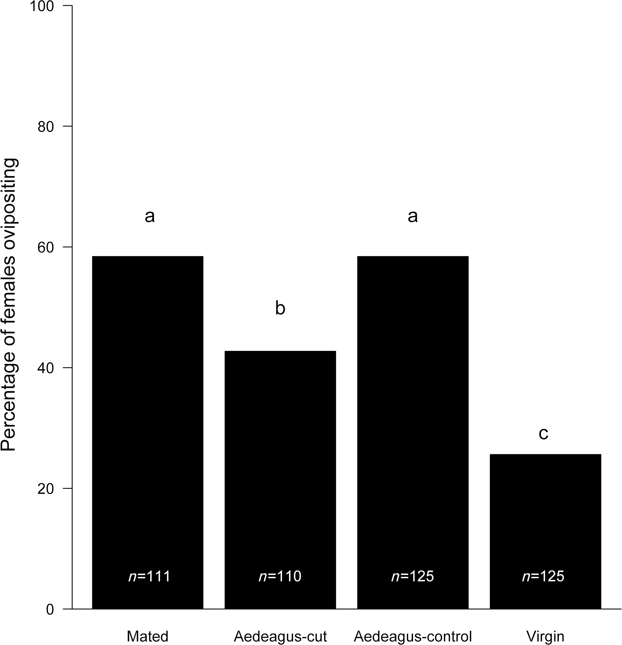
As predicted, mating increased the probability of ovipositing, whereas females that mated but did not receive an ejaculate were more likely to oviposit than virgin females were (Fig. 1; χ 2 = 55.5, df = 3, P < 0.001). Replicate also had a significant effect (χ 2 = 184.2, df = 4, P < 0.001).
Percentage of Anastrepha ludens females ovipositing after mating with normal, aedeagus-cut (no ejaculate received), or a sham (aedeagus-control) males. A final group of virgin females were kept without males (did not mate or receive an ejaculate). Numbers inside bars represent sample size. Different letters represent significant differences after post hoc tests.

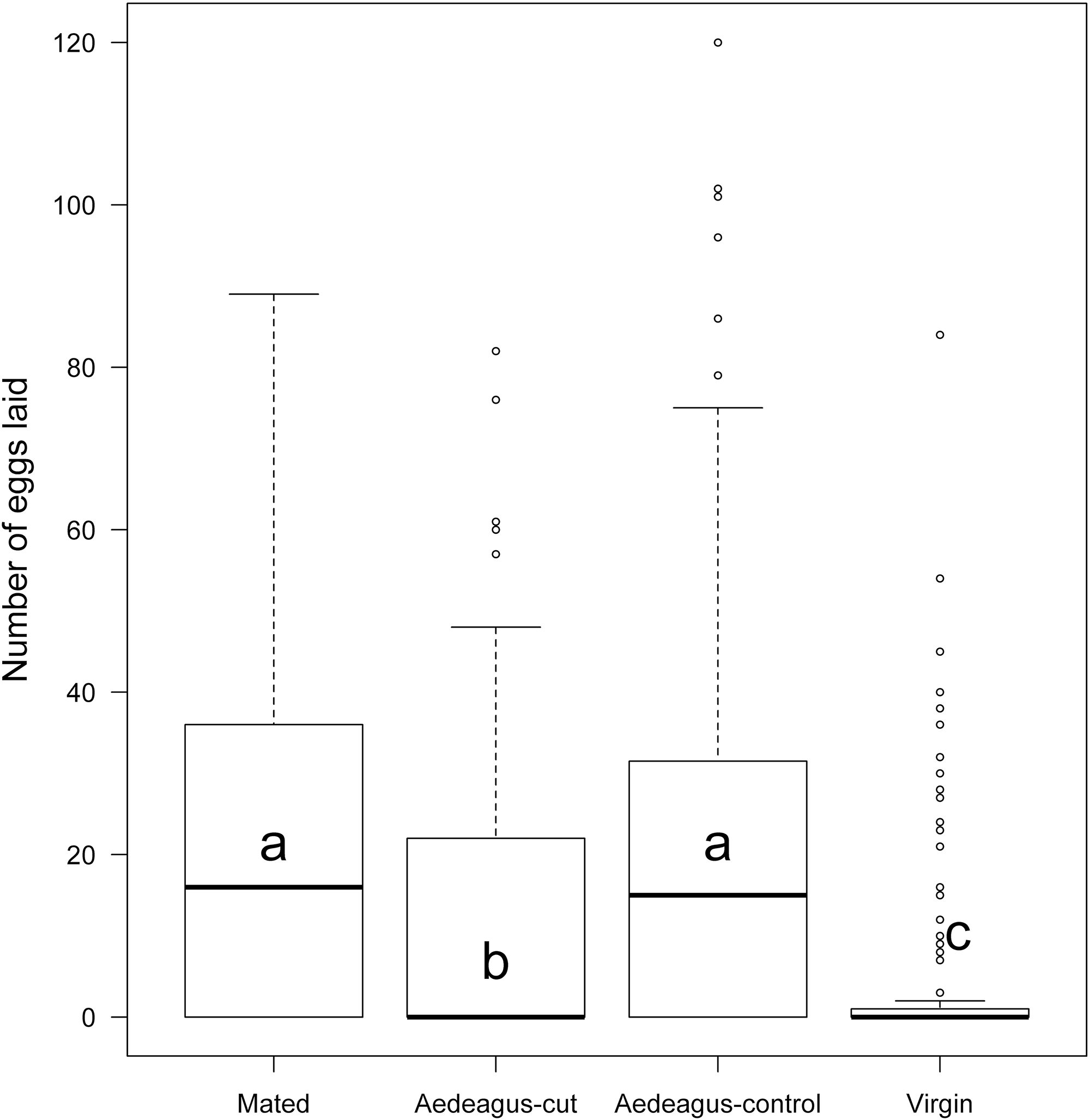
In support of the mechanical stimulation hypothesis, we found that females mated to aedeagus-cut males laid more eggs than virgin females did. The number of eggs laid in all cases was significantly greater than that observed for virgins (Fig. 2; treatment, χ 2 = 55.5, df = 3, P < 0.001; replicate, χ 2 = 184.2, df = 4, P < 0.001).
Number of eggs laid by Anastrepha ludens females after mating with normal, aedeagus-cut (no ejaculate received), or sham (aedeagus-control) males. A final group of females were kept without males (did not mate or receive an ejaculate). Horizontal lines inside boxes represent the median. Circles outside whiskers represent outliers. Different letters represent significant differences after post hoc tests.

Effect of male accessory gland secretions on ovarian development
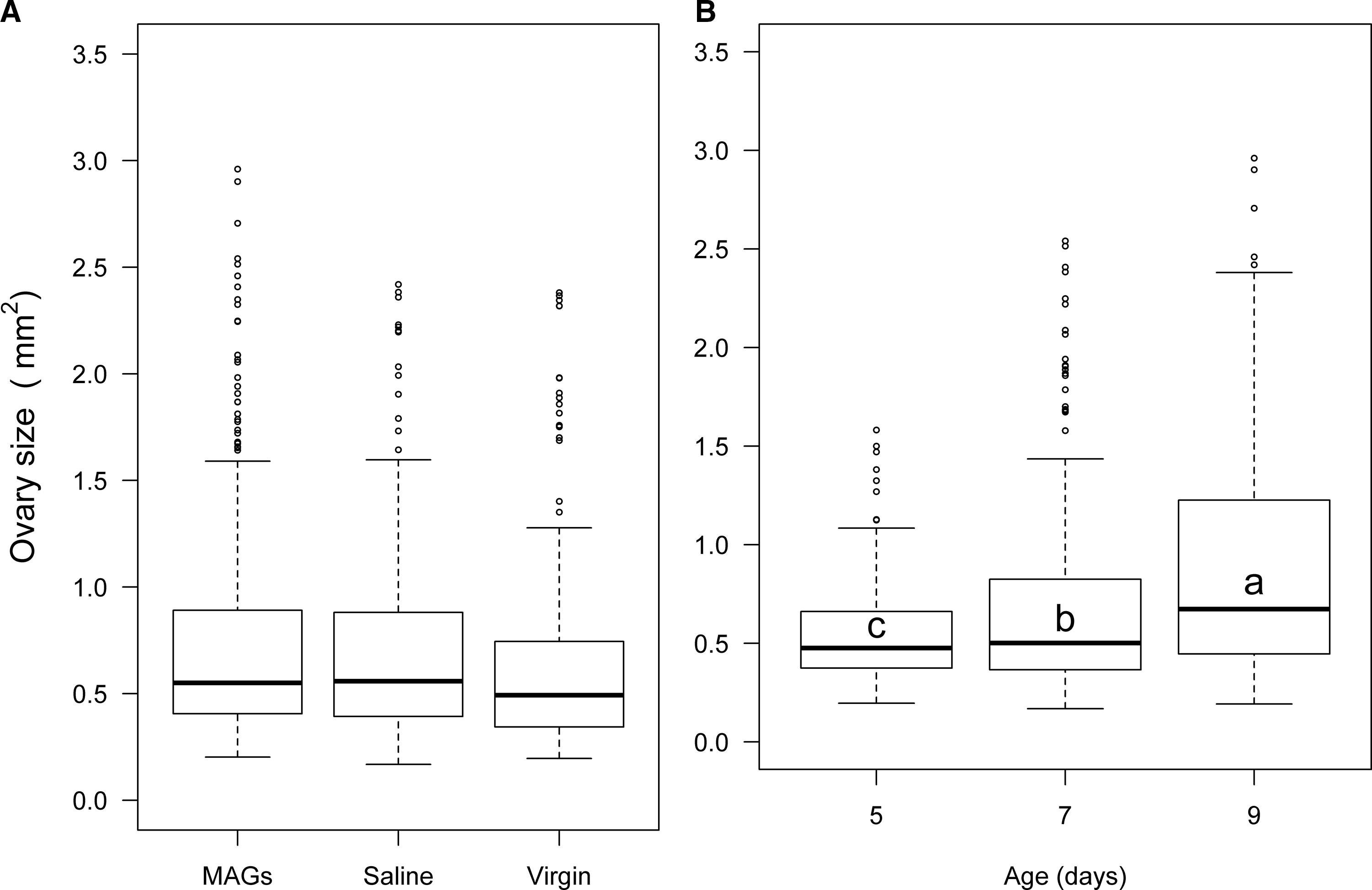
Ovary development was measured for a total of 324 females injected with male accessory gland secretions, 160 injected with saline, and 168 virgin females (without treatment) across all ages. Contrary to our prediction, secretion injection did not increase ovary development (Fig. 3A). There was no significant difference in ovary size according to treatment (χ 2 = 1.1, df = 2, P = 0.578) or the interaction between treatment and age (χ 2 = 2.11, df = 4, P = 0.715). Larger females had larger ovaries (χ 2 = 10.6, df = 1, P = 0.001). Female age was also significant (χ 2 = 74.3, df = 2, P < 0.001), as was replicate (χ 2 = 45.7, df = 3, P = < 0.001). As females aged, the size of the ovaries increased (Fig. 3B).
Ovary sizes of developing Anastrepha ludens females. A, Ovary size of females injected with mature male accessory gland secretions (MAGs), or saline, compared to virgin females; B, ovary size five to nine days of age after emergence. Horizontal lines inside boxes represent the median. Circles outside whiskers represent outliers. Different letters represent significant differences after Tukey’s post hoc test.

Effect of male accessory gland secretions on oviposition
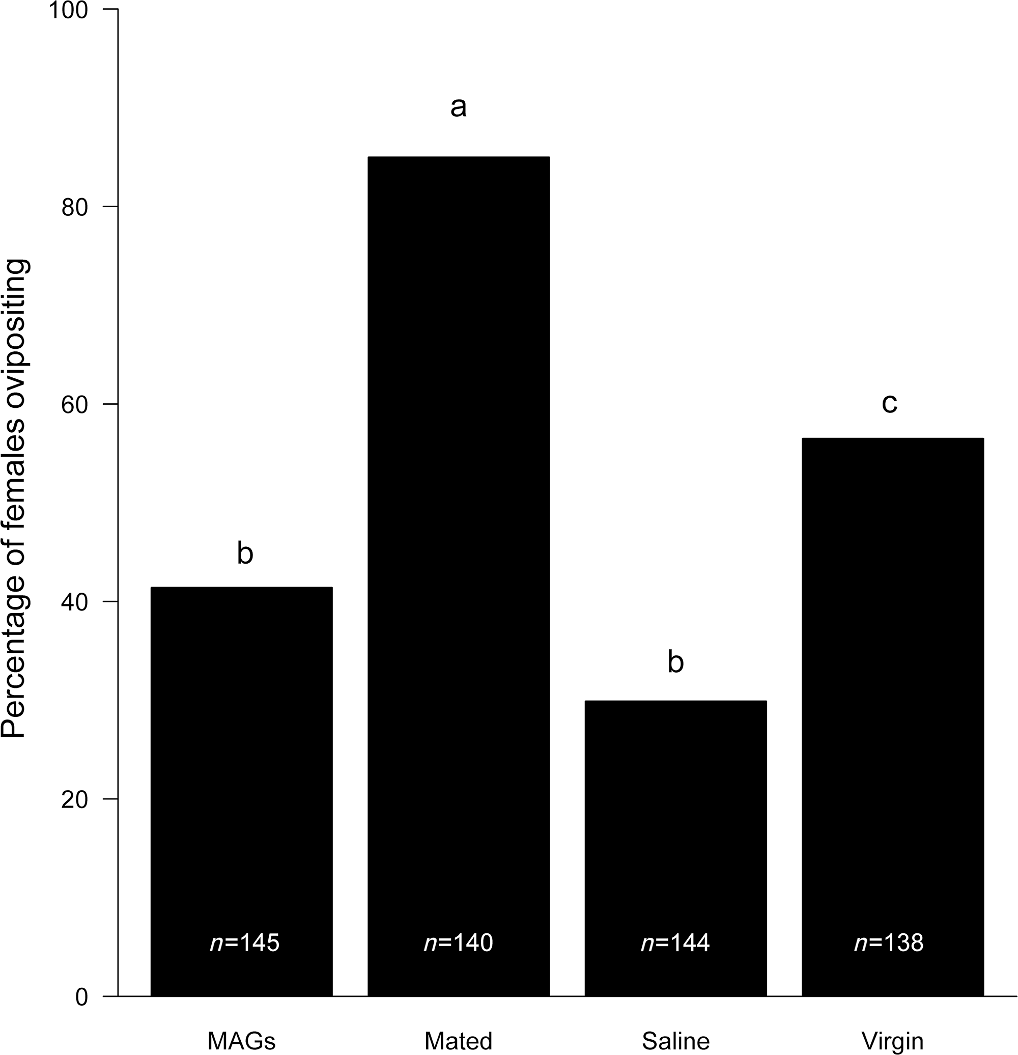
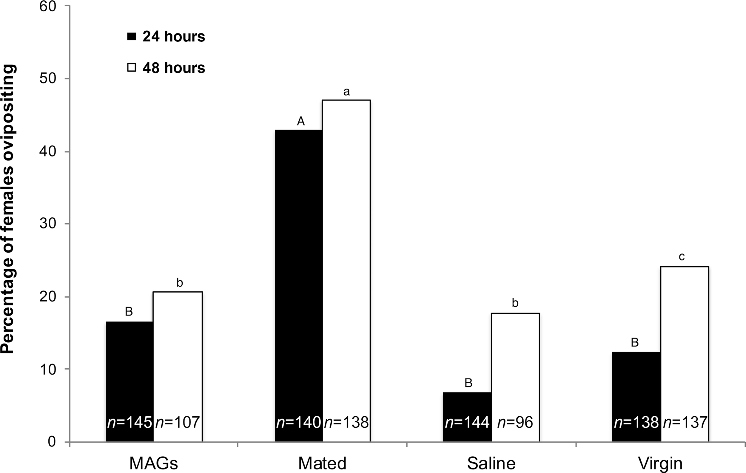
Contrary to our predictions, male accessory gland secretions did not increase the likelihood of females ovipositing, how fast females oviposited, or the total number of eggs females laid. The probability of egg laying differed significantly according to treatment (χ 2 = 116.7, df = 3, P < 0.001) and replicate (χ 2 = 37.7, df = 4, P = < 0.001). However, only mated females were more likely to lay eggs. Females that received only the secretions were not more likely to oviposit than virgin females were (Fig. 4).
Percentage of Anastrepha ludens females ovipositing after mating, injected with male accessory gland secretions (MAGs), injected with saline, or maintained as virgins (no injection, no mating). Numbers inside bars represent the sample size. Different letters represent significant differences after post hoc tests.

Females receiving the secretions were not faster at ovipositing than virgin females were. The probability of females ovipositing at 24 hours differed significantly between treatments (χ 2 = 67.7, df = 3, P < 0.001) and replicates (χ 2 = 32.5, df = 4, P = < 0.001). The probability of females ovipositing at 48 hours also varied according to treatment (χ 2 = 54.4, df = 3, P < 0.001) and replicate (χ 2 = 14.8, df = 4, P = 0.005). However, for both timelines used in the study, only mated females oviposited faster than the females from the other treatments (Fig. 5).
Percentage of Anastrepha ludens females ovipositing after 24 or 48 hours after mating, injected with male accessory gland secretions (MAGs), injected with saline, or maintained as virgins (no injection, no mating). Numbers inside bars represent the sample size. Different letters (capitals or lowercase) represent significant differences across treatments after post hoc tests.

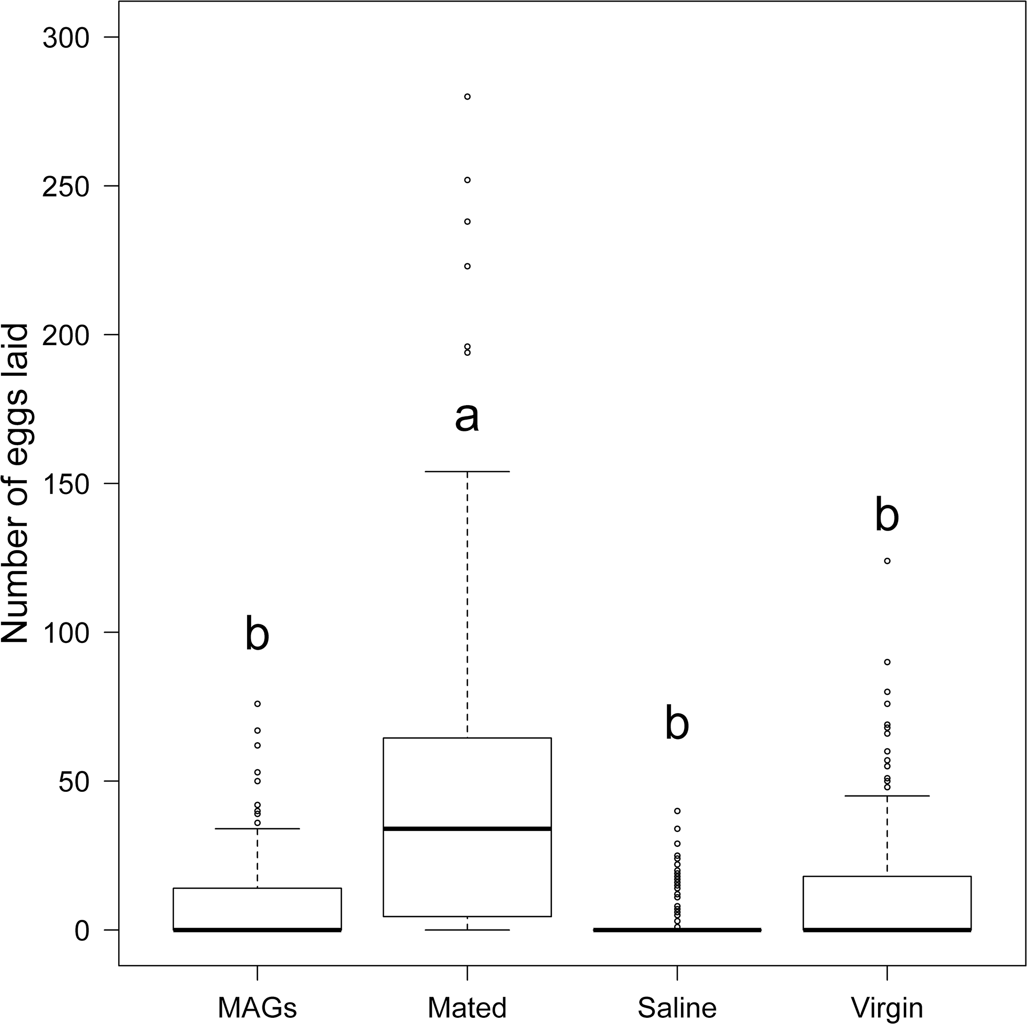
Contrary to the hypothesis that male accessory gland secretions increase egg-laying behaviour, we found no significant increase in the number of eggs laid by secretion-injected females. Mated females laid more eggs over the four days than did females from all the other treatments (Fig. 6). Replicate had a significant effect (χ 2 = 40.0, df = 4, P < 0.001) on the number of eggs deposited.
Number of eggs laid by Anastrepha ludens females after mating with a normal male, injected with male accessory gland secretions (MAGs), injected with saline, or maintained as virgins (no injection, no mating). Eggs were collected for four days. Horizontal lines inside boxes represent the median. Circles outside whiskers represent outliers. Different letters represent significant differences after Tukey’s post hoc test.

Discussion
Mating and receiving an ejaculate can affect the postmating behaviour and physiology of females. Here, we observed that, for A. ludens, mating increased the probability of an egg being laid, how fast oviposition occurred, and the number of eggs laid. However, contrary to our predictions, aqueous injections of male accessory gland secretions increased neither egg development nor oviposition. We found support for the mechanical stimulus hypothesis in similar egg-laying results when comparing females that had received or had not received an ejaculate. Although a biological effect of male accessory gland secretions on female behaviour still needs to be found, these results highlight how the act of mating per se can modify female behaviour.
Mating stimulates oviposition in several species, either through chemosensation or mechanosensation. Reception of male accessory gland secretions and sperm can trigger multiple chemical signals, whereas the filling of sperm storage organs would affect stretch receptors (Cury et al. Reference Cury, Prud’homme and Gompel2019). For example, mechanosensitive neurons in the female reproductive tract can generate stimuli that ready the female for egg laying (Gou et al. Reference Gou, Liu, Guntur, Stern and Yang2014). Tephritids store sperm in a ventral receptacle and in spermathecae, and female contractions and muscle fibres around the ventral receptacle aid in sperm storage (Fritz and Turner Reference Fritz and Turner2002; Mossinson and Yuval Reference Mossinson and Yuval2003; Pérez-Staples et al. Reference Pérez-Staples, Córdova-García and Aluja2014; Thomas et al. Reference Thomas, Leal and Conwat2014). However, the present study supported the mechanical stimulus hypothesis in that mating per se – regardless of whether an ejaculate was received – increased the likelihood that a female would oviposit and the number of eggs laid compared to virgin females. This suggests that the mechanosensation that triggers postmating oviposition responses could be independent of the stretch receptors that would be stimulated when receiving an ejaculate. The aedeagus has three sections: a small basiphallus, a long and sclerotised distiphallus, and at the end of the organ, a complex structure called the glans, which tends to inflate itself as a balloon and induce movements due to the pressure of the liquid inside it (Eberhard Reference Eberhard2005). Even though aedeagus-cut males did not have a glans, some amount of internal courtship by the distiphallus might have taken place, alongside complex external courtship, during mating (Briceño et al. Reference Briceño, Orozco, Luis, Hanson and Hernández2011), thereby stimulating oviposition even if an ejaculate was not received.
Few studies have tested the effect of mating on female postcopulatory behaviour independently of females receiving an ejaculate, and the role of mechanosensation in eliciting oviposition has also been little studied (Cury et al. Reference Cury, Prud’homme and Gompel2019). In female wolf spiders, Schizocosa malitiosa Tullgren (Araneae: Lycosidae), when females were mated with males whose palps were sealed to prevent sperm transfer, no difference in egg sac production or female remating propensity was observed compared to females that had mated with normal males (Aisenberg and Costa Reference Aisenberg and Costa2005). Likewise, in A. ludens and C. capitata, mating without receiving an ejaculate did not inhibit female remating compared to females mating with normal males (Miyatake et al. Reference Miyatake, Chapman and Partridge1999; Abraham et al. Reference Abraham, Lara-Pérez, Rodríguez, Contreras-Navarro, Núñez-Beverido, Ovruski and Pérez-Staples2016). In Gryllus texensis (Orthoptera: Grillidae), females mating with males without testes laid fewer eggs than did females receiving a full ejaculate (Worthington et al. Reference Worthington, Jurenka and Kelly2015). Postcopulatory consequences of mating without an ejaculate have usually been studied in the context of sperm depletion or ablation of testes, yet this approach considers other behaviours that occur during copulation beyond the transfer of sperm. The present study shows that mating without receiving an ejaculate stimulates oviposition and egg production and highlights the importance of the occurrence of internal stimulus mechanisms and mechanosensations during mating, regardless of sperm or male accessory gland secretions. Experiments that manipulate internal and external courtship will help to elucidate the relative importance of these mating behaviours in stimulating oviposition.
The increased oviposition by mated females that did not receive ejaculate in comparison to virgin females has important implications for the control and management of this species. Anastrepha ludens is a pest of quarantine interest that is controlled through the sterile insect technique (Orozco-Dávila et al. Reference Orozco-Dávila, Quintero, Hernández, Solis, Artiaga and Hernández2017). This technique is based on the massive breeding and liberation of irradiated sterile flies in specific areas where sterile males need to mate with wild females, transfer an ejaculate, and induce infertility (Knipling Reference Knipling1955). For most tephritids controlled through this technique, both sterile males and females are released into infected areas. Thus, when sterile females are released, they will also mate and lay eggs in fruit (Orozco et al. Reference Orozco, Hernández, Meza and Quintero2013). In A. ludens, as in other tephritids, sterile males transfer a smaller ejaculate than do wild males and may become sperm depleted after a few matings (e.g., Kuba and Ito Reference Kuba and Ito1993; Radhakrishnan et al. Reference Radhakrishnan, Pérez-Staples, Weldon and Taylor2009; Abraham et al. Reference Abraham, Lara-Pérez, Rodríguez, Contreras-Navarro, Núñez-Beverido, Ovruski and Pérez-Staples2016). Our results suggest that, even if females do not receive ejaculate, the effect of mating per se will increase egg laying compared to virgin females. Although this would need to be verified in sterile females, our results imply that, once released, mated sterile females will increase oviposition relative to virgin females, thereby exacerbating the detrimental effects to fruit quality and its commercial value that are known to occur from sterile female stings (Hendrichs et al. Reference Hendrichs, Franz and Rendon1995). Thus, our results support the use of unisexual strains in sterile insect technique programmes, whereby only males are sterilised and released in affected areas. In the case of A. ludens, a unisexual genetic sexing strain (Tapachula-7) is currently being produced and released (Orozco-Dávila et al. Reference Orozco-Dávila, Adriano-Anaya, Quintero-Fong and Salvador-Figueroa2015).
Here, as with the Caribbean fly, Anastrepha suspensa (Loew) (Diptera: Tephritidae), injections from the accessory glands did not increase the number of eggs laid (Lentz et al. Reference Lentz, Miller, Spencer and Keller2009). However, in Lentz et al. (Reference Lentz, Miller, Spencer and Keller2009), a very high dose of accessory gland secretions was used, and the absence of effects could have been due to the overstimulation of females (Radhakrishnan and Taylor Reference Radhakrishnan and Taylor2007). In contrast, injections of male accessory gland secretions in the moth Spodoptera litura (Fabricius) (Lepidoptera: Noctuidae) triggered oviposition and promoted egg development independently of receiving sperm or of the mechanical stimuli during mating (Yu et al. Reference Yu, Li, Xu, Liu and Ye2014). Similarly, injections of an extract obtained from the combined reproductive tract of males, accessory glands, seminal vesicles, and testes have been shown to induce oviposition in Callosobruchus beetles (Coleoptera: Chrysomelidae) (Yamane and Miyatake Reference Yamane and Miyatake2010). Research into the function of accessory gland–secretion molecules in A. ludens is ongoing, yet these and previous results suggest that specific peptides such as the sex peptide or ovulin may not be found in A. ludens (Abraham et al. Reference Abraham, Núñez-Beverido, Contreras-Navarro and Pérez-Staples2014). Oviposition could be induced by receiving a full ejaculate alongside mechanical stimulation. On the other hand, oviposition-inducing peptides may be found in other areas of the male reproductive system, such as the ejaculatory bulb (Imamura et al. Reference Imamura, Haino-Fukushima, Aigaki and Fuyama1998). Specifically, in Drosophila, the protein PEBme may contribute to egg laying, and its absence results in failed oviposition (i.e., unfertilised eggs; Markow Reference Markow2015). We lack information on the effect of the ejaculatory bulb in stimulating oviposition in A. ludens. Further research on the function of accessory gland secretions in this species is needed to determine, for example, whether they provide defence against pathogens or modulate sperm use by females. Our results suggest there may be varying roles for the ejaculate and for male and female interactions during mating in modifying postcopulatory female physiology and behaviour.
Acknowledgements
The authors thank Solana Abraham, Dinesh Rao, Martha Baena Hurtado, and Luis Esparza for comments made regarding the manuscript and the experiments. The authors also thank Evaristo Calihua Chipahua, Carlos Manuel Bonilla Carmona, Cruz Raúl Perea Castellanos, Nicolás Núñez Beverido, Helena Ajuria Ibarra, and Ricardo Macías Díaz del Castillo for their technical support and suggestions, and they extend thanks to the owners of the sour orange farms in Tuzamapan, Veracruz, Mexico, for their permission to collect material. This study was funded by CONACyT Mexico (Ciencia Básica 179741), awarded to DPS. MRH and GC thank CONACyT for Ph.D. scholarships.