Introduction
Since the report of piglets following the transfer of in vitro-produced (IVP) embryos (Yosida et al., 1993), many trials have been attempted to obtain offspring derived from IVP embryos (Funahashi et al., Reference Funahashi, Kim, Stumpf, Cantley and Day1996; Day et al., Reference Day, Abeydeera, Johnson, Welch, Wang, Cantley and Rieke1998) and somatic cell nuclear transfer (SCNT) embryos (Onishi et al., Reference Onishi, Iwamoto, Akita, Mikawa, Takeda, Awata, Hanada and Perry2000; Polejaeva et al., Reference Polejaeva, Chen, Vaught, Page, Mullins, Ball, Dai, Boone, Walker, Ayares, Colman and Campbell2000). In particular, offspring derived from embryos reconstituted with a variety of somatic cell types as nuclear donors gave rise to conclusive evidence that nuclei from differentiated cells inserted into a recipient enucleated ooplasm have the ability for reprogramming, which is an essential prerequisite and characterizes the dispersion of nucleoli and swelling of the transferred nucleus. Porcine cloning constitutes an integral part of animal models in developing the fields of organ xenotransplantation and bioreactor production for use in industry (Prather, Reference Prather2000). In spite of valuable advantages, porcine embryos produced from IVP system appeared to have a low developmental competence and few cell numbers (Kikuchi et al., Reference Kikuchi, Kashiwazaki, Noguchi, Shimada, Takahashi, Hirabayashi, Shino, Ueda and Kaneko1999; Pomar et al., Reference Pomar, Teerds, Kidson, Colenbrander, Tharasanit, Aguilar and Roelen2005), which might be due to differences in in vitro and in vivo culture environments (Kitagawa et al., Reference Kitagawa, Suzuki, Yoneda and Watanabe2004; Pomar et al., Reference Pomar, Teerds, Kidson, Colenbrander, Tharasanit, Aguilar and Roelen2005). An improved technology to develop IVP embryos has been of major interest to developmental biologists for decades, but the developmental retardation of the embryos has still not been fully elucidated. Developmental retardation in porcine embryos has been demonstrated both in quantitative terms, such as cell number and the rate of trophoblast and inner cell mass (Pomar et al., Reference Pomar, Teerds, Kidson, Colenbrander, Tharasanit, Aguilar and Roelen2005) and in qualitative terms, such as developmental delay and apoptotic incidence (Kidson et al., Reference Kidson, Rubio-Pomar, Van Knegsel, Van Tol, Hazeleger and Ducro-Steverink2004). Thus, the selection of suitable medium, additives, co-culture system and gas atmosphere has been a focus in the successful development of embryos in vitro (Kikuchi et al., Reference Kikuchi, Onishi, Kashiwazaki, Iwamoto, Noguchi, Kaneko, Akita and Nagai2002; Karja et al., Reference Karja, Medvedev, Onishi, Fuchimoto, Iwamoto, Otoi and Nagai2004; Medvedev et al., Reference Medvedev, Onishi, Fuchimoto, Iwamoto and Nagai2004).
In general, important factors to judge quality of embryos were total cell number, the rate of trophoblast and inner cell mass and apoptosis rate (Pomar et al., Reference Pomar, Teerds, Kidson, Colenbrander, Tharasanit, Aguilar and Roelen2005). Use of an in vitro culture system could be an important factor in apoptosis levels in preimplantation embryos of cattle (Gjorret et al., Reference Gjorret, Wengle, Maddox-Hyttel and King2005), horses (Moussa et al., Reference Moussa, Tremoleda, Duchamp, Bruyas, Colenbrander, Bevers and Daels2004), sheep (Rizos et al., Reference Rizos, Ward, Duffy, Boland and Lonergan2002) and pigs (Park et al., Reference Park, Hong, Yong, Hwang, Lim and Lee2005). It is well known that in vivo embryos, compared with in vitro embryos, appeared to have a low apoptosis rate. One of the important reasons for this might be a low oxygen concentration to prevent excessive formation of reactive oxygen species (ROS). The ROS, such as hydrogen peroxide, superoxide anions and hydroxyl ions, could damage cell membranes, DNA, protein and lipid close to their sites of generation and might play a pivotal role in apoptosis. Therefore, embryos from cattle (Pomar et al., Reference Pomar, Teerds, Kidson, Colenbrander, Tharasanit, Aguilar and Roelen2005) and horse (Moussa et al., Reference Moussa, Tremoleda, Duchamp, Bruyas, Colenbrander, Bevers and Daels2004) cultured in a high oxygen concentration appeared to have low development competence and high apoptosis rates. However, in porcine, some researchers have reported that oxygen concentration did not significantly affect the development competence (Kikuchi et al., Reference Kikuchi, Onishi, Kashiwazaki, Iwamoto, Noguchi, Kaneko, Akita and Nagai2002, Park et al., Reference Park, Hong, Yong, Hwang, Lim and Lee2005).
In pigs, SCNT embryos have shown low developmental rates and few cell numbers when compared with IVF embryos (Hao et al., Reference Hao, Lai, Mao, Im, Bonk and Prather2003) and only 1–5% of the resulting SCNT embryos that were transferred into surrogates developed to term (De Sousa et al., Reference De Sousa, Dobrinsky, Zhu, Archibald, Ainslie, Bosma, Bowering, Bracken, Ferrier, Fletcher, Gasparrini, Harkness, Johnston, Ritchie, Ritchie, Travers, Albetini, Dinnyes, King and Wilmut2002). Culture conditions have apparently contributed to this low developmental rate (Prather, Reference Prather2000). In addition, oxygen concentration in the embryo culture system has been considered to affect development and apoptosis (Olson & Seidel, Reference Olson and Seidel2000). In porcine, fragmentation is one of the most important problems reported in embryos derived from IVF, intracytoplasmic sperm injection and SCNT. Cytoplasmic fragmentation resulted in developmental arrest and finally cells underwent apoptosis (Hao et al., Reference Hao, Lai, Mao, Im, Bonk and Prather2003). So far, most researchers have reported that apoptosis rate affected embryo quality, as investigated by independent embryo production methods such as IVF (Mateusen et al., Reference Mateusen, Soom, Maes, Donnay, Duchateau and Lequarre2005, Pomar et al., Reference Pomar, Teerds, Kidson, Colenbrander, Tharasanit, Aguilar and Roelen2005), parthenote (Cui et al., 2004) and SCNT (Hao et al., Reference Hao, Lai, Mao, Im, Bonk and Prather2003), but reports comparing apoptosis rates among embryo production methods have been few.
The present study, therefore, investigated the most important factors in deciding the quality of in vitro embryos produced by different in vitro culture systems, such as oxygen concentration and supplementation time of major energy substrates, such as glucose, pyruvate and lactate and embryo production methods, such as IVF, parthenote and SCNT. To confirm in vitro embryo quality, the total cell number, development and apoptosis rates were investigated.
Materials and methods
Chemicals and media
All chemicals used in this study were purchased from the Sigma Chemical Company and media were from Gibco (Life Technologies) unless otherwise stated. For all media, pH was adjusted to 7.4 and the osmolality to 280 mOsm/kg.
Preparation of porcine fetal fibroblasts
Porcine fetal fibroblasts were isolated from a female fetus (∼3 cm of crown rump length). After being dissected, tissues were then incubated in Ca2+- and Mg2+-free Dulbecco's phosphate-buffered saline (D-PBS) supplemented with 0.25% trypsin–0.04% EDTA solution at 39 °C for 15 min, followed by dispersal in Dulbecco's modified Eagle's medium (DMEM) containing 10% (v/v) fetal calf serum (FCS) by pipetting through a 100 µl micropipette tip to make a single cell suspension. The dispersed cells were washed twice by centrifugation at 300 g for 10 min with DMEM supplemented with 10% FCS. The cells were then incubated in DMEM supplemented with 10% FCS and 1% (v/v) penicillin–streptomycin (100 IU and 125 µg/ml, respectively, Pen–Strep, Gibco) for up to 6–8 days until confluent, at 38.5 °C in a humidified atmosphere of 5% CO2 in air. The cells, at five to seven passages, were grown to confluence and dissociated by trypsinization with 0.25% trypsin–0.04% EDTA solution immediately before SCNT.
Preparation of oocytes
Oocytes were obtained from abattoir ovaries and subjected to maturation based on the protocol of Fan & Sun (Reference Fan and Sun2004) with minor modifications. Briefly, cumulus–oocyte complexes (COCs) were aspirated from antral follicles of 3–6 mm in diameter with an 18G needle fitted to a 10 ml syringe. Sets of 50 COCs were matured in 500 µl TCM199 with Earle's salts supplemented with 10 ng/ml EGF, 0.5 µg/ml FSH, 0.5 µg/mL LH, 0.57 mM cysteine and 0.91 mM Na pyruvate in each well of a 4-well multidish (Nunc) for 24 h and further cultured in the same medium without FSH and LH for 20 h at 38.5 °C, 5% CO2 in air. At 38 h and 44 h of culture, COCs were taken off their cumulus cells by vortexing in D-PBS containing 0.1% (w/v) hyaluronidase for 1 min. Oocytes at 38 h of culture were used for SCNT, whereas oocytes at 44 h were divided into two groups, for IVF and activation procedures. Oocytes that possessed a polar body (PB) and even cytoplasm as seen under the microscope (×200) were used for SCNT and activation.
Production of embryos
For the production of SCNT embryos, the oocytes were transferred into micromanipulation drops for the SCNT process. Medium used for micromanipulation was HEPES-buffered TCM199 supplemented with 10% FBS and 7.5 µg/ml cytochalasin B (CCB) (Funahashi et al., Reference Funahashi, Kim, Stumpf, Cantley and Day1996). The first polar body and metaphase II plate with a small volume of surrounding cytoplasm were removed by aspiration with a 20 µm internal diameter pipette. The enucleated oocytes were labelled with 0.5 µg/ml bisbenzimide (Hoechst-33342) in HEPES-TALP medium for 2 min at room temperature and ensuring that the nucleus was completely removed. After being trypsinized, a single intact cell was transferred by micropipette into the perivitelline space of each enucleated oocyte. For fusion and activation of the cytoplast and donor cell, the egg was oriented exactly in a BTX Electro chamber (BTX, Inc.) filled with 0.28 M mannitol solution containing 0.01% (w/v) BSA, 0.05 µM CaCl2 and 0.01 µM MgSO4 (Fan & Sun, Reference Fan and Sun2004), with the help of holding and injection pipettes under the micromanipulator and pulsed twice with 2.0 kV/cm DC for 30 µs using a BTX Electro-Cell Manipulator 200. After 1 h, fusion of cytoplasm and donor cell was examined under microscopy and only oocytes that did not exhibit donor cells were transferred into 5.5 mM glucose-free NCSU-23 medium containing 7.5 µg/ml CCB and were cultured for 3 h at 38.5 °C in a humidified atmosphere of 5% CO2 and 5% O2 in air (day 0).
For production of parthenote embryos, cumulus-free oocytes were transferred into a BTX Electro chamber filled with 0.28 M mannitol solution containing 0.01% (w/v) BSA, 0.05 µM CaCl2 and 0.01 µM MgSO4 and pulsed twice with 2.0 kV/cm DC for 30 µs using a BTX Electro-Cell Manipulator 200 and were cultured in 5.5 mM glucose-free NCSU-23 medium containing 7.5 µg/ml CCB for 3 h at 38.5 °C in a humidified atmosphere of 5% CO2 and 5% O2 in air or 5% CO2 and 20% O2 in air (day 0).
As IVF controls, sets of 20 cumulus-free oocytes were transferred into 50 µl drops of modified Tris-buffered medium (mTBM) consisting of 113.1 mM NaCl, 3.0 mM KCl, 7.5 mM CaCl2·2H2O, 20.0 mM Tris crystallized free base, 11.0 mM glucose and 5.0 mM sodium pyruvate, supplemented with 2 mM caffeine and 0.03 g BSA (fatty acid-free, fraction V). Oocytes were inseminated with frozen-thawed sperm prepared by Percoll (Pharmacia, Uppsala, Sweden) density gradient (45 vs. 90%) as described by Matas et al. (Reference Matas, Coy, Romar, Marco, Gadea and Ruiz2003). The final sperm concentration was adjusted to 1 × 10⁵ sperm/ml. Co-incubation of oocytes with sperm was carried out for 5 h at 38.5 °C in a humidified atmosphere of 5% CO2 in air.
Culture of embryos
Culture of all eggs was performed by modified method of Kikuchi et al. (Reference Kikuchi, Onishi, Kashiwazaki, Iwamoto, Noguchi, Kaneko, Akita and Nagai2002). Eggs were cultured in sets of 20 in 50 µl drops of 5.55 mM d-glucose-free NCSU-23 medium (NCSU−) supplemented with 0.17 mM Na pyruvate, 2.73 mM Na lactate and 0.4% BSA for 58 h and subsequently cultured in NCSU-23 with 5.55 mM glucose (NCSU+) until day 6 (NUSU −/+), or were cultured NCSU-23 with 5.55 mM d-glucose until day 6 (NCSU +/+) at 38.5 °C, 20% CO2, 5% O2 and 90% N2 or 5% CO2, 5% O2 in air. The rates of cleavage and blastocyst formation were assessed on days 2 and 6, respectively.
Cytological evaluations
To count total cell numbers, day 6 blastocysts fixed in 3.7% formaldehyde for overnight were stained with 40 µg/ml propidium iodide (PI) for 10 min. After being mounted onto a precleaned microscope slide, the nuclei of the embryos were counted under a Nikon fluorescence microscope.
Analysis of apoptosis rate was performed using terminal deoxynucleotidyl transferase-mediated dUTP nick-end labelling (TUNEL) assay as described previously by Hao et al. (Reference Hao, Lai, Mao, Im, Bonk and Prather2004) with minor modifications. Briefly, blastocysts were fixed in 3.7% formaldehyde for 4 h at room temperature and permeabilized by incubation in 0.5% Triton X-100 for 1 h. The embryos were then incubated with fluorescein-conjugated dUTP and terminal deoxynucleotidyl transferase enzyme (Roche) for 1 h at 37 °C in the dark, treated with 50 µg/ml RNase at room temperature for 1 h and counterstained with 40 µg/ml PI for 1 h at 37 °C. All samples were examined under a Nikon fluorescence microscope and those that stained red were considered to be nuclei and those stained green and yellow were apoptotic bodies. Total apoptotic indices were calculated for each embryo as follows: apoptotic index = (number of TUNEL-positive nuclei/total number of nuclei blastocyst) × 100. Apoptosis-generating rate was calculated as follows: the number of apoptosis generating blastocyst/total blastocyst number.
Experimental design
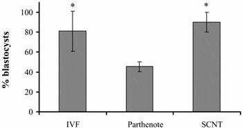
This study comprised of five experiments for evaluating the developmental rate, quality and apoptosis of porcine embryos under different culture systems and embryo production methods. Embryos were assigned to four treatment groups: Group 1, embryos were cultured in NCSU+ until day 6 (NCSU +/+) on 20% O2, or 5% O2 (Group 2). Group 3, embryos were cultured in NCSU– with pyruvate/lactate for 58 h and subsequently cultured in NCSU+ until day 6 (NCSU –/+) on 20% O2, or 5% O2 (Group 4). In Experiment 1, the rates of cleavage and blastocyst development of IVF embryos produced by four treatment groups were compared (Table 1). In total, 1123 oocytes were used in five replicates. In Experiment 2, 135 day 6 IVF blastocysts produced by groups 3 and 4 were assessed for their total cell number and apoptosis index (Table 2). In Experiment 3, the rates of cleavage and blastocyst development of parthenotes and SCNT embryos were compared (Table 3). In total, 443 oocytes were used in five replicates. In Experiment 4, 30 parthenotes and 54 SCNT blastocysts produced on day 6 were assessed for their total cell number and apoptosis index (Table 4). In Experiment 5, apoptosis-generating rate from IVF, parthenote and SCNT blastocysts produced by treatment group 4 were compared (Fig. 1).
The rate of apoptosis generated from total blastocysts that were produced at 5% O2 concentration. *p < 0.05. Bars represent SEM. IVF, in vitro fertilization; SCNT, somatic cell nuclear transfer.

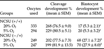
Development of porcine embryos in different O2 concentrations and media

‘+’ means with 5.5 mM d-glucose and ‘–’ means without 5.5 mM d-glucose.
NCSU (+/+) refers to culture in NCSU-23 with 5.55 mM d-glucose until day 6 and NCSU (–/+) refers to culture in NCSU-23 with pyruvate/lactate for 58 h and subsequently cultured in NCSU-23 with 5.55 mM d-glucose until day 6.
Data represent the mean of five replicates.
a,b Within a column, percentages with different superscripts differ significantly, p < 0.05.
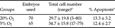
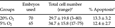
Apoptosis of IVF blastocysts cultured in NCSU (−/+) under different O2 concentrations on day 6
‘+’ means with 5.5 mM d-glucose and ‘−’ means without d-glucose.
NCSU (+) refers to culture in NCSU-23 with 5.55 mM d-glucose until day 6 and NCSU (−/+) refers to culture in NCSU-23 with pyruvate/lactate for 58 h and subsequently cultured in NCSU-23 with 5.55 mM d-glucose until day 6.
a Mean ± SEM.
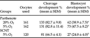
Development of parthenote and SCNT embryos on day 6

Data are derived from five replicates.
a,b Within a column, percentages with different superscripts differ significantly, p < 0.05.
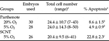
Apoptosis of parthenotes and SCNT blastocysts on day 6

a Mean ± SEM.
b,c Within a column, percentages with different superscripts differ significantly, p < 0.05.
Statistical analysis
Differences among groups were analysed using one-way analysis of variance (ANOVA) by SPSS after arc–sine transformation of proportional data. Data were expressed as mean ± SEM. Comparisons of mean values among treatments were performed using Duncan's & Tukey's multiple comparisons test. Differences were considered to be significant when p < 0.05.
Results
Effect of glucose and O2 concentration in culture medium
Development rates of porcine embryos by glucose and O2 concentration in culture medium were compared in Table 1. Cleavage rates were not significantly different between 20% and 5% O2 of NCSU (+/+) and 20% and 5% O2 of NCSU (−/+) (76.5 ± 9.0, 80.5 ± 5.1 vs. 77.5 ± 7.3, 81.9 ± 13.5). Although cleavage rates between groups at the appropriate time of glucose supplementation were not significantly different, use of a low O2 concentration showed a slightly higher cleavage rate than for high O2 concentration. Blastocyst rates were significantly different (p < 0.05) between NCSU (+/+) and NCSU (−/+) (5.3 ± 2.1, 5.3 ± 3.2 vs. 27.1 ± 7.2, 27.9 ± 8.0), but were not significantly different in O2 concentrations among groups. The results suggested that porcine IVF embryos required an appropriate time of glucose supplementation into culture medium. Hence, for further studies the NCSU (−/+) in vitro culture system was used.
Total cell numbers and apoptosis of IVF blastocysts cultured in NCSU (−/+)
Apoptosis rates and total cell numbers of blastocysts cultured under different O2 concentrations were estimated (Table 2). Although no significant difference in O2 concentrations was observed, the low O2 concentrations group showed slightly higher total cell numbers with a lower apoptosis index when compared with the high O2 concentration group (34.7 ± 15.8 vs. 29.7 ± 19.8 and 12.4 ± 2.7 vs. 13.3 ± 3.2). In addition, the range of total cell number showed low variation in the low O2 concentration group when compared with the high O2 concentration group (17–75 vs. 3–80).
Development of parthenotes and SCNT embryos produced by different O2 concentration
Table 3 shows the development of parthenotes and SCNT embryos produced by different O2 concentrations. In parthenotes, the rates of cleavage and blastocyst development showed no significant difference between high O2 concentration (82.7 ± 9.8% and 38.9 ± 7.5%) and low O2 concentration (82.8 ± 11.4% and 47.5 ± 8.2%), respectively. SCNT embryos when compared with parthenotes showed significantly (p < 0.05) lower cleavage (66.5 ± 4.1% vs. 82.8 ± 11.4%) and blastocyst (24.0 ± 4.0% vs. 47.5 ± 8.2%) rates.
Apoptosis of parthenotes and SCNT blastocysts produced by different O2 concentration
The results of total cell number and apoptosis index of parthenotes and SCNT blastocysts produced by different O2 concentration are presented in Table 4. Although total cell numbers (24.4 ± 10.7 vs. 24 ± 14.3) of parthenotes produced by high and low O2 concentrations did not differ, apoptosis index (4.9 ± 0.9%) at the low O2 concentration was significantly (p < 0.05) lower than that at high O2 concentration (8.6 ± 1.5%). In addition, the range of total cell number between parthenote groups had a similar trend (7–43 vs. 8–50). SCNT blastocysts when compared with parthenotes showed low total cell numbers (20.4 ± 9.5) and range (6–41) and a significantly high apoptosis index (22.8 ± 2.3%).
Apoptosis generated from total blastocysts produced in low O2 concentration
Figure 1 shows apoptosis-generating rate of embryos produced in low O2 concentrations by different methods. Although no significant difference (81.1 ± 20.1 vs. 90.0 ± 10.0) between IVF and SCNT groups was observed for apoptosis-generating rate, this was slightly high in the SCNT group. The apoptosis rate in the parthenogenote group (45.5 ± 5.0) was significantly lower (p < 0.05) when compared with IVF (81.1 ± 20.1) and SCNT (90.0 ± 10.0) groups, respectively.
Discussion
In general in vitro-cultured embryos compared with in vivo-cultured embryos appeared to have a low development rate, smaller total cell number and high apoptosis rate in many animals (Booth et al., Reference Booth, Holm and Callesen2005; Gjorret et al., Reference Gjorret, Wengle, Maddox-Hyttel and King2005; Pomar et al., Reference Pomar, Teerds, Kidson, Colenbrander, Tharasanit, Aguilar and Roelen2005). In addition, offspring rate after transfer of porcine embryos cultured in vitro have been reported to be low (Kikuchi et al., Reference Kikuchi, Onishi, Kashiwazaki, Iwamoto, Noguchi, Kaneko, Akita and Nagai2002). One of the causes could be an incomplete in vitro-culture system and many researchers have used a standard such as total cell number and apoptosis rate to evaluate suitable culture systems (Kikuchi et al., Reference Kikuchi, Onishi, Kashiwazaki, Iwamoto, Noguchi, Kaneko, Akita and Nagai2002; Pomar et al., Reference Pomar, Teerds, Kidson, Colenbrander, Tharasanit, Aguilar and Roelen2005).
The present study assessed the quality of embryos cultured by different in vitro-culture systems, using parameters such as oxygen concentration and appropriate time of major energy substrate supplementation (glucose, pyruvate and lactate) during IVC and differing embryo production methods such as IVF, parthenogenetic activation and SCNT procedures.
Glucose was known as an important energy source in late preimplantation embryos, whereas pyruvate and lactate are important during early stages in mice (Gardner & Leese Reference Gardner and Leese1988) and cattle (Kim et al., Reference Kim, Niwa, Lim and Okuda1993). Contrary to the observations, glucose-added culture medium inhibited the development of full preimplantation hamster embryos (Seshagiri & Bavister Reference Seshagiri and Bavister1989). Therefore, glucose for preimplantation embryos depended on the embryo stage, had varied effect on the embryonic development and was species specific (Barnett and Bavister, Reference Barnett and Bavister1996; Swian et al., 2002; Karja et al., Reference Karja, Medvedev, Onishi, Fuchimoto, Iwamoto, Otoi and Nagai2004; Medvedev et al., Reference Medvedev, Onishi, Fuchimoto, Iwamoto and Nagai2004). Kikuchi et al. (Reference Kikuchi, Onishi, Kashiwazaki, Iwamoto, Noguchi, Kaneko, Akita and Nagai2002) and Karja et al. (Reference Karja, Medvedev, Onishi, Fuchimoto, Iwamoto, Otoi and Nagai2004) reported that late-stage embryos required glucose as a major energy substrate and early-stage embryos required pyruvate/lactate and their culture system could accomplish high development and cell numbers in porcine IVP embryos. The present IVF experiment result also agrees that groups cultured in pyruvate/lactate for 58 h with additional supplementation of glucose until day 6 had a significantly high blastocyst rate when compared with groups cultured in glucose until day 6.
Many studies have reported that the culture of embryos in low oxygen concentrations, similar to the in vivo environment, could improve development in cattle (Pomar et al., Reference Pomar, Teerds, Kidson, Colenbrander, Tharasanit, Aguilar and Roelen2005); but the results in IVP porcine embryos have been inconsistent (Kitagawa et al., Reference Kitagawa, Suzuki, Yoneda and Watanabe2004; Booth et al., Reference Booth, Holm and Callesen2005; Park et al., Reference Park, Hong, Yong, Hwang, Lim and Lee2005). Although variable results in embryo development were observed, total cell number of blastocysts improved when using low oxygen concentration during in vitro culture (Kikuchi et al., Reference Kikuchi, Onishi, Kashiwazaki, Iwamoto, Noguchi, Kaneko, Akita and Nagai2002; Booth et al., Reference Booth, Holm and Callesen2005; Park et al., Reference Park, Hong, Yong, Hwang, Lim and Lee2005). Similar to the observations, the present study demonstrated that embryos cultured at a low oxygen concentration and compared with a high, did not differ in cleavage and blastocyst rate but had slightly higher total cell numbers with a lower apoptosis rate. Recently porcine have been highlighted as being advantageous for methods such as xenotransplantation and for animal models for human diseases, etc., thereby satisfy several requirements for the production of SCNT embryos (Prather et al., 2000). However, porcine blastocyst rates by SCNT have shown poor success and the cause might be an unsuitable culture environment (Lee et al., Reference Park, Lai, Cheong, Im, Sun, Wu, Day and Prather2003b) and activation method (Park et al., Reference Lee, Kim, Hyun, Kim, Lee, Nam, Jeong, Kim, Kang, Lee and Hwang2001; De Sousa, 2002). So, the present study estimated developmental capacity under modified culture systems and parthenote and SCNT embryos on the basis of IVF results and accomplished higher blastocyst rates (39–48%, 24%). Lee et al., (Reference Lee, Hyun, Kim, Kim, Lee, Lim, Lee, Kang, Lee and Hwang2003a, Reference Lee, Kim, Hyun, Kim, Lee, Nam, Jeong, Kim, Kang, Lee and Hwangb) reported that embryos fused and activated with a single DC pulse 2.0 kV/cm for 50 µs after SCNT and using fetal fibroblasts gave about 19% blastocyst rate on day 7. When treated additionally with CCB for 2 h after fusion and activation under the same electric treatment gave about 15% blastocyst rate on day 6. Cheong et al., (Reference Cheong, Park, Im, Lai, Sun, Day and Prather2002) reported that porcine embryos with same fusion time and activation with two DC pulses (1.25 kV/cm for 30 µsec) gave a 16.4% blastocyst rate on day 6, whereas independent fusion and activation gave about 20%. Above reports used NCSU-23 base culture medium, with low development rate when compared with the present study. In contrast, Lee et al., (Reference Lee, Kim, Hyun, Kim, Lee, Nam, Jeong, Kim, Kang, Lee and Hwang2003b) reported that embryos cultured in lactate/pyruvate-supplemented NCSU-23 accomplished better results when compared with glucose supplemented until day 6. Contrasting results might be due to culture systems deficient in a major energy substrate that are dependent on the cell stage, from 8-cell to 16-cell. Swain et al., (Reference Swain, Bormann, Clark, Walters, Wheeler and Krisher2002) reported significantly increased glycolytic activity after the 8-cell stage in an in vitro culture system. Although pyruvate had minimal effect throughout porcine embryos of all stages, this was not found with NCSU-23 medium and had antioxidant protection against external stress. The present results were in agreement with other reports (Kikuchi et al., Reference Kikuchi, Onishi, Kashiwazaki, Iwamoto, Noguchi, Kaneko, Akita and Nagai2002; Swain et al., Reference Swain, Bormann, Clark, Walters, Wheeler and Krisher2002; Karja et al., Reference Karja, Medvedev, Onishi, Fuchimoto, Iwamoto, Otoi and Nagai2004). In addition, the developmental result for parthenotes also agreed and this showed a high blastocyst rate (Cheong et al., Reference Cheong, Park, Im, Lai, Sun, Day and Prather2002; Lee et al., Reference Lee, Kim, Hyun, Kim, Lee, Nam, Jeong, Kim, Kang, Lee and Hwang2003b; Hao et al., Reference Hao, Lai, Mao, Im, Bonk and Prather2004; Im et al., Reference Im, Yang, Lai, Liu, Hao and Prather2005).
The results of the present study on development and quality of embryos between parthenote groups were not significantly affected by oxygen concentration, but low oxygen concentration when compared with high could increase development ability (Booth et al., Reference Booth, Holm and Callesen2005) in porcine. In the present study the modified culture system used might account for the discrepancy, because of the absence of significant difference prior to IVF.
High apoptosis rate resulted in embryonic loss and reduced embryo quality especially in SCNT embryos (Gjorret et al., Reference Gjorret, Wengle, King, Schellander and Hyttel2002; Hao et al., Reference Hao, Lai, Mao, Im, Bonk and Prather2003), because of inappropriate reprogramming in the cell cycle of the donor nucleus and ooplasm through reconstruction into enucleated oocytes (Otoi et al., 2002). The present study observed similar findings, with high apoptosis rate in SCNT embryos when compared with parthenotes cultured under the same activation and culture system. This result clearly indicated that apoptosis rate was influenced by the embryo-producing method. Hao et al. (2003, 2004) reported that the apoptosis rate on day 6 was approximately 2.5 times that in morphologically normal SCNT blastocyst compared with IVF and was approximately 1.9 times that in parthenote compared with their IVF counterpart. Although there was no direct comparison, the present study results showed that apoptosis rate in SCNT embryos was approximately 1.9 times when compared with that of IVF, whereas parthenote embryos, compared with IVF, was approximately 0.4 times and NT embryos, compared with parthenote, was approximately 4.6 times. Disagreement in the results of parthenotes compared with IVF embryos might be attributed to the incomplete culture system and embryo-producing methods. This fact was evidenced in the apoptosis-generating rates (the total amount of the blastocysts generated apoptosis/the total amount of the blastocysts) of IVF, SCNT and parthenote embryos on day 6 cultured under the same culture treatments (Fig. 1). In the present study, the rising tendency of apoptosis-generating rate in IVF, parthenote and SCNT embryos differed among embryo-producing methods. In addition, rate was affected by environment, such as the medium and culture method. The percentage of porcine embryos with DNA-fragmented nuclei in IVF and SCNT blastocyst was more than 90%, as reported earlier in bovine (Fahrudin et al., Reference Fahrudin, Otoi, Karja, Mori, Murakami and Suzuki2002).
Based on the above results, in vitro development of porcine embryos was not affected by oxygen concentration, but was affected by major energy source and embryo-producing methods, suggesting that porcine embryos had species specificity. Even though, the concentration of major energy source and the timing of its inclusion in culture could accomplish relatively high embryo development, the apoptosis rate indicates that more work still needs to be done in developing better defined culture conditions that could support NT embryos with equivalent success to in vivo preimplantation porcine embryos.
Acknowledgements
The financial support of BioGreen 21 (grant no. 1000520040020000) and KOSEF (grant no. R05–2004–000–10702–0) in Korea are gratefully acknowledged.