INTRODUCTION
Edible snails are a highly prized food in many European countries, but mainly in France where consumption reaches 40 000 tonnes/year (Daguzan, Reference Daguzan and Henderson1989; Kiebre-Toe et al. Reference Kiebre-Toe, Borges, Maurin, Richard and Kodjo2003). In Spain, current consumption is estimated to be about 16 000 tonnes/year showing a clear rising trend (Ministerio de Medio Ambiente y Medio Rural y Marino, 2008). However, similar to France and other European countries, most of the snails consumed are collected from the wild and only about 3% of the total consumption comes from heliciculture, which is an emerging zootechnic activity. Two rearing systems are mainly used in Spanish farms: the extensive system, in which snails are reared in outdoor enclosures, and the mixed system, which combines indoor breeding and outdoor fattening (Arrébola and Álvarez, Reference Arrébola Burgos and Álvarez Halcón2001). As in other European countries (Lazaridou-Dimitriadou et al. Reference Lazaridou-Dimitriadou, Alpoyanni, Baka, Brouziotis, Kifonidis, Milhaloudi, Sioula and Vellis1998; Dupont-Nivet et al. Reference Dupont-Nivet, Mallard, Bonnet and Blanc2000), the main species produced in Spain is Helix aspersa. This species includes the autochthonous subspecies Helix aspersa aspersa (petit-gris), more appreciated by Spanish consumers, and Helix aspersa maxima (gros-gris), a subspecies originating from Algeria, which exhibits a higher genetic potential for growth and fertility in indoor and outdoor systems (Chevallier, Reference Chevallier1990). Production of autochthonous snails, however, is especially problematic since severe mortalities are often observed during intensive reproduction in the mixed rearing system. Although the causes of mortality have not yet been precisely identified, preliminary studies conducted in our laboratory demonstrated that certain parasite species, including a renal tetrahymenid ciliate, were more abundant in reproducers from intensive indoor conditions than in those maintained outdoors under extensive conditions (unpublished observations).
Numerous parasite species have been detected in wild and farmed H. aspersa (Morand, Reference Morand1985, Reference Morand1986; Morand and Daguzan, Reference Morand and Daguzan1986; Morand and Petter, Reference Morand and Petter1986; Cabaret et al. Reference Cabaret, Morand, Aubert and Yvore1988). In particular, nematodes belonging to the genera Alloionema, Rhabditis, Angiostoma, and Nemhelix, trematodes, and the mite Riccardoella limacum are considered potential pathogens for farmed snails (Cooper and Knowler, Reference Cooper and Knowler1991). Ciliates, by contrast, have been reported only once in snails from a French farm, but no information on the species identity and its microhabitat was provided by Cabaret et al. (Reference Cabaret, Morand, Aubert and Yvore1988). Since Tetrahymena rostrata has been found to date in the renal organ of 7 species of slugs and 7 species of non-helicid snails (Van As and Basson, Reference Van As, Basson and Barker2004), and it is considered highly pathogenic and even lethal to slugs (Brooks, Reference Brooks1968; Barker, Reference Barker1993; Wilson et al. Reference Wilson, Coyne and Glen1998), we have conducted morphological and molecular studies to determine whether the renal ciliates infecting both H. aspersa subspecies in Galician farms belong to this pathogenic species. In addition, we have also characterized the ciliates parasitizing the kidney of the slug Deroceras reticulatum, which is usually found invading the greenhouses and outside enclosures where first growing and fattening of snails are accomplished.
MATERIALS AND METHODS
Collection and dissection of gastropods
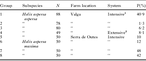
A total of 345 Helix aspersa aspersa and 150 Helix aspersa maxima adults belonging to 8 different groups were collected between 2004 and 2007 from 2 mixed rearing system-based heliciculture farms located in Galicia (NW Spain) and examined for the presence of renal ciliates (Table 1). One farm was located in Valga (42°42′N, 8°38′W) and the other one in Serra de Outes (42°54′N, 8°51′W). According to the persons in charge of both farms, snails belonging to each group had different origins: those of group 4 were collected from an outdoor enclosure where they were reared from juveniles produced in the same farm; the remaining snails, all collected from buildings where reproduction is carried out at high densities and under controlled environmental conditions, were originally obtained from other minor farmers or collectors. All snails from reproduction buildings were collected at the beginning of the last month of the reproduction phase (a 4-month reproduction period is used in both farms).
Main characteristics of the 8 groups of snails examined and prevalence (P %) of ciliate infection for each group

1 Snails were maintained at high densities and under controlled environmental conditions in reproduction buildings.
2 Adults were reared from juveniles in an outdoor enclosure.
In addition to snails, 47 Deroceras reticulatum collected from greenhouses used for first growing and fattening of both Helix aspersa subspecies were also examined (25 slugs were collected from Valga and 22 from Serra de Outes).
Each gastropod was anaesthetized by injection of a sterile 50 mm MgCl2 solution. The kidney was resected under aseptic conditions, transferred into a Petri dish, and bisected. A touch preparation (imprint) was prepared by touching one cut surface of the bisected organ to a glass slide. The preparation was immediately covered with a 22×22 mm cover-slip and examined as a wet mount under the light microscope. The total number of ciliates under the cover-slipped area was used as a measure of parasite density according to Bush et al. (Reference Bush, Lafferty, Lotz and Shostak1997). If ciliates were present, the renal tissue was used for culturing and/or staining of parasitic trophonts. In the case of snails, the surface of the pulmonary plexus facing the mantle cavity was also scraped with a scalpel blade onto a glass slide. The preparation thus obtained was examined as a wet mount for the presence of ciliates.
Axenic culturing of ciliates
The infected renal tissues from the gastropod hosts were thoroughly washed in autoclaved distilled water and then introduced into 25 cm2 tissue-culture flasks with filter-vented caps (IWAKI Brand, Asahi Techno Glass, Chiba, Japan) containing 10 ml of ATCC 357 medium (American Type Culture Collection, Manassas, VA) supplemented with 1% (v/v) of an antibiotic-antimycotic suspension (10 000 units/ml penicillin G, 10 mg/ml streptomycin sulfate, and 25 μg/ml amphotericin B (Sigma-Aldrich Chemie, GmbH, Steinheim, Germany)). Primary cultures were incubated at 18°C and controlled daily for bacterial or fungal growth. Axenic cultures were then maintained by subculturing to 100 ml of fresh antibiotic-free ATCC 357 medium every 14 days using 250 ml Erlenmeyer flasks.
Three cultures of the Tetrahymena species were established using 1 isolate from each host species: TRO1 was collected from a Helix aspersa aspersa from Valga (isolation date: 14 June 2005); TRO2 was collected from a Helix aspersa maxima from Serra de Outes (isolation date: 14 September 2007); and TRO3 was collected from a Deroceras reticulatum from Serra de Outes (isolation date: 7 September 2007). Although these strains are not clonal, there was no indication of nucleotide polymorphisms within each strain in either the mitochondrial cytochrome c oxidase 1 (cox1) or the nuclear small subunit (SSU) rRNA genes (data not shown).
Morphological and morphometrical studies
Ciliates freshly liberated from the renal organ of gastropods and from axenic cultures were studied in vivo using a 100× oil immersion objective and bright-field and phase-contrast microscopy. The ciliates were also silver impregnated by the Chatton-Lwoff technique (Foissner, Reference Foissner1991). The nuclear apparatus of trophonts and encysted ciliates was stained by the Feulgen reaction (Pearse, Reference Pearse1968) and observed in an Olympus BX41 microscope equipped with an Olympus U-RFL-T ultraviolet light power supply unit and a green excitation U-MWG2 mirror unit.
Measurements were taken from silver- and Feulgen-stained ciliates using a calibrated ocular micrometer. All determinations reported are based on the observation of 50 cells. Photographs were taken using an Olympus DP11 digital camera.
For scanning electron microscopy, ciliates from axenic cultures were collected by centrifugation at 500 g for 5 min and fixed with 2·5% (v/v) glutaraldehyde in 0·1 m cacodylate buffer, pH 7·2 for 30 min at 4°C. After 3 washes in the same buffer, ciliates were post-fixed in 1% (w/v) osmium tetroxide in cacodylate buffer for 15 min at 4°C, washed thoroughly, and adhered to poly-L-lysine-coated cover-slips for 2 h. Cells were chemically dried with hexamethyldisilazane (Sigma-Aldrich Chemie, GmbH, Steinheim, Germany). Cover-slips were then mounted on metal stubs, gold-coated with a sputter coater, and observed and photographed with a Philips XL30 scanning electron microscope.
Histological studies
Ten H. aspersa aspersa snails with the same origin as those included in group 1 were dissected and their renal organs fixed for 24–48 h in 10% (v/v) buffered formalin and embedded in paraffin following standard histological procedures. Sections (4–5 μm) were obtained using a microtome Microm HM-340E (Microm, Walldorf, Germany) and stained with haematoxylin-eosin (H&E).
Production of encysting organisms
Encystment was induced in every isolate by using sterilized low mineral content water (Mondariz™) as starvation medium. Trophonts from axenic cultures at early stationary phase were washed 3 times with mineral water by centrifugation at 500 g for 5 min. Ciliate density was adjusted to 2×10⁴/ml and 1 ml of this suspension was distributed to each well of a non-treated 24-well culture plate (IWAKI Brand, Asahi Techno Glass, Chiba, Japan). Ciliates were incubated at 18°C in the dark, and monitored at days 1, 2, 3, 4, 7, and 9 using an inverted light microscope. After microscopic observation, the ciliates in each well were fixed with formalin (final concentration 10% (v/v)) and the encysted and non-encysted cells were counted in a haemocytometer. Four replicate wells were counted for each time-point. The percentage of encystment for each replicate was determined by dividing the number of encysted ciliates by the total number of ciliates (encysted and non-encysted) multiplied by 100.
To study the process of autogamy, encysted ciliates were also stained with Feulgen stain at different days following starvation.
Molecular genetic analyses
Ciliates were collected from axenic cultures by centrifugation (500 g, 5 min) and fixed in 70% ethanol. DNA was extracted from ethanol-fixed cells using the MasterPure™ DNA Purification Kit (Epicentre, Madison, WI, USA). Two μl of extracted DNA were added to tubes containing puReTaq Ready-To-Go PCR beads (Amersham Biosciences, Baie d'Urfé, PQ, Canada) along with 0·5 μl of 25 mm MgCl2, 2·5 μl each of 10 μm forward and reverse primers, and 17·5 μl of dH2O to bring the total reaction volume to 25 μl. The PCR reaction was performed in a PerkinElmer GeneAmp 2400 thermal cycler (PE Applied Biosystems, Mississauga, ON, Canada). Forward primer CO1-FW (5′-ATGTGAGTTGATTTTATAGAGCAGA-3′; Chantangsi et al. Reference Chantangsi, Lynn, Brandl, Cole, Hetrick and Ikonomi2007) and reverse primer CO1-2003 (5′-CCTGGAATACCATTCATTTTAGC-3′; C. Chantangsi, personal communication) were used to amplify the cox1 gene. Forward primer A (5′-CAACCTGGTTGATCCTGCCAGT-3′; Medlin et al. Reference Medlin, Elwood, Stickel and Sogin1988) and reverse primers C (5′-TTGGTCCGTGTTTCAAGACG-3′; Jerome and Lynn, Reference Jerome and Lynn1996) or LSU-R (5′-CCTCACGGTACTTGTTTGCT-3′; Strüder-Kypke, personal communication) were used to amplify the SSUrRNA gene. The PCR products were purified using the QIAGEN MinElute Gel Extraction kit (QIAGEN, Mississauga, ON, Canada) and sequencing was performed in both directions with a 3730 DNA Analyzer using the BigDye Terminator Version 3.1 Cycle Sequencing kit (Applied Biosystems Inc., Foster City, CA, USA). Amplification primers were used in the sequencing reaction along with one additional forward and reverse internal SSUrRNA primer (Elwood et al. Reference Elwood, Olsen and Sogin1985).
Electropherograms for the DNA sequences were trimmed, assembled, and edited using the program Sequencher™ version 3.1 (Gene Codes Corporation, Ann Arbor, MI, USA). The sequences were added to our existing cox1 and SSUrRNA alignments containing sequences published by Chantangsi et al. (Reference Chantangsi, Lynn, Brandl, Cole, Hetrick and Ikonomi2007). These alignments were created in MEGA version 4.0 (Molecular Evolutionary Genetics Analysis; Kumar et al. Reference Kumar, Tamura and Nei2004) and were aligned by eye. Genetic distances for both cox1 and SSUrRNA sequence datasets were calculated using the Kimura 2-parameter (K2P) model, implemented in the DNADIST program in PHYLIP version 3.65 (Kimura, Reference Kimura1980; Felsenstein, Reference Felsenstein2005). The neighbour joining (NJ) algorithm was then employed to construct a phylogenetic tree based on these distances with the NEIGHBOR program in the PHYLIP package (Saitou and Nei, Reference Saitou and Nei1987). SEQBOOT was used to resample 1000 datasets out of the original dataset by the bootstrapping method and CONSENSE in PHYLIP was used to create consensus trees (Felsenstein, Reference Felsenstein1985).
RESULTS
Ciliate infections
Parasitic ciliates were found predominantly in the nephridium of the 3 gastropods (Fig. 1 A and B) although they were also detected, often with a lower density, in the pulmonary plexus of snails. In a few cases, the latter was the only tissue that was found parasitized. Prevalences were very variable depending on the group of snails examined, reaching values higher than 40% in both subspecies but especially in H. aspersa maxima (Table 1). Maximum densities were about 6300 ciliates for H. aspersa aspersa and 15 700 ciliates for Helix aspersa maxima. Forty-two H. aspersa aspersa snails belonging to group 1 exhibited swelling of the mantle collar and a marked inability to retract the body into the shell. Prevalence (47·6%) and mean density (400) in these snails were higher than in the other 46 snails belonging to the same group (34·8% and 118). Mortalities in all groups excepting group 4 were, in general, high. Unfortunately, no precise values were provided by the farmers. Prevalences in Deroceras reticulatum were markedly different in both farms (Valga, 84%; Serra de Outes, 4·5%).
(A–O) Morphological characterization of stages in the life cycle of Tetrahymena rostrata isolated from the snail Helix aspersa aspersa. (A) Five parasitic trophonts swimming between the urate concretions contained in the renal tissue of the gastropod. (B) Histological section of a heavily infected nephridial sac showing extensive areas of tissue destruction that are occupied by numerous ciliates (arrows). Note the presence of zones of inflammatory infiltration (asterisks) (H&E). (C) Formalin-fixed trophont from culture showing the typical ‘rostrate’ anterior end. (D) Preoral suture onto which most of the somatic kineties converge (Chatton-Lwoff stain). (E) Unfixed trophont bearing the caudal cilium (arrow) at the antapical pole. The unfixed ciliate is rounded and vacuolized as a result of cover-slip pressure (phase-contrast microscopy). (F) Antapical region showing the polar basal body complex (arrow) corresponding to the caudal cilium (Chatton-Lwoff stain). (G) Detail of the oral apparatus showing the 3 ciliary membranelles and the paroral membrane (Chatton-Lwoff stain). (H) Posterior end of a cultured trophont showing 3 contractile vacuole pores (arrowheads) and the cytoproct (arrow). Four CVP's are shown in the posterior end of a parasitic trophont (inset) (Chatton-Lwoff stain). (I) Scanning electron micrograph demonstrating 3 pores of the contractile vacuole. (J) Nuclear apparatus of the trophont stage. Note the large spheroid macronucleus and the characteristic ovoid-ellipsoid micronucleus (arrow) lying adjacent to the former (Feulgen stain). (K) Swimming tomont, soon to separate as 2 tomites. (L) Resting cyst. (M) Scanning electron micrograph of the resting cyst. (N) Reproductive cyst with 2 tomites inside. (O) Theront stage, which is typically narrower than the trophont. Note the ‘rostrate’ anterior end. All scale bars=10 μm, except (A)=50 μm, (B)=100 μm and (I)=2 μm.

Histological examinations revealed the presence of abundant ciliates embedded in the renal epithelium of the saccular portion of the kidney (Fig. 1B). Some ciliates, however, were also visible in the lumen. In heavily infected hosts, extensive areas of the epithelium were destroyed by the parasites. In a few cases, a marked inflammatory response was also observed (Fig. 1B).
Morphological description
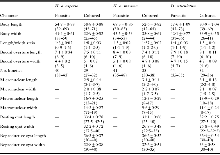
Ciliates from the 3 hosts exhibited an identical polymorphic life cycle consisting of 4 stages: trophont, tomont, tomite, and theront (Fig. 1C–O). Morphological and morphometric characteristics of the different stages are quite similar in the 3 isolates (Table 2), and so they are treated together.
Morphometric characterization of trophonts and cysts of 3 isolates of Tetrahymena rostrata derived from the gastropods Helix aspersa aspersa, Helix aspersa maxima, and Deroceras reticulatum
(Body and buccal overture dimensions as well as the number of somatic kineties are given for both parasitic and cultured ciliates, while nuclear dimensions are only from cultured cells. Cysts used for measuring were obtained between 2 and 4 days after starvation in mineral water. The mean±s.e. and range (in parentheses) of 50 determinations are given for each character. All measurement values are in μm.

Trophonts from the kidney and from axenic cultures are broadly ovoid to pyriform, with a rounded posterior end and a pointed, occasionally rostrate, anterior end (Fig. 1C). They ranged in size from 39 to 83 μm in length, with cultured forms being, in general, more slender and slightly shorter than parasitic forms (Table 2). The ciliates are highly plastic, burrowing through host tissues. They swim slowly, sometimes by rotating around the longitudinal axis. The cytoplasm, especially in the parasitic stages, contains densely packed inclusions that make the ciliates conspicuously opaque in transmitted light.
Parasitic ciliates have 35–55 somatic kineties depending on the host, but the range of kineties drops to 27–38 after a period as short as 4 days under axenic culture conditions. The somatic kineties run pole to pole except where truncated by the oral apparatus, where there are typically 2 postoral kineties. Anteriorly, they converge upon the apex forming a preoral suture, which is often ‘rostrate’ in appearance (Fig. 1D). Incomplete rows, however, are not infrequent, and some specimens showing a disorganized ciliature present a considerable number of interdigitated, incomplete ciliary rows. Intrameridional cross-fibrils extending from the primary meridians are often observed. Weakly stained secondary meridians were rarely detected in our silver-impregnated ciliates.
A single motionless caudal cilium, slightly longer than somatic cilia, while difficult to observe, is more easily seen in slow swimming trophonts under phase-contrast microscopy (Fig. 1E). A polar basal body-complex, composed of either 1 or 2 ‘granules’ and corresponding to this cilium complex, is visualized in the centre of the antapical region in most of silver-stained specimens (Fig. 1F), but it is usually very difficult to distinguish it from the posterior somatic kinetids of some kineties.
The oral apparatus is situated near the end of the first one-fourth or one-third of the body. The buccal overture is always oval to elliptical, ranging from 6 to 10 μm in length by 3 to 7 μm in width (Table 2). The buccal organelles are typically ‘tetrahymenal’ with 3 ciliary membranelles on the left and a single paroral membrane on the right (Fig. 1G). The anterior or first membranelle is the largest and the posterior one is clearly the smallest.
The contractile vacuole is near the posterior end of the ciliate. Three to five pores (normally 4 in parasitic forms, and 3 in cultured ciliates) are associated with 2–4 adjacent kineties between meridians 7 and 13 (Fig. 1H and I). Kineties bearing pores do not reach the antapical pole.
An ovoid to spherical macronucleus resides at the mid-body, ranging from 8 to 21 μm in length by 7 to 19 μm in width (Fig. 1J, Table 2). A smaller ovoid to ellipsoidal micronucleus lies adjacent to the macronucleus, sometimes partially embedded in a depression of the latter (Fig. 1J). It ranges in length from 2·2 to 4·0 μm in length by 1·5 to 3·5 μm in width (Table 2).
Both parasitic and cultured trophonts reproduce by transverse binary fission while swimming freely (Fig. 1K). Although resting cysts (Fig. 1 L and M) are mostly formed upon starvation, a few reproductive cysts containing 2 (Fig. 1N), rarely 3, intracystic tomites are occasionally observed (<1% of total cysts). No active division was, however, detected inside these cysts. Resting cysts are spherical to subspherical, ranging from 25 to 42·5 μm in length by 22·5 to 40 μm in width (Table 2). Reproductive cysts are chiefly subspherical, ranging from 30 to 40 μm in length by 30 to 40 μm in width (Table 2). Active theronts (Fig. 1O) emerging from cysts are more slender than trophonts, almost cylindrical, and swim extremely rapidly, changing direction continually.
Encystment and autogamy
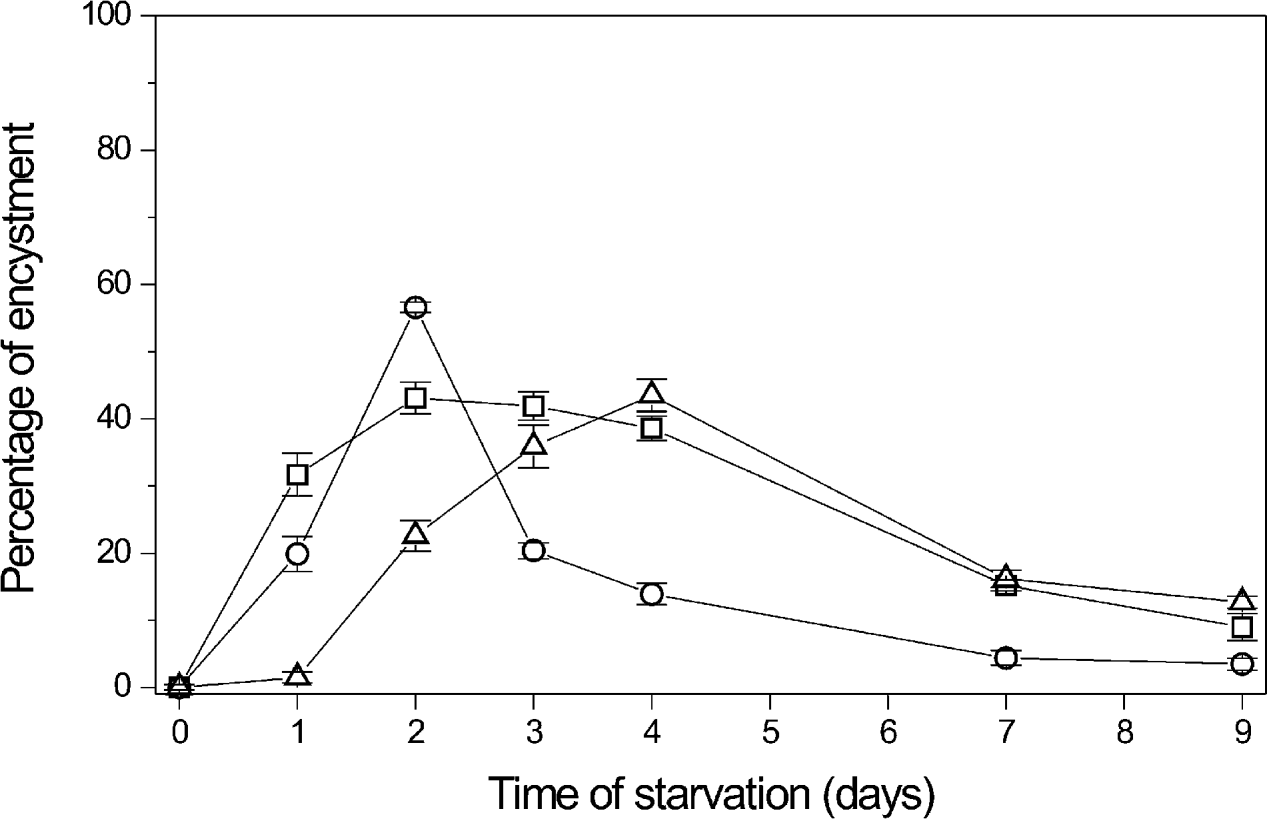
Upon starvation, individuals from all 3 isolates encysted with the first encysting individuals appearing within 2 h. By 12 h, about 4% of the ciliates were encysted and 40% were in various pre-cystic stages. While the highest encystment response differed only slightly among the isolates (i.e. 56·6%, 43·8%, and 43·1% for ciliates isolated from H. aspersa maxima, H. aspersa aspersa, and D. reticulatum, respectively), it was reached earlier (2 days post-starvation) in ciliates from the 2 snail subspecies compared to those isolated from the slug (4 days post-starvation) (Fig. 2). Even though starvation conditions were maintained, spontaneous excystment was almost complete by day 9 in all 3 isolates (Fig. 2).
Encystment kinetics of 3 isolates of Tetrahymena rostrata from Helix aspersa aspersa (square), Helix aspersa maxima (circle), and Deroceras reticulatum (triangle) under starvation in mineral water. Values are means±s.e. of 4 replicates.

Autogamy occurs in resting cysts (Fig. 3A–O), but also in reproductive cysts (Fig. 3P). The process observed in our ciliates is similar to that previously described by Corliss (Reference Corliss1965). The first evidence of this meiotic process is the appearance of the micronucleus in the ‘crescent’ stage (Fig. 3B), which is conspicuously different from a mitotic micronucleus (Fig. 3G). This yields the 2 haploid division products of Meiosis I (Fig. 3D), which divide mitotically (Fig. 3E) to yield the 4 products of Meiosis II (Fig. 3F). One of these products, the gametic nucleus, divides mitotically (Fig. 3G), and the resulting pronuclei (Fig. 3H) fuse to form the diploid zygotic nucleus or synkaryon (Fig. 3I). The remaining 3 haploid products, the ‘relic’ nuclei, are normally present when synkaryon is formed but they degenerate and finally disappear (Fig. 3H–K). The zygotic nucleus divides twice mitotically to produce 4 diploid nuclei (Fig. 3K). Immediately after the second post-zygotic division, 2 nuclei begin differentiation as 2 new macronuclei (Fig. 3L and M), while the old parental macronucleus and 1 of the 2 remaining products are degraded (Fig. 3M and N). At the final stage of the process, each encysted ciliate bears 2 new macronuclei and the micronucleus located between them (Fig. 3N). This nuclear configuration is also present in the theront stage emerging from the cyst (Fig. 3O). The vegetative nuclear system composed of 1 macronucleus and 1 micronucleus is restored after a post-excystment division (data not shown).
(A–P) Main nuclear events occurring during autogamy of Tetrahymena rostrata as revealed by Feulgen stain. (A) Resting cyst showing the micronucleus still adjacent to the macronucleus. (B) Micronucleus in the characteristic ‘crescent’ stage at the prophase of the first pre-zygotic (meiotic) division. Note that the crescent is close to the macronucleus. (C) Micronuclear chromatin condensing into chromosomes. (D) Two daughter haploid nuclei resulting from the first meiotic division. (E) Nuclear products entering into the second pre-zygotic (meiotic) division. (F) Four haploid nuclei resulting from the second meiotic division. (G) Third pre-zygotic (mitotic) division occurring in one of the meiotic products. (H) Pronuclei (arrows) resulting from the third pre-zygotic division accompanied by the other 3 ‘relic’ pycnotic nuclei. (I) Synkaryon (arrow) formed by fusion of the 2 pronuclei. Note that 2 of the 3 nuclear ‘relics’ begin to disappear. (J) Synkaryon undergoing its first post-zygotic mitotic division. A nuclear ‘relic’ has already disappeared while 1 of the other 2 still visible ‘relic’ nuclei is soon to do so. (K) Four daughter nuclei resulting from the second post-zygotic mitotic division. No nuclear ‘relics’ are observed at this time. (L) Two macronuclear anlagen (arrows) are visible below the original macronucleus. Note that this pair of nuclei is larger and more faintly stained than the other pair. (M) Two enlarged and diffuse macronuclear anlagen (arrows) accompanied by 2 brightly stained micronuclei and the pycnotic parental macronucleus. (N) ‘Long-lasting’ cyst resulting from autogamy. Note that the parental macronucleus has finally disappeared and only 1 micronucleus is present. (O) Spontaneously excysted theront showing the new micronucleus between the 2 new macronuclei. (P) Reproductive cyst with 2 tomites undergoing autogamy. Scale bar in (A)=10 μm is the same for all figures.

Molecular genetic analyses
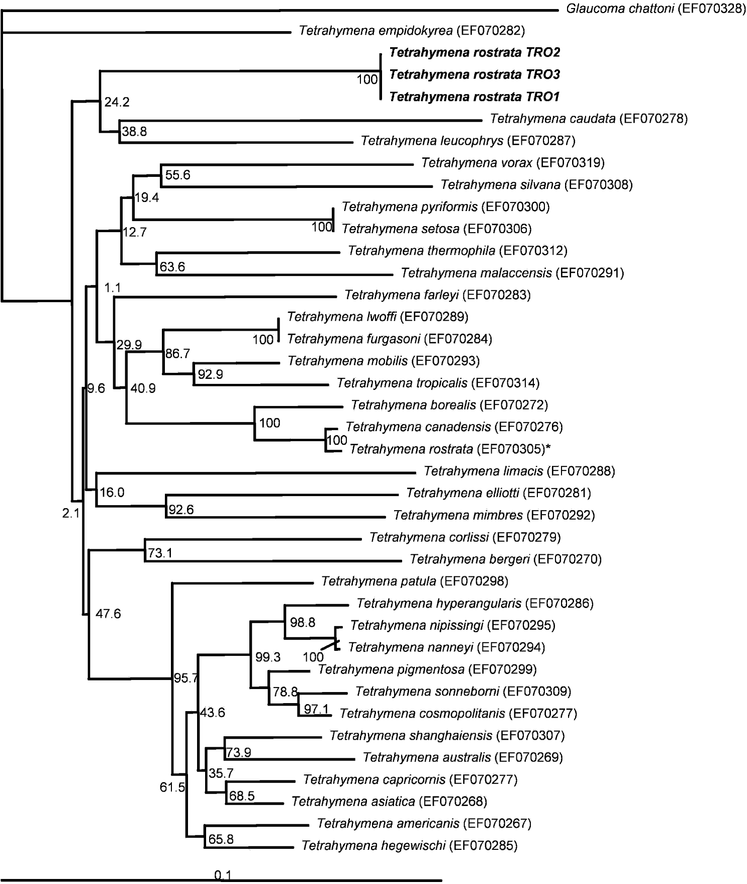
The 3 strains cluster together on both cox1 (Fig. 4) and SSU rRNA (data not shown) gene sequences. There is <0·4% genetic divergence of the cox1 genes and <0·2% divergence of the SSU rRNA genes among the three. These isolates are also ~14% divergent in cox1 sequence from the ATCC® 30770™ T. rostrata and ~2% divergent from this strain on the SSU rRNA gene. Moreover, they robustly cluster at the base of the borealis clade of Tetrahymena species, based on the SSU rRNA gene (data not shown). On the other hand, ATCC® 30770™ T. rostrata clusters robustly well within the australis clade, closely adjacent to Tetrahymena canadensis; ATCC® 30770™ shows very low divergence based on the cox1 gene to isolates of T. canadensis (Fig. 4).
Dendrogram of relationships among Tetrahymena species based on the mitochondrial cytochrome c oxidase 1 gene. Note that the 3 isolates of Tetrahymena rostrata (TRO1, TRO2, TRO3) from Spanish gastropods are almost identical and very genetically distinct from the ATCC 30770 strain of T. rostrata. The tree was generated using the Kimura 2-parameter measure of genetic distance and the neighbour-joining algorithm.

DISCUSSION
Tetrahymena rostrata was first described as Paraglaucoma rostrata by Kahl (Reference Kahl1926), who isolated it from moss in Germany. Later, Corliss (Reference Corliss1952a) isolated ciliates with identical characteristics from moss samples collected in Paris and definitely considered this species to be a member of the genus Tetrahymena. Later studies based on morphology and life-cycle features demonstrated that ciliates belonging to this species are widely distributed both as free-living protists in edaphic habitats (principally in moss, litter and soil) and as facultative parasites in terrestrial gastropods and other invertebrates (Stout, Reference Stout1954; Kozloff, Reference Kozloff1957; Thompson, Reference Thompson1958; Corliss, Reference Corliss1970; Van As and Basson, Reference Van As, Basson and Barker2004). This Tetrahymena species, as its name suggests, is characterized by a ‘rostrate’ anterior end, a unique ovoid micronucleus, an inconspicuous caudal cilium, a bimodal number of kineties depending on whether it is in a free-living or parasitic state, and a polymorphic life cycle with trophonts, resting and reproductive cysts, tomites, and theronts (Corliss, Reference Corliss1970, Reference Corliss, Elliott and Stroudsburg1973). In addition, T. rostrata is the only species in the genus that undergoes an autogamic process following encystment (Corliss, Reference Corliss1952b, Reference Corliss1965; Gutierrez, Reference Gutierrez1985).
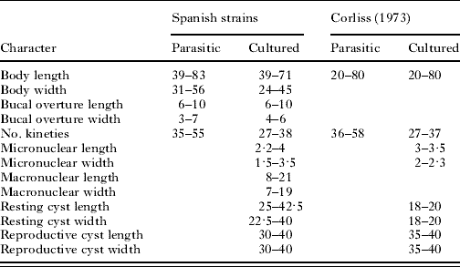
The results of our morphological and morphometrical studies as well as the confirmation that autogamy occurs within resting cysts demonstrate that the ciliates isolated from the renal organ of the snails Helix aspersa aspersa and H. aspersa maxima and the slug Deroceras reticulatum collected in 2 heliciculture farms of Galicia (NW Spain) belong to the species Tetrahymena rostrata (Kahl, Reference Kahl1926) Corliss, Reference Corliss1952. Using the summary data provided by Corliss (Reference Corliss, Elliott and Stroudsburg1973), our Spanish isolates clearly fall within the ranges reported for this species (Table 3). In addition, our molecular genetic data confirm that they are almost identical based on the cox1 and SSU rRNA genes, indicating that they can be placed in the same genetic species. Interestingly, our isolates are genetically ~14% divergent based on cox1 gene and ~2% on the SSU rRNA gene from the ‘T. rostrata’ ATCC® 30770™ strain. Considering that (i) levels of genetic divergence of these magnitudes on these two genes have been considered sufficient to distinguish species of Tetrahymena and establish new species within this genus (Jerome et al. Reference Jerome, Simon and Lynn1996; Lynn and Strüder-Kypke, Reference Lynn and Strüder-Kypke2006; Chantangsi et al. Reference Chantangsi, Lynn, Brandl, Cole, Hetrick and Ikonomi2007), and (ii) the life-history and life-cycle characters of the ATCC® 30770™ strain have not been completely described but it are apparently not able to undergo encystment (Chantangsi et al. Reference Chantangsi, Lynn, Brandl, Cole, Hetrick and Ikonomi2007), we believe that this commercial strain is not an isolate of T. rostrata (in order to give a solution to this problem, one of our isolates is currently in the process of being submitted to ATCC). Indeed, ATCC® 30770™ is quite closely related to strains of T. canadensis, and might probably better be assigned to that species.
Comparison of the Spanish strains of Tetrahymena rostrata with the summary measurements provided for the species by Corliss (Reference Corliss, Elliott and Stroudsburg1973)
(All measurements are in μm.)

Tetrahymena rostrata has been found to date in the nephridium of 14 gastropod species, including Deroceras reticulatum (reviewed by Van As and Basson, Reference Van As, Basson and Barker2004). However, as far as we know, this is the first report of this species parasitizing helicid snails. Prevalences under intensive reproduction conditions reached values close to 50% in both Helix aspersa subspecies, but especially in H. aspersa maxima. In the case of H. aspersa aspersa, prevalence only exceeded 10% in 1 of the 5 groups examined. According to the information provided by the farmers, the 8 groups of snails analysed in our study came from different sources. Since the 2 farms included in the study habitually use breeders coming from different wild locations or from other small farms that are dedicated almost exclusively to fattening, it is probable that the variations in the prevalence between groups are related to their origin. Although the precise causes determining heavier or lighter infections in wild snails are difficult to determine, some factors directly associated with heliciculture practices could facilitate the propagation of the ciliate under farming conditions. Thus, the population density and the hygienic and handling practices used during intensive reproduction could influence the infection dynamics throughout the reproductive period. In addition, the use of breeders coming from greenhouses or outdoor pens where invasive slugs (or other snail species) are abundant may be not advisable since, as it has been demonstrated in the present study and as it is reflected in the existing literature (reviewed by Van As and Basson, Reference Van As, Basson and Barker2004), these gastropods can act as reservoirs of T. rostrata.
In spite of T. rostrata being considered an important pathogen for slugs (Brooks, Reference Brooks1968; Barker, Reference Barker1993; Wilson et al. Reference Wilson, Coyne and Glen1998), we have not been able to establish a direct relationship between ciliate infection and the morbimortality observed. The concomitant presence of other well-known pathogenic parasites in most of the snails examined (data not shown) makes it impossible to draw definitive conclusions concerning the precise pathogenic role of this ciliate in heliciculture. However, some evidence suggests that T. rostrata may have deleterious effects on Helix aspersa. First, the prevalence of infection and parasite densities were especially high in the snails of group 1 exhibiting mantle swelling. Similar signs have been reported in Deroceras reticulatum infected experimentally with T. rostrata (Brooks, Reference Brooks1968). Second, our histological studies revealed that these histophagous ciliates, as previously described in slugs (Brooks, Reference Brooks1968), cause severe renal damage in heavily infected snails. Experimental infections using snails reared in a parasite-free environment are currently underway in our laboratory. These studies will help to determine how pathogenic this parasitic ciliate might be to both subspecies of Helix aspersa.
We wish to express our gratitude to Dr Michaela Strüder-Kypke for assistance with phylogenetic analyses, and to J. Méndez and I. Pazos from C.A.C.T.I. (Centro de Apoio Científico e Tecnolóxico á Investigación; Universidade de Vigo) for technical assistance in SEM studies. This work was partially supported by grant PGIDIT04RAG310015PR (Xunta de Galicia).