Introduction
Post-traumatic stress disorder (PTSD) and traumatic brain injuries (TBI) can have detrimental effects on the cognitive and emotional functioning of U.S. veterans returning from Operation Enduring Freedom (OEF) and Operation Iraqi Freedom (OIF). Impairments in executive control functions are frequently observed in this population (Vasterling, Verfaellie, & Sullivan, Reference Vasterling, Verfaellie and Sullivan2009). Although the effects of PTSD on executive functions have not received as much attention as the well-documented changes in memory and fear learning, many studies have found that impairments do occur (Koso & Hansen, Reference Koso and Hansen2006; Leskin & White, Reference Leskin and White2007; Vasterling, Brailey, Constans, & Sutker, Reference Vasterling, Brailey, Constans and Sutker1998). Recent reviews have suggested that deficits in attention and executive control can be evident even when the experimental stimuli are emotionally neutral, as opposed to trauma-related (Vasterling & Verfaellie, Reference Vasterling and Verfaellie2009; Vasterling et al., Reference Vasterling, Verfaellie and Sullivan2009; Qureshi et al., Reference Qureshi, Long, Bradshaw, Pyne, Magruder, Kimbrell and Kunik2011). Subtle impairments in executive function could hinder the effectiveness of PTSD treatments that rely on the retrieval of autobiographical memories and cognitive reappraisal techniques, such as prolonged exposure and cognitive processing therapy (Vasterling & Verfaellie, Reference Vasterling and Verfaellie2009). Furthermore, executive control over thought and behavior is necessary for effective disengagement from an overwhelming preoccupation with traumatic stimuli (Aupperle, Melrose, Stein, & Paulus, Reference Aupperle, Melrose, Stein and Paulus2012).
The lateral prefrontal cortex (PFC) is thought to implement cognitive control by exerting top-down influences over sensory and motor processing (Miller & Cohen, Reference Miller and Cohen2001). In addition, the anterior cingulate cortex (ACC) has been implicated in a variety of cognitive tasks that require executive control processes (Botvinick, Cohen, & Carter, Reference Botvinick, Cohen and Carter2004; Fan, McCandliss, Fossella, Flombaum, & Posner, Reference Fan, McCandliss, Fossella, Flombaum and Posner2005; Swick & Turken, 2002). Response inhibition, or the ability to inhibit prepotent responses, is thought to rely on the integrity of specific regions in the lateral and medial PFC (Aron, Fletcher, Bullmore, Sahakian, & Robbins, Reference Aron, Fletcher, Bullmore, Sahakian and Robbins2003; Picton et al., Reference Picton, Stuss, Alexander, Shallice, Binns and Gillingham2007; Swick, Ashley, & Turken, Reference Swick, Ashley and Turken2008). It is a core executive control function that has been dissociated from other higher cognitive processes such as task switching and working memory updating (McNab et al., Reference McNab, Leroux, Strand, Thorell, Bergman and Klingberg2008; Miyake et al., Reference Miyake, Friedman, Emerson, Witzki, Howerter and Wager2000; Nee, Wager, & Jonides, Reference Nee, Wager and Jonides2007). In PTSD, functional alterations have been observed in the ACC and other medial frontal regions (Etkin & Wager, Reference Etkin and Wager2007; Shin, Rauch, & Pitman, Reference Shin, Rauch and Pitman2006), as well as in lateral PFC (Morey, Petty, Cooper, Labar, & McCarthy, Reference Morey, Petty, Cooper, Labar and McCarthy2008). These alterations could account for some of the observed deficits in emotion regulation and inhibitory control functions.
The Go/NoGo (GNG) task has been used extensively to assess response inhibition in both animals (Petrides, Reference Petrides1986) and humans (Swick, Ashley, & Turken, Reference Swick, Ashley and Turken2011). In this task, a motor response is given to one stimulus class and withheld to another. The NoGo stimuli are typically infrequent to establish a prepotent tendency to respond. Impairments in the GNG task have been observed in clinical populations with inhibitory deficits, such as attention deficit hyperactivity disorder (ADHD), substance abuse, schizophrenia, and borderline personality disorder (Chambers, Garavan, & Bellgrove, Reference Chambers, Garavan and Bellgrove2009; Donohoe et al., Reference Donohoe, Reilly, Clarke, Meredith, Green, Morris and Robertson2006; Fisher, Aharon-Peretz, & Pratt, Reference Fisher, Aharon-Peretz and Pratt2011; Rentrop et al., Reference Rentrop, Backenstrass, Jaentsch, Kaiser, Roth, Unger and Renneberg2008). These disorders are thought to involve dysfunctions of frontal inhibitory processes, which can lead to increases in impulsive behavior. In line with these observations, a recent meta-analysis of 48 GNG imaging studies in controls revealed that two major foci of activation included the right middle frontal gyrus (MFG) and the ACC/pre-supplementary motor area (pre-SMA) region (Swick et al., Reference Swick, Ashley and Turken2011). Both of these frontal areas have been implicated in PTSD. Indeed, a group of civilian participants with PTSD showed an increase in false alarm errors in a GNG task and reduced activation in these same regions, relative to controls (Falconer et al., Reference Falconer, Bryant, Felmingham, Kemp, Gordon, Peduto and Williams2008).
The OEF/OIF patient population differs from many other populations because PTSD and mild TBI (mTBI) frequently co-occur. The estimated prevalence of this co-morbidity has ranged from 33% to 39% in the largest studies of OEF/OIF veterans (Carlson et al., Reference Carlson, Kehle, Meis, Greer, Macdonald, Rutks and Wilt2011). Therefore, it is important to determine the extent of inhibitory control deficits in these patients, who are at increased risk for substance abuse and other impulsive behaviors (Jakupcak et al., Reference Jakupcak, Cook, Imel, Fontana, Rosenheck and McFall2009).
Studies in civilians with mTBI commonly observe executive dysfunction and memory impairments (Mathias, Beall, & Bigler, Reference Mathias, Beall and Bigler2004), although these deficits tend to resolve within one to three months (Belanger, Curtiss, Demery, Lebowitz, & Vanderploeg, Reference Belanger, Curtiss, Demery, Lebowitz and Vanderploeg2005). There is considerable disagreement, however, in the characterization of mTBI as a minor contributor to post-deployment problems in OEF/OIF veterans (Sigford, Cifu, & Vanderploeg, Reference Sigford, Cifu and Vanderploeg2009). Nonetheless, the overlap with PTSD symptoms is extensive (Stein & McAllister, Reference Stein and McAllister2009), and disentangling the effects of each has been challenging. It is becoming increasingly apparent that PTSD makes a substantial contribution to the persistent post-concussive symptoms (PCS) reported by OEF/OIF veterans (Hoge et al., Reference Hoge, McGurk, Thomas, Cox, Engel and Castro2008). In one recent study of 339 OEF/OIF veterans with positive mTBI histories, PTSD symptoms uniquely accounted for 46.6% of the variance in self-reported PCS, while loss of consciousness accounted for only 1.6% (Lippa, Pastorek, Benge, & Thornton, Reference Lippa, Pastorek, Benge and Thornton2010).
The cumulative impact of mTBI and PTSD on neurocognitive function has not been extensively explored in soldiers who have served in OEF and OIF, who are typically exposed to chronic stressors and threats to safety. Previous neuropsychological results in this population using standardized tests have been mixed, with some reporting deficits (Marx et al., Reference Marx, Brailey, Proctor, Macdonald, Graefe, Amoroso and Vasterling2009; Nelson, Yoash-Gantz, Pickett, & Campbell, Reference Nelson, Yoash-Gantz, Pickett and Campbell2009) while others have not (Brenner et al., Reference Brenner, Terrio, Homaifar, Gutierrez, Staves, Harwood and Warden2010; Gordon, Fitzpatrick, & Hilsabeck, Reference Gordon, Fitzpatrick and Hilsabeck2011). However, no study has yet examined response inhibition in OEF/OIF veterans with mTBI and PTSD using the sensitive GNG task.
The current experiment tested veterans with PTSD and mTBI primarily due to blast injury, and veterans with PTSD only. Because our population had a paucity of OEF/OIF veterans with TBI but without PTSD, these individuals were excluded. Determining the effects of PTSD and mTBI on inhibitory control functions is critical to providing appropriate cognitive therapies and rehabilitation programs. After returning from Iraq and Afghanistan, many veterans face difficulties returning to work and maintaining relationships, even if deficits on standardized neuropsychological tests are not observed. Therefore, the development of more sensitive experimental designs is critical in evaluating potential tendencies toward impulsive behaviors.
The major question posed by the present study was whether OEF/OIF veterans with PTSD would show impairments in motor response inhibition. False alarm errors on NoGo trials were used as the primary measure of inhibitory control abilities. To manipulate the prepotency of responding, and hence the need for inhibitory control, the probability of Go to NoGo stimuli alternated between 50/50 (“easy”) and 90/10 (“difficult”) in different blocks. If the function of lateral and medial PFC regions is altered in the patients, one might predict that their performance in the GNG task would be impaired. Although the majority of patients (75%) had both PTSD and mTBI, a secondary question was whether the presence of a mild TBI would result in further deficits in those with PTSD.
Participants also completed standardized questionnaires to assess the severity of PTSD and depressive symptoms. We predicted that response inhibition performance would be related to scores on the PTSD checklist (PCL), with higher error rates in those with higher PCL scores. If the addition of a mild TBI is associated with a further decline in inhibitory control, then the combination of blast-related mTBI with PTSD could ultimately hinder recovery, from both the post-concussive symptoms and the psychiatric sequelae.
Methods
Participants
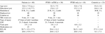
The participants were 40 combat veterans diagnosed with PTSD (39 male, 1 female) and 33 age-matched veteran controls (31 male, 2 female). Among the PTSD patients, 30 had sustained one or more mTBIs (primarily due to blast injury while serving in the military), while 10 had no history of mTBI (see Table 1 for details). Participants with evidence of significant medical disease, severe psychiatric problems (such as schizophrenia or bipolar disorder), active substance abuse, visual deficits, or history of other neurological events were excluded. Another 6 participants (4 patients, 2 controls) were initially enrolled, then excluded when additional information was revealed (childhood TBI; non-military PTSD; moderate TBI; other psychiatric disorder; not OEF/OIF). Most of the patients were identified and diagnosed in the TBI clinic of the consulting neurologist. A semi-structured clinical interview was conducted, and mild TBI was diagnosed based on patient self-report of the following criteria from the VA/DoD Clinical Practice Guidelines—loss of consciousness (LOC) 30 min or less or altered mental status (e.g., feeling dazed, disoriented, or confused), with post-traumatic amnesia less than 24 hr (The Management of Concussion/mTBI Working Group, 2009). PTSD diagnosis was based on semi-structured clinical interview using DSM-IV criteria. The diagnoses of mTBI and PTSD were corroborated with available VA medical records, to the fullest extent possible.
Demographic information and self-rating scores for the PTSD patients, the patient subgroups with and without mTBI, and the controls

Note. The mean ± standard deviation are given for age, education, estimated years post-event(s), PCL-M, and BDI. The patient subgroups did not differ from each other for age, education, years post-event, PCL-M, and BDI.
n.s. = not significantly different from controls; *** Significantly different from controls at p < .001.
R = right; L = left; ambi = ambidextrous; LOC = loss of consciousness (of 30 patients with mTBI, 21 had LOC, 5 did not, and 4 were not sure whether they had LOC); Medications = number on psychoactive medications; PCL-M = post-traumatic stress disorder checklist, military version; BDI = Beck Depression Inventory; mTBI = mild traumatic brain injury.
The diagnosis of PTSD was based on a review of the VA's Computerized Patient Record System (CPRS) for each enrolled patient. The initial PTSD diagnosis was made when the veteran sought help through the VA. The majority (36 of 40) were diagnosed by VA mental health providers. The presence of PTSD was confirmed by the consulting neurologist in 10 of these 36 patients upon entry into the study. One patient was diagnosed solely by the neurologist, and 3 patients were not enrolled in the VA system. A small number of participants were recruited from the local Vet Center, which provides services for PTSD but does not share diagnostic information with the VA.
Controls were recruited primarily through advertisements. Potential control subjects were screened for exclusionary criteria (described above) and history of mTBI or PTSD through an initial telephone interview, and further assessed at the first visit. Demographic information is shown in Table 1. The groups were matched for age but not education level. This could be due to the inability of many of the patients to return to school after their military service, and is typical of earlier studies on veterans with PTSD (e.g., McNally, Kaspi, Riemann, & Zeitlin, Reference McNally, Kaspi, Riemann and Zeitlin1990; Vrana et al., Reference Vrana, Roodman and Beckham1995). However, another possibility is that low education serves as a risk factor for developing PTSD (Iversen et al., Reference Iversen, Fear, Ehlers, Hacker Hughes, Hull, Earnshaw and Hotopf2008; Larson, Booth-Kewley, Highfill-McRoy, & Young, Reference Larson, Booth-Kewley, Highfill-McRoy and Young2009); thus, those with lower educational attainment were at greater risk for PTSD. Level of education did not influence the outcome, however, as will be discussed in the Results section.
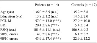
Wechsler Test of Adult Reading (WTAR) data (Wechsler, Reference Wechsler2001) were available for a subset of the participants (14 patients and 17 controls). The estimated full-scale IQ (FSIQ) did not differ between the groups [t(1,29) = 1.44; p = .16], who were well-matched and representative of the entire sample (Table 2).
Demographic information, self-rating scores, estimated full-scale IQ based on Wechsler Test of Adult Reading (WTAR) scores, and NoGo errors in the GNG task for a subset of the participants
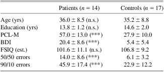
Note. The mean ± standard deviation are given for age, education, PCL-M, and BDI. n.s. = not significantly different from controls; *** significantly different from controls at p ≤ .001.
PCL-M = post-traumatic stress disorder checklist, military version; BDI = Beck Depression Inventory; GNG = Go/NoGo task; FSIQ = full-scale IQ.
English was the primary language for all participants. The subjects signed informed consent statements approved by the Institutional Review Board of the VA Northern California Health Care System and were paid for their participation. All procedures were in compliance with the Declaration of Helsinki.
Go-NoGo Task
We implemented the experimental design used in a previous study on patients with frontal lobe lesions (Swick et al., Reference Swick, Ashley and Turken2008). Stimuli consisted of single uppercase letters printed in a large black font (248 pt) on a white background. The stimuli were presented on a 16 inch ViewSonic monitor using a PC that ran Presentation® software (Neurobehavioral Systems, Inc., http://www.neurobs.com/). Stimuli were rapidly and serially presented at the center of a computer screen for 200 ms duration once every 1500 ms. Subjects were instructed to respond as quickly as possible to every letter except for “X” by pressing a button on the keyboard with the index finger of the dominant hand. In four separate blocks of trials, the proportion of “Go” to “NoGo” trials alternated between 50/50 and 90/10. There were 140 trials per block, with short rest breaks between each block. A short practice set of 30 trials (15 Go and 15 NoGo, randomly intermixed) preceded the experimental trials.
Questionnaires
At the end of the session, all subjects completed three self-report questionnaires: the Barratt Impulsiveness Scale (BIS), the PTSD Checklist, Military Version (PCL-M), and the Beck Depression Inventory (BDI). The BIS is a 30-item self-report measure thought to assess the personality construct of “impulsiveness” (Patton, Stanford, & Barratt, Reference Patton, Stanford and Barratt1995). Results from the BIS will be reported in a separate publication. The PCL-M for DSM-IV (Weathers, Litz, Huska, & Keane, Reference Weathers, Litz, Huska and Keane1994) is an accepted diagnostic tool for measuring PTSD (Blanchard, Jones-Alexander, Buckley, & Forneris, 1996). The PCL-M is a 17-item self-report tool that establishes the presence and degree of PTSD symptoms in military personnel. It has three clusters or subsets: re-experiencing, numbing, and hyperarousal. PTSD is indicated in a veteran population with a score of 50 or greater (Forbes, Creamer, & Biddle, Reference Forbes, Creamer and Biddle2001). The PCL-M score of one control participant who had not yet sought clinical care placed them in the PTSD group. This individual was subsequently diagnosed with PTSD by a psychiatrist. Another veteran recruited via an advertisement initially self-identified as having PTSD but had a low score on the PCL-M. Omitting these two individuals did not affect the results, so they are included in all analyses. In addition, a clinical neuropsychologist reviewed information from both patients and determined that their PCL scores reflected current symptomotology (or lack thereof). The BDI is one of the most commonly used self-report screens for major depressive disorder (MDD) and has been validated with well-established psychometric properties (Beck, Steer, & Gabin, Reference Beck, Steer and Gabin1988). The BDI is a 21-item test which measures the presence and degree of depression in adolescents and adults.
Data Analysis
Error data were characterized as missed responses to Go stimuli and false alarm responses to NoGo stimuli. The mean reaction time (RT) was calculated for each subject and sorted into correct responses to Go stimuli and incorrect responses to NoGo stimuli. Statistical analyses were carried out using repeated measures analyses of variance (ANOVAs) with factors of group (patients, controls) and probability (50/50, 90/10). Secondary analyses compared patients with mTBI and PTSD to those with PTSD only. The correlations between self-report measures and errors in the difficult 90/10 condition were determined using the Spearman rank-order statistic, with a Bonferroni correction for multiple comparisons (p < .005). Effect sizes are reported as partial eta-squared (ηp2) for ANOVA and Cohen's d for follow-up comparisons.
Results
Accuracy
An initial ANOVA with factors of group (controls, patients), probability (50/50, 90/10), and error type (misses, false alarms) revealed that every main effect and interaction was highly significant, including group × error type [F(1,71) = 26.11; p < .0001; ηp2 = .26]. Thus, separate ANOVAs were performed for errors of omission on Go trials (misses) and errors of commission on NoGo trials (false alarms). In general, the rate of misses was very low and did not differ by probability (p = .19). The percentage of missed responses for the 50/50 and 90/10 probability conditions was 0.65% and 0.28%, respectively, for controls; and 1.93% and 1.55% for patients. Although floor effects are a concern, the percentage of misses was greater in the patients than in controls [F(1,71) = 5.20; p = .03; ηp2 = .07], which did not interact with probability (p > .9).
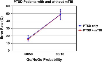
In contrast, NoGo errors (Figure 1, top) showed a highly significant effect of group [F(1,71) = 26.44; p < .0001; ηp2 = .27], probability [F(1,71) = 93.97; p < .0001; ηp2 = .73], and an interaction between the two [F(1,71) = 14.03; p = .0004; ηp2 = .17]. The PTSD patients made more false alarm errors than controls for both the 50/50 [F(1,71) = 22.83; p < .0001; d = 1.12] and the 90/10 [F(1,71) = 23.35; p < .0001; d = 1.14] probability conditions. Although the effect sizes are nearly equivalent, the significant interaction suggests the patients’ difficulty with inhibiting inappropriate responses was exacerbated in the difficult 90/10 condition, when responding was prepotent. A secondary ANOVA was conducted to compare PTSD patients with and without mTBI (Figure 2), revealing that patients with both PTSD and mTBI did not differ from those with PTSD only. The main effect of group [F(1,38) = 0.2; p = .89] and the group by probability interaction [F(1,38) = 0.2; p = .88] were not significant.
Top: False alarm errors (percentage of NoGo errors) for the patients (n = 40) and controls (n = 33) in the easy (50/50) and difficult (90/10) conditions. Bottom: Reaction times (RTs) on correct Go trials (in milliseconds) in the easy (50/50) and difficult (90/10) conditions. The error bars depict standard errors.

False alarm errors (percentage of NoGo errors) for patients with both mild traumatic brain injury (mTBI) and post-traumatic stress disorder (PTSD; n = 30) and patients with PTSD only (n = 10) in the easy (50/50) and difficult (90/10) conditions. The error bars depict standard errors.

Reaction Times
The initial comparison examined RTs on correct Go trials only (Figure 1, bottom), and revealed no differences between the patients and controls in the speed of responding (p > .7). All subjects were faster to respond to targets in the 90/10 condition than in the 50/50 condition, which was reflected in a highly significant main effect of probability [F(1,71) = 200.59; p < .0001]. Probability did not interact with group (p > .7). The secondary analysis showed that patients with both PTSD and mTBI did not differ from those with PTSD only (p > .7).
An additional ANOVA compared response times for correct and error trials. All participants had faster RTs on incorrect NoGo trials (308 ms ± 70 ms) than on correct Go trials (376 ms ± 86 ms), suggesting that impulsive responding led to the majority of errors in performance. This result was indicated by a main effect of accuracy [F(1,70) = 479.30; p < .0001]Footnote 1 that did not interact with group (p > .3). This speeding up on error trials was numerically greater for the 50/50 condition (80 ms) than for the 90/10 condition (57 ms), as indicated by the probability by accuracy interaction [F(1,70) = 11.28; p = .001].
Correlations Between Experimental and Self-Report Measures
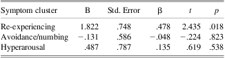
The associations between scores on the self-report questionnaires and false alarm errors in the difficult 90/10 condition were determined using Spearman Rank Correlations (corrected at p < .005). Scores on the PCL-M and BDI showed a strong correlation with performance: more severe levels of PTSD symptoms (rho = .52; p = .0001) and depression (rho = .53; p < .0001) were both associated with higher error rates. All three PTSD symptom clusters produced a correlation with error rates: re-experiencing (rho = .54; p < .0001), avoidance/numbing (rho = .47; p < .0001), and hyperarousal (rho = .49; p < .0001). However, when these three variables were entered into a standard multiple regression analysis to control for shared variance (see Vasterling et al., Reference Vasterling, Brailey, Constans and Sutker1998), re-experiencing was the only significant predictor of errors in the 90/10 condition (p = .02; see Table 3). Finally, a striking correlation between PCL-M and BDI scores was observed (rho = .90; p < .0001), indicating that PTSD and depression symptoms showed a high level of co-morbidity in these OEF/OIF veterans. As clearly expected based on clinician diagnosis, the patients reported higher PCL-M and BDI scores than the control group, but there were no differences between PTSD patients with and without mTBI (Table 1).
Relationship of false alarm errors in the 90/10 condition to the three PTSD symptom clusters, based on self-reported PCL-M scores
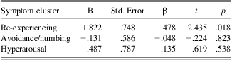
Note. R = .557; Adjusted R 2 = .281; F(3,69) = 10.36, p < .0001.
PTSD = post-traumatic stress disorder; PCL-M = post-traumatic stress disorder checklist, military version.
Effects of Education, Estimated IQ, Diagnostic Certainty, and Medications
Two additional analyses established that the patients’ deficits in accuracy were unrelated to education level. In the first, the less educated half of the control group (n = 17) was compared to the entire patient group (now matched for education: 13.4 vs. 13.3 years, respectively). The same results for false alarm errors were obtained: a main effect of group [F(1,55) = 14.27; p = .0004], and an interaction between group and probability [F(1,55) = 6.72; p = .01]. In the second, the groups were more closely matched in number. We compared the lower educated half of controls (n = 17) to the upper half of patients (n = 20), so now the patients were significantly more educated (13.4 vs. 14.3 years, respectively; p = .001). Again, the same impairment was observed in the patients: a main effect of group [F(1,35) = 14.01; p = .0007], and an interaction between group and probability [F(1,35) = 7.55; p = .009].
Thus, group differences in education level did not influence the outcome. Another question is whether there were group differences in IQ which might have affected the results. WTAR data were available for a subset of the participants to provide an estimate of pre-morbid IQ (Wechsler, Reference Wechsler2001). As reported previously, the estimated FSIQ did not differ between the groups, who were well-matched and representative of the entire sample (Table 2). This subset of patients made significantly more false alarm errors than controls for both the 50/50 and 90/10 conditions (p's ≤ .001). Furthermore, errors on the 90/10 condition were not at all correlated with estimated FSIQ (r = .017; p = .92).
Although the PCL-M was not used for diagnostic purposes, eight patients with a formal diagnosis of PTSD from semi-structured clinical interview had scores below 50 (range, 31–49) on the day they were tested. Removing these patients and any other clinically discrepant participants from the analyses did not affect the results (p's ≤ .0001 for false alarm errors in both the 50/50 and 90/10 conditions), nor did it change group demographics (mean age for all 40 patients = 32.6 years and for 32 patients = 32.6 years; mean education for all 40 patients = 13.3 years and for 32 patients = 13.1 years).
To examine the effects of prescription drugs on performance, the 23 patients taking psychotropic medication(s) of any class (sedative/hypnotics, antidepressants, mood stabilizers, atypical antipsychotics, opioids, or alpha adrenergic blockers) were compared to the 17 patients who were not. Medication use did not affect RTs (main effect p = .20 and interaction p = .11, with the trend being faster RTs in those taking medications) or NoGo error rate (main effect p = .28 and interaction p = .31).
Role of Deployment, Loss of Consciousness, and Number of Events
Among the veterans in the control group, 19 of 33 were deployed. An additional ANOVA compared these deployed controls (n = 19) to the patients (n = 40) for NoGo errors. Results were similar to the main analysis: a highly significant effect of group [F(1,57) = 14.13; p = .0004] and a group by probability interaction [F(1,57) = 6.56; p = .01] were observed.
As stated earlier, the secondary analysis comparing PTSD patients with and without mTBI found no differences in performance. However, the definition of mTBI includes individuals with altered mental status but no loss of consciousness (LOC). Self-reported LOC occurred in 21 of 30 patients with mTBI. To examine whether PTSD+mTBI patients with self-reported LOC (n = 21) might differ from those with PTSD only (n = 10), another ANOVA was run. Again, there were no significant main or interactive effects of group (both p's > .9). Finally, the group with mTBI was restricted further to those with both LOC and more than two events (n = 15), and compared to the PTSD only group. These two patient subgroups did not differ significantly in their PCL-M scores (59.1 vs. 56.6 respectively; p = .62). There were still no differences for NoGo errors (main effect of group, p > .9; interaction: p > .8).
Discussion
The present study demonstrated that OEF/OIF veterans with PTSD were impaired at inhibiting inappropriate motor responses. A speed-accuracy trade-off could not account for this result, as RTs in the patient and control groups were virtually identical. As well, the severity of PTSD and depressive symptoms were both highly correlated with performance. These results suggest that response inhibition is compromised in participants with PTSD, which is consistent with previous results in civilians (Falconer et al., Reference Falconer, Bryant, Felmingham, Kemp, Gordon, Peduto and Williams2008; Wu et al., Reference Wu, Ge, Shi, Duan, Wang, Sun and Zhang2010) and Gulf War veterans (Vasterling et al., Reference Vasterling, Brailey, Constans and Sutker1998). A deficit in inhibitory control could have detrimental effects on daily activities such as driving (Lew, Amick, Kraft, Stein, & Cifu, Reference Lew, Amick, Kraft, Stein and Cifu2010), and may hinder recovery from traumatic events (Aupperle et al., Reference Aupperle, Melrose, Stein and Paulus2012).
In addition, the inhibitory control deficit occurred whether or not the patient had reported a mild TBI in addition to PTSD. Although this finding is preliminary, the fact that mTBI did not add to the cognitive deficits seen in those with PTSD suggests that in the current population, where loss of consciousness was brief (less than 1–2 min in most patients) and where no clear LOC occurred in 30% (with dazed/altered mental status), PTSD was the primary driver of performance. Further restriction of the mTBI group to those with self-reported LOC and more than two events did not alter this outcome. Furthermore, the severity of PTSD symptoms did not differ in patients with and without mTBI, in agreement with Romesser and colleagues (Reference Romesser, Shen, Reblin, Kircher, Allen, Roberts and Marchand2011). There has been considerable controversy over the diagnosis of mTBI in OEF/OIF veterans, with some questioning the impact of mTBI on post-deployment functioning relative to PTSD, depression, and other psychiatric disorders (e.g., Hoge et al., Reference Hoge, McGurk, Thomas, Cox, Engel and Castro2008; Hoge, Goldberg, & Castro, Reference Hoge, Goldberg and Castro2009). Results could differ in military personnel with more “severe” mTBIs, such as those with a combination of blast injury and secondary head trauma, for example, the group of U.S. military personnel airlifted to Landstuhl Medical Center in Germany (Mac Donald et al., Reference Mac Donald, Johnson, Cooper, Nelson, Werner, Shimony and Brody2011). Those subjects showed evidence of white matter abnormalities on diffusion tensor imaging (DTI) scans.
On a related note, the co-morbidity between PTSD and depression symptoms was striking, with a very high correlation between the severity of self-reported symptoms on the two scales. Although the two disorders share the overlapping construct of negative affect, the symptom cluster of re-experiencing is unique to PTSD (Cloitre, Koenen, Gratz, & Jakupcak, Reference Cloitre, Koenen, Gratz and Jakupcak2002). Increased scores on both the BDI and the PCL-M were strongly associated with a higher percentage of false alarm errors in the difficult condition. All three PTSD symptom clusters (re-experiencing, avoidance/numbing, and hyperarousal) were correlated with performance individually, but when entered into a multiple regression, re-experiencing was the only significant predictor of error rate. This finding replicates Vasterling et al. (Reference Vasterling, Brailey, Constans and Sutker1998) and suggests that the symptom cluster most unique to PTSD was specifically related to the decline in inhibitory control.
The strong correlation between PCL-M scores and error rates is in agreement with previous results. Falconer and colleagues (2008) also found a positive correlation between false alarm errors and PTSD severity as measured by the Clinician-Administered PTSD Scale (CAPS). In their imaging study, civilian PTSD patients showed reduced activity in the right lateral PFC and the ACC/pre-SMA regions relative to controls. Furthermore, more severe PTSD symptoms were associated with less activation in bilateral PFC and medial frontal areas in the patients (Falconer et al., Reference Falconer, Bryant, Felmingham, Kemp, Gordon, Peduto and Williams2008). This is in accord with what would be predicted on the basis of meta-analytic studies of the GNG task in controls (Swick et al., Reference Swick, Ashley and Turken2011), because those regions were uniformly recruited for response inhibition across a large number of experiments. The activation foci showing the greatest overlap across GNG imaging studies included the right anterior insula and right MFG (e.g., Zheng, Oka, Bokura, & Yamaguchi, Reference Zheng, Oka, Bokura and Yamaguchi2008) and dorsomedial areas such as the SMA, pre-SMA, and ACC (e.g., Li, Huang, Constable, & Sinha, Reference Li, Huang, Constable and Sinha2006; Mostofsky & Simmonds, Reference Mostofsky and Simmonds2008). As mentioned previously, individuals with PTSD have smaller ACC volumes (Hamner, Lorberbaum, & George, Reference Hamner, Lorberbaum and George1999; Rauch et al., Reference Rauch, Shin, Segal, Pitman, Carson, McMullin and Makris2003; Woodward et al., Reference Woodward, Kaloupek, Streeter, Martinez, Schaer and Eliez2006). It is now becoming more apparent that dorsolateral PFC function may be compromised in PTSD as well (Aupperle et al., Reference Aupperle, Melrose, Stein and Paulus2012; Simmons & Matthews, Reference Simmons and Matthews2012). Difficulties in recruiting the MFG during a cognitive task were associated with higher levels of PTSD symptoms (Morey et al., Reference Morey, Petty, Cooper, Labar and McCarthy2008).
Disentangling the effects of mTBI, PTSD, and depression on cognitive performance and brain function has not been a straightforward endeavor. In a structural imaging study of individuals with both PTSD and depression, common areas of volume reduction were located in the PFC (Kroes, Rugg, Whalley, & Brewin, Reference Kroes, Rugg, Whalley and Brewin2011). An fMRI study demonstrated that veterans with both mTBI and MDD showed greater activity in the amygdala, and less activity in dorsolateral PFC, than veterans with mTBI only during an emotional face matching task (Matthews et al., Reference Matthews, Strigo, Simmons, O'Connell, Reinhardt and Moseley2011).
Robertson, Manly, Andrade, Baddeley, and Yiend (Reference Robertson, Manly, Andrade, Baddeley and Yiend1997) have argued that in addition to motor response inhibition, the Go/NoGo task is a measure of sustained attention. Both motor response inhibition and/or lapses of attention can produce high NoGo error rates. In our experiment, the 90/10 blocks might have been more monotonous than the 50/50 blocks, so sustained attention was required to a greater degree in the former. Thus, it is noteworthy that the patients showed substantially elevated false alarm rates in both conditions. In addition, omitted responses on Go trials were not greatly increased (mean of 1.7% in the patients), as might be expected if distractibility and sustained attention had been the primary difficulties. Although a significant difference was observed, this finding should be interpreted with caution because the controls showed a floor effect, with the rate of misses below 1%. Finally, the pattern of RTs on correct Versus incorrect Go trials indicated that errors were due to impulsive responding. Therefore, an inhibitory control deficit remains the best explanation for the patients’ performance.
Previous Go/NoGo results in TBI patients with moderate to severe injuries have been mixed, but a recent meta-analysis of 20 response inhibition studies in adults found a moderate effect size (Dimoska-Di Marco, McDonald, Kelly, Tate, & Johnstone, Reference Dimoska-Di Marco, McDonald, Kelly, Tate and Johnstone2011). Although many papers have reported deficits (e.g., Robertson et al., Reference Robertson, Manly, Andrade, Baddeley and Yiend1997), others have not (Swick et al., Reference Swick, Ashley and Turken2008; Whyte, Grieb-Neff, Gantz, & Polansky, Reference Whyte, Grieb-Neff, Gantz and Polansky2006). Our prior study demonstrated that patients with severe TBIs and large bilateral lesions in the orbitofrontal cortex were not impaired on the GNG task (Swick et al., Reference Swick, Ashley and Turken2008). On the other hand, stroke patients with focal lesions in the left inferior frontal gyrus and left anterior insula showed a pattern of impairment similar to that reported here (Swick et al., Reference Swick, Ashley and Turken2008). However, the present group of OIF/OEF veterans had an even greater deficit in motor response inhibition, which can have important implications for daily life. Since performance did not differ in patients with and without mTBI, these results suggest that PTSD symptoms interfere with effective response inhibition.
The present study has several limitations. PTSD was diagnosed by semi-structured clinical interview instead of the CAPS, which is considered the “gold standard” (Blake et al., Reference Blake, Weathers, Nagy, Kaloupek, Gusman, Charney and Keane1995). Nonetheless, a strong correlation between false alarm errors and PCL-M scores was observed, suggesting a relationship between inhibitory control deficits and self-reported PTSD symptom severity that was independent of formal diagnosis. Furthermore, there is a very high correlation between the PCL and the CAPS: diagnostic efficiency of the PCL is 0.900 versus the CAPS (Blanchard et al., Reference Blanchard, Jones-Alexander, Buckley and Forneris1996). The difficult issue of making an accurate mTBI diagnosis pertains to most veterans of OEF/OIF, as it is dependent on recollection and self-report. Medical records from Iraq and Afghanistan were not available for the patients, as they had no medical treatment at the time. Brief losses of consciousness or altered mental status may not always be caused by blast exposure itself, but can be due to acute stress, confusion, or sleep deprivation (Hoge et al., Reference Hoge, Goldberg and Castro2009). Nevertheless, all current participants with mTBI were diagnosed by a neurologist.
Other limitations include the fact that the control veterans were not all deployed or exposed to combat. Future studies should attempt to better match the groups on these factors, as deployment and combat exposure may have detrimental effects on their own. However, an analysis restricted to only those controls who were deployed revealed that the patients were still impaired relative to this group. The controls and patients were not matched for years of education, although subgroup analyses convincingly demonstrated this did not affect the pattern of results. Since all patients were highly motivated to participate in the study, we did not believe that effort was an issue. However, we did not use a measure of effort or malingering to verify this. Another difficult issue is separating the effects of PTSD and depressive symptoms on cognitive performance (Cloitre et al., 2002), due to their high co-morbidity in this population. The current study was not designed to address this question. The recruitment and selection of patients was not completely random, but was primarily focused on those who attended a specialty TBI clinic. Additional efforts were made to recruit from mental health clinics and veterans organizations as well. However, there were fewer patients with PTSD only, so the comparisons between this group and the mTBI + PTSD group were low in power. Finally, due to the difficulty of finding patients with pure mTBI in isolation from PTSD, we were not able to include this population in the current study. Inclusion of this group in future studies will allow stronger conclusions about the effects of mTBI on response inhibition.
Conclusions
The present results indicated that OEF/OIF veterans with PTSD were impaired at inhibiting motor responses in a Go/NoGo task. The inhibitory control deficit was exacerbated when responding was more prepotent, suggestive of more impulsive responding in the patients. False alarm error rates were strongly correlated with self-reported symptoms of PTSD and depression. Furthermore, the combination of mTBI and PTSD did not result in worse performance than PTSD alone in the present population. Taken together, the current findings suggest that OEF/OIF veterans with PTSD show impairments in response inhibition. Additional studies are needed to verify that these findings are independent of mTBI. Since neurocognitive impairments may hinder the effectiveness of PTSD therapies that rely on cognitive reappraisal and disengagement from traumatic stimuli (Aupperle et al., Reference Aupperle, Melrose, Stein and Paulus2012; Vasterling & Verfaellie, Reference Vasterling and Verfaellie2009), incorporating treatments that strengthen executive functions might be considered in the future.
Acknowledgments
We thank Dr. Andrew Kayser for patient referrals, Dr. Juliana Baldo for providing WTAR data, Timothy Heron for statistical advice, and all participants for taking part in the study. This work was supported by the U.S. Army Medical Research and Materiel Command under W81XWH-08-2-0086 and a VA Merit Review grant. The authors declare that they have no conflicts of interest. The information in this manuscript and the manuscript itself has never been published either electronically or in print. The contents do not represent the views of the Department of Veterans Affairs or the United States Government.