Introduction
The standard treatment of locally advanced cancer cervix is external radiation with concurrent chemotherapy along with intracavitary brachytherapy. Although three-dimensional (3D) external radiation planning has been largely implemented, planning in brachytherapy is still widely based on traditional 2D point-based systems. Studies have shown that the traditionally used point A is a poor surrogate for individual target dose and magnetic resonance imaging (MRI)-guided brachytherapy using the GEC-ESTRO guidelines has shown a superiority over 2D point-based systems in treatment planning and dose prescription in cervical cancer.Reference Tanderup, Nielsen and Nyvang 1 The superiority of MRI over computed tomography (CT) lies in its greater soft-tissue resolution, which enables the precise delineation of the tumour extension.Reference Boss, Barentsz and Massugen 2 Hence, MRI considered the gold standard for image-based brachytherapy and the clinical results using the same have been very encouraging.Reference Potter, Georg and Dimopoulos 3 However, despite its distinct proven advantage, MRI-guided brachytherapy has not gained wide implementation owing to various reasons. MRI at each brachytherapy is labour intensive as majority of MRI units are located away from the radiotherapy departments. The longer time required for MRI sequences and treatment planning limits the number of patients that can be treated in a day. The MRI-compatible applicators, which are made of titanium and polymer fibres, are fragile and expensive as compared with the standard stainless steel applicators and thus are not easily affordable by most centres.
In a progress update of implementation of image-guided brachytherapy for cancer cervix in the United Kingdom in 2011, of the 42 centres that responded, 23 (51%) used CT for brachytherapy planning and 9 (20%) had access to MRI for brachytherapy planning.Reference Tan 4 In a survey of 3D imaging in gynaecological brachytherapy by the American Brachytherapy Society in 2010, 55% centres reported using CT, 43% used X-ray films and only 2% reported using MRI for dose specification to the cervix. Despite the large availability of CT, 76% centres used point A as the prescription method, whereas only 14% prioritised the use of 3D volume-based dose prescriptions.Reference Viswanathan and Erickson 5
In a similar survey of the Gynecologic Cancer Intergroup on brachytherapy practices in Japan, Korea, Australia, New Zealand, Europe and North America, of the 72 responders, 57% reported using CT compared with 25% who reported using MRI as the imaging modality. In terms of dose prescription, 78% prescribed to point A and only 11% followed the GEC-ESTRO guidelines alone, 21% followed the GEC-ESTRO and reported dose to point A, 4 (6%) followed the ABS guidelines alone, and 8 (11%) use both the ABS and point A.Reference Viswanathan, Creutzberg and Craighead 6 This was attributed to lack of acceptance and perceived benefit of image-guided brachytherapy, lack of time and limitations pertaining to infrastructure, and set-up required in terms of imaging and software.Reference Viswanathan and Erickson 5 Thus, adoption and applicability of MRI-guided brachytherapy remains limited and alternate imaging modalities need to be explored.
Nowadays, CT scanners are widely available in most radiotherapy departments and may be an alternative when MRI at brachytherapy is not possible. Studies comparing CT and MRI contouring have shown that, although the organs at risk (OAR) can be as accurately contoured on CT as that of MRI, contours of high-risk clinical target volume (HRCTV) tend to be overestimated with CT.Reference Viswanathan, Dimopoulos and Kirisits 7 , Reference Krishantry, Patel and Singh 8
Thus, in an effort to provide the benefit of image-guided brachytherapy in a resource-constrained setting, in the present study we decided to explore the possibility of using a diagnostic MRI for contouring on CT at brachytherapy instead of an MRI at brachytherapy. In the present study, we compared two sets of contours on CT with the standard contours on MRI. In the first set, contouring was done on the CT with the help of clinical findings at brachytherapy (CT-HRCTV). In the second set, the contouring was done on CT with the help of the MRI at diagnosis and the clinical findings at brachytherapy (CT-HRCTVdmri). These were then compared with the reference HRCTV contours delineated on the MRI (MR-HRCTV). The end point of this study was to assess the feasibility of using CT (with and without the diagnostic MRI) for defining the HRCTV compared with the reference MRI-based contours.
Materials and methods
Fifteen patients with cervical cancer undergoing image-based brachytherapy were included in the study. All patients underwent a baseline diagnostic MRI before treatment. A detailed clinical pelvic examination was conducted and clinical drawings were recorded. External beam radiotherapy (EBRT) planning was done using CT-based computerized 3D conformal or IMRT plans A dose of 46 Gy in 2 Gy per fraction was planned over four and a half weeks along with concurrent weekly Cisplatin (40 mg/m2). The patients were assessed for brachytherapy in the 3rd week of EBRT and brachytherapy was performed towards the 4th or 5th week. Two brachytherapy applications were performed at 1-week interval and two fractions were delivered at each application. The second brachytherapy fraction at each application was delivered the next day ensuring a minimum gap of 8 hours between the two fractions. An MRI along with a CT scan was done at the first brachytherapy fraction followed by a CT scan only at subsequent fractions The dose per fraction was 7 Gy (HDR).
Brachytherapy procedure
The intracavitary brachytherapy application was performed under general anaesthesia with the patient in the lithotomy position. The bladder was catheterised and all patients underwent bowel preparation before the procedure. The brachytherapy procedure was performed under ultrasound guidance using CT/MRI-compatible ring tandem applicators (Nucletron, an Elekta company, Elekta AB, Stockholm, Sweden), with nominal dimensions of 40·60 mm in length for the tandem (diameter 6 mm) and 26 and 30 mm for the diameter for the ring with 45 and 60° curvature. Vaginal packing was done with roller gauze anterior and posterior to the applicator to push the bladder and rectum, respectively.
CT scan and MRI at brachytherapy
After the procedure, the patients underwent a CT scan followed by an MRI with the applicator in situ. The CT was acquired on the CT simulator Light Speed; VFX-16 (GE Healthcare, Chalfont St. Giles, UK). A standardised bladder filling was done before the CT scan, MRI and before treatment. The bladder was first drained and 50 mL of saline was instilled in the bladder via the urinary catheter. The urine was then allowed to drain with gravity to attain a constant bladder filling. CT images were taken (without contrast) with slice thickness of 2·5 mm starting from the level of 3–4 cm above the fundus superiorly to the level of ischial tuberosities inferiorly. After the CT scan, the patient was taken for MRI in the radio diagnosis department.
MRI imaging was performed in accordance with the protocol described by Dimopoulos et al.Reference Dimopoulos, Schard and Berger 9 Images were acquired on a 3-T Magnetron Veiro System (Siemens AG, Healthcare Sector, Erlangen, Germany) with pelvic surface coils with the patient in the supine position. The section thickness was 5 mm with no intersection gap. The axial images were obtained from the level above uterine fundus to the inferior border of symphysis pubis or below in case of vaginal tumour extension. The sagittal images were obtained between the internal obturator muscles. The coronal, paracoronal and para-axial images included the tumour, entire cervix, corpus uteri, parametrium and vagina.
Contouring and brachytherapy treatment planning
The contours were first defined on the CT without any information of the brachytherapy MRI. A single radiation oncologist determined all the contours on CT and MRI. This was done to avoid any interobserver variability. The contours on the CT were delineated in accordance with the GEC-ESTRO recommendations.Reference Haie-Meder, Potter and Limbergen 10 , Reference Pötter, Haie-Meder and Limbergen 11 As the GTV cannot be defined on CT, only the HRCTV was contoured. The contouring was done on the axial slices and reformatted sagittal images were used to determine the height of the HRCTV. Two sets of contouring were defined on CT. In the first set, contours were defined on CT set with the help of clinical findings at brachytherapy (CT-HRCTV). In the second set, the contouring was done on CT with the help of the MRI at diagnosis along with the clinical findings at brachytherapy (CT-HRCTVdmri). The contours of the HRCTV began at the superior level of the ring. Superiorly the contours continued until the cervical–uterine junction (level where there is expansion of the contours indicating the presence of the uterine tissue). Two additional slices with narrower diameters were contoured around the uterine tandem to cover the conical apex. For the involved parametrium and vaginal extension of the disease, the contours were extended according to clinical findings and findings of the diagnostic MRI. For the corpus extension, the contours were extended superiorly into the uterus according to the findings of the diagnostic MRI. The OARs were contoured in accordance with GEC-ESTRO protocol. After contouring on the CT, the contouring was done on the MRI data sets in accordance with the GEC-ESTRO guidelines.Reference Haie-Meder, Potter and Limbergen 10 , Reference Pötter, Haie-Meder and Limbergen 11 For the MRI data sets, the MRI images were transferred to Oncentra master plan version 4·3 (Nucletron, an Elekta company). The GTV, HRCTV and the OARs were defined on axial T2-weighted MRI images taken at the time of brachytherapy with the applicator in situ.
Applicator reconstruction was performed on MRI images with the help of CT image data using rigid registration technique. For the first application, the tandem of the applicator was contoured on both the CT and MR image data sets using the pearl contouring tool of 3 mm diameter sphere. These applicator contours were aligned by rotating and translating the 3D CT and MR images data set interactively. After verification of the fusion at different important landmark points, digitisation for applicator reconstruction on MRI image was performed with the help of the spy glass tool along the centre of the applicator axis of CT images. The standard dwell positions were used and the dose was initially normalised to point A. The standard dwell positions for 6 cm uterine tandem were 3, 6, 9, 12, 15, 18, 21 and 24 and for 3·4 cm ring 7, 8, 9, 10, 25, 26, 27 and 28 with a step size of 2·5 mm. The doses to the HRCTV and the OARs were then evaluated, and optimisation was done keeping point A as the starting reference point. The target dose per fraction for the HRCTV was to achieve a minimum dose of 7 Gy to D90. The constraints for the 2 cc dose to the bladder, rectum and sigmoid were kept at 6·2, 5 and 5 Gy, respectively. The total planned EQD2 for HRCTV was ≥85 Gy α / β = 10 and for the 2 cc bladder, rectum and sigmoid were limited to ≤90 Gy, ≤75 Gy and ≤75 Gy α / β = 3, respectively.Reference Lang, Kirisits and Dimopoulos 12 , Reference Pötter, Dimopoulos and Georg 13
The dimensions, volumes and the dose volume histograms (DVH) parameters were compared for MR-HRCTV, CT-HRCTV and CT-HRCTVdmri. The doses to 0·1 and 2 cc volumes of the OARs of the MRI and CT were evaluated. Comparison between the different sets of contours was made using a two-sided paired t-test. A ‘p’-value of ≤0·05 was considered significant.
Results
The median age of the patients was 48·2 years and ranged from 32 to 68 years. Of the 15 patients, 11 had stage IIB disease and 4 had stage IIIB disease. All the patients received external radiotherapy 46 Gy in 23 fractions with concurrent weekly cisplatin along with brachytherapy 7 Gy × 4 fractions. In six patients, the first application was carried out in the 4th week of external radiotherapy, and in nine patients the brachytherapy was performed after the completion of external radiation. In all the patients, the treatment was completed within 50 days.
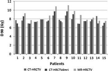
The mean values with standard deviation for the height, maximum width, width at point A, maximum thickness, thickness at point A, and volumes for CT-HRCTV, CT-HRCTVdmri and MR-HRCTV are shown in Table 1. The difference in the volumes and doses were calculated for all three pairs, that is, CT-HRCTV compared with CT-HRCTVdmri; CT-HRCTV compared with MR-HRCTV; and CT-HRCTVdmri compared with MR-HRCTV. The two-sided paired t-test showed that CT significantly overestimated the maximum width and the width at point A in both the CT-HRCTV and CT-HRCTVdmri contours compared with the MR-HRCTV (Table 2). The overestimation of width was more with CT-HRCTV compared with the CT-HRCTVdmri, although the difference was statistically not significant. The height was significantly underestimated in patients with CT-HRCTV (p = 0·05) as compared with CT-HRCTVdmri and MR-HRCTV. This is because two patients had extension of the disease into the corpus that could not be identified on CT alone. The contours were extended superiorly for the CT-HRCTVdmri according to the findings on diagnostic MRI. For patients without corpus extension, the height of CT-HRCTV was comparable to CT-HRCTVdmri and MR-HRCTV. The total thickness, thickness at point A and total volumes were comparable for CT-HRCTV, CT-HRCTVdmri and MR-HRCTV. On comparing the DVH parameters for the HRCTV, the D90 values were significantly lower for the CT-HRCTV and CT-HRCTVdmri as compared with MR-HRCTV (p = 0·03; 0·01), respectively. The mean difference with a standard deviation of −0·82 ± 1·29 Gy and −0·69 ±0·81 Gy (− sign denotes lower values) was observed for the CT-HRCTV and CT-HRCTVdmri as compared with HRCTV-MRI.
Mean dimensions with standard deviations of volumes for HRCTV on CT with clinical findings alone (CT-HRCTV), with MRI at diagnosis (CT-HRCTVdmri) and with MRI at brachytherapy (MR-HRCTV)

Abbreviations: CT-HRCTV, high-risk clinical target volume defined on CT with clinical findings at brachytherapy alone; CT-HRCTVdmri, high-risk clinical target volume defined on CT with MRI at diagnosis; MR-HRCTV, high-risk clinical target volume defined on MRI at brachytherapy; MRI, magnetic resonance imaging.
Mean difference with standard deviations in HRCTV dimensions and DVH for CT-HRCTV; CT-HRCTVdmri and MR-HRCTV

Notes: Data presented as mean and standard deviation.
a Normalised to 7 Gy/fraction.
b Statistically significant ‘p’ value (≤0·05).
Abbreviations: V100, volume treated to 100% of prescription dose; D98, dose received by 98% volume; D90, dose received by 90% of volume.
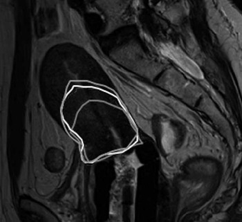
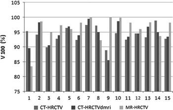
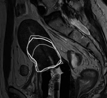
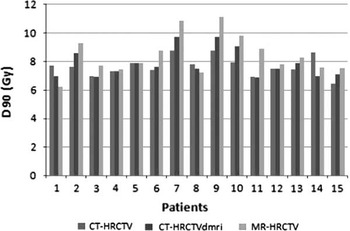
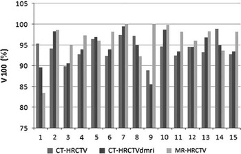
On analysis of individual patient data, it was observed that in two patients there was gross underestimation of height for the CT-HRCTV, as there was an extension of the disease into the uterine corpus on MRI that could not be assessed with clinical examination and CT scan (Figure 1). This resulted in a higher V100 and D90 values and an apparent superior coverage of target on CT-HRCTV and a relatively inferior coverage CT-HRCTVdmri and HRCTV-MRI as compared with the CT-HRCTV, whereas in the actual picture the adequate DVH parameters could not be achieved because of a large volume of CT-HRCTVdmri and HRCTV-MRI (Figures 2, 3). One patient had a large uterine fibroid in the lower uterine segment that could not be differentiated from disease on CT. This led to a large difference in the HRCTV volumes on the CT contours.
Sagittal view of overlapped HRCTV contours of CT and MRI. Note the underestimation of height with CT-HRCTV contours in a patient with intrauterine disease extension.
D90 for HRCTV for a single fraction for CT-HRCTV, CT-HRCTVdmri and MR-HRCTV.
V100 for HRCTV for a single fraction for CT-HRCTV, CT-HRCTVdmri and MR-HRCTV.
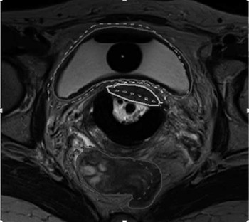
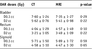
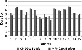
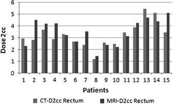
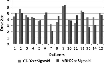
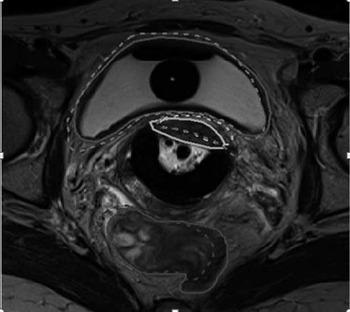
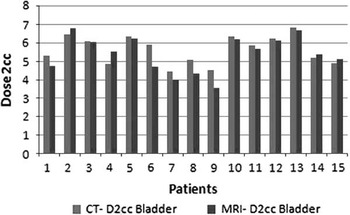
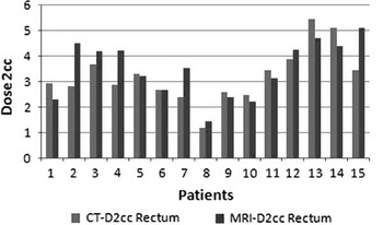
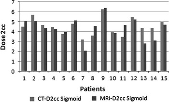
On comparison of the OAR contours, the contours of the bladder and rectum appeared to overlap nicely on both CT and MRI (Figure 4). No statistical difference was observed for the doses to 0·1 cc and 2 cc volumes for the bladder, rectum and sigmoid for CT and MRI (Table 3; Figures 5–7).
Figure showing the overlapped contours for the HRCTV and the OAR. While the width of the CT contours of the HRCTV is larger, the contours of the OAR seem to nicely overlap.
Mean 2 cc doses with standard deviation for bladder rectum and sigmoid with CT and MRI
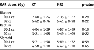
Abbreviations: OAR, organ at risk; CT, computed tomography; MRI, magnetic resonance imaging; D0·1 cc, dose to 0·1 cm3; D2 cc, dose to 2 cm3.
D2cc Bladder for a single fraction with CT and MRI.
D2cc Rectum for a single fraction with CT and MRI.
D2cc Sigmoid for a single fraction with CT and MRI.
Discussion
In the present study, we wanted to evaluate whether CT could be used instead of MRI in a context of limited access to MRI-guided brachytherapy facilities. As MRI is superior to CT and clinical examination in determining the tumour size and intrauterine extensionReference Dimopoulos, Schard and Berger 9 , we tried to incorporate the information from the diagnostic MRI along with the clinical findings in delineation of HRCTV on CT.
The results in our study were similar to that reported in literature and a significant overestimation of width on CT was observed in our study. However, the width of HRCTV was less with the CT-HRCTVdmri as compared with the CT-HRCTV. The height was underestimated for CT-HRCTV owing to disease extension into the uterine corpus and was comparable for CT-HRCTVdmri. Thus, CT contours could be improved with the integration of information from the diagnostic MRI.
Vishwanathan et al. compared the contours and DVH of the tumour and OARs with CT and MRI. Although significant overestimation of HRCTV width was reported with CT, the height, thickness and volume did not differ significantly. The contours and doses to the OARs were also comparable. In a similar study, Krishnatry et al. compared the volumes and doses for the tumour and OARs with CT and MRI. Besides an overestimation of width on CT, a significant underestimation of height was also reported. No statistical difference was observed between the volumes and DVH with CT and MRI.Reference Viswanathan, Dimopoulos and Kirisits 7 , Reference Krishantry, Patel and Singh 8
Whereas the overestimation of width on CT may result in large HRCTV volumes, the underestimation of height may lead to a geographical miss and thus offset the advantage of image-guided brachytherapy. Thus, methods to improve HRCTV definition on CT are required. In a recent study by Hegazy et al., CT-based HRCTV delineation using information from FIGO stage with or without 3D documentation of clinical gynaecological examination (CGE) was compared with MRI-based HRCTV. The use of standardised uterine heights was also investigated. A large overestimation of width and volume was observed with the CT-based HR CTV contouring based on FIGO stage alone. The authors concluded that target delineation accuracy could be improved by incorporation of additional information from 3D documentation of repetitive gynaecological examination in the contouring protocol and could help in improving the accuracy of dose optimisation in settings with limited access to imaging facilities at the time of brachytherapy. Although the authors recommended a minimum of second/third uterine height on CT as a good surrogate for HRCTV height, inclusion of the entire length of the uterine cavity was suggested at an advanced stage. For this, traditional loading of the uterine tandem from tip was suggested.Reference Hegazy, Pötter and Kirisits 14 In our study, both the patients with intrauterine disease extension had FIGO stage IIb disease, and keeping a standard HRCTV height of second/third intrauterine length would have led to a geographical miss. On the other hand, in a given clinical situation, loading the tandem until the tip alone may still result in inadequate coverage incase of a large width of the HRCTV. In our study, the intrauterine disease extension could easily be covered and there was no geographical miss with CT contours defined with the information from diagnostic MRI.
In a study by Fedrico et al., HRCTV was retrospectively contoured on CT, with the help of diagnostic pre-BT MRI (HRCTV-CTpreBTMRI). This was compared with the planning MRI (HRCTV-MRI) contour. The mean HRCTV-CTpreBTMRI to HRCTV-MRI volume ratio was 1·04 for Stage IB1-IB2 patients, 1·08 and 1·17 for Stage II and III–IVA, respectively. The volumes for CT-based contouring of HRCTV with information from pre-BT MRI were comparable to MRI-based contouring. The best agreement was achieved for stage I patients and the maximum geographical miss of CT contours was observed for stage IVa patients.Reference Fedrico, Fotina and Hegazy 15
The dosimetric effect of CT contouring was also evaluated in our study. With increase in HRCTV volumes, decrease in D90 values is expected and this was reflected in our study. Lower D90 values were observed both for the CT-HRCTV and CT-HRCTVdmri contours. Although the width of the CT-HRCTV contours was larger and as a result larger HRCTV volumes would be expected, the mean volumes of CT-HRCTV were found to be smaller compared with CT-HRCTV. This was because of the underestimation of height in CT-HRCTV in two patients that resulted in lower mean HRCTV volume compared with CT-HRCTVdmri. The difference in the HRCTV volumes did not impact the dosimetry and the D90 values were comparable for both the CT contours. Besides using different imaging modalities, interobserver variation is another factor that may influence the HRCTV contouring. Hellibust et al. studied the dosimetric impact of interobserver delineation variability (IODV) in MRI-based cervical brachytherapy for six patients distributed over ten experienced observers. A mean relative standard deviation of 8–10% was reported for GTV and HRCTV for a single fraction and for the whole treatment the IODV resulted in a dose uncertainty of ±5Gy α / β = 10 for HRCTV.Reference Hellebust, Tanderup and Lervåg 16 In our study, the mean difference in D90 for HRCTV was <1 Gy for a single fraction and the interobserver variability was limited, as all the contours were delineated by a single observer.
Overall, our study shows that CT contouring can be improved with the incorporation of information from diagnostic MRI. The role of MRI in image-guided brachytherapy cannot be underestimated, and even in situations with limited MRI access, at least one MRI is required to passably assess the tumour extension. In addition, residual disease at brachytherapy may be an important factor leading to concordance and discordance between CT and MRI. Thus, a pre-brachytherapy MRI may give a better estimation of residual disease and may result in better delineation of HRCTV on CT. The drawback of our study is a small cohort of patients and the results need to be validated in larger studies. Further improvement in the quality of CT contouring is required and minimising the artefacts using various algorithms and modern contrast-enhancing techniques may help in better delineation of the target.Reference Xia, Roeske and Yu 17
Conclusion
The role of MRI in image-guided brachytherapy in cervical cancer cannot be undermined. However, in centres without access to MRI-guided brachytherapy facilities, CT may be used for contouring of tumour and OAR, provided there is at least one MRI available before brachytherapy.
Acknowledgements
None.
Financial Support
None.
Conflicts of Interest
None.