Introduction
Nasopharyngeal carcinoma (NPC)Reference Liu, He, Wang, Peng, Chen and Chi1 is a distinct epithelial malignancy originating from the nasopharyngeal mucosa; it exhibits a unique geographic distribution, with particularly high incidence in Southeast Asia and southern China. Despite significant advances in radiotherapy techniques and the widespread use of concurrent chemoradiotherapy, which remains the standard curative treatment for NPC, long-term outcomes for many patients remain unsatisfactory. Locoregional recurrence and distant metastasis continue to be the primary causes of treatment failure and disease-related mortality. Therefore, identifying reliable biomarkers capable of predicting recurrence risk, monitoring disease progression and guiding individualised follow-up strategies remains a critical clinical need.
Tumour recurrence in nasopharyngeal carcinoma
Nasopharyngeal carcinoma is generally considered a radiosensitive malignancy, and most patients achieve an initial favourable response to concurrent chemoradiotherapy.Reference Chen, Zhu, Liang, Li, Qu and Chen2 However, some patients experience disease recurrence or distant metastasis during long-term follow up, which compromises long-term survival.
Current prognostic assessment in NPC mainly relies on tumour–node–metastasis (TNM) staging and plasma Epstein–Barr virus DNA load.Reference Chan, Lee, Hong, Ahn, Chong and Spreafico3, Reference Neo, Ong, Zhang, Chow, Wee and Fong4 These indicators reflect tumour burden and viral activity but do not directly assess extracellular matrix remodelling. Laminin-dependent epithelial polarity and matrix organisation are relevant to tumour invasion.Reference Matlin, Myllymäki and Manninen5 Additional microenvironmental biomarkers may therefore improve prognostic stratification and enable earlier detection of recurrence.
Tumour microenvironment and extracellular matrix
The tumour microenvironment contributes to cancer progression, immune evasion and therapeutic resistance. Basement membrane proteins within the extracellular matrix can change with tumour development and treatment response.Reference Jiang, Kumar, Yu, Shipman, Wang and McKay6 The extracellular matrix also provides structural support and an active signalling platform that regulates cell adhesion, migration and tumour–stromal interactions.
Experimental models also show that extracellular proteolysis can promote tumour development and metastasis.Reference Giebeler, Schönefuß, Landsberg, Tüting, Mauch and Zigrino7 Breach of the native basement membrane, the specialised extracellular matrix structure separating epithelial cells from underlying stroma, is the defining event distinguishing invasive carcinoma from carcinoma in situ. Beyond this initial breach, tumour nests within the stroma can show variable, often discontinuous, peri-tumoural laminin deposition; this residual matrix organisation has been linked to invasive behaviour in squamous cell carcinomas.Reference Rahman, Rao, Tippu, Patil, Agarwal and Srivastava8, Reference Berndt, Gaßler and Franz9
Basement membranes are primarily composed of laminin, collagen IV and other structural proteins that collectively maintain epithelial architecture and tissue integrity. However, compared with studies focusing on immune regulation and viral oncogenesis in NPC, the structural remodelling of basement membranes and its clinical implications have received relatively limited attention.
Laminin and peri-tumoural matrix integrity
The laminin N-terminal protein LaNt α31 has been linked to altered tumour invasion mode in breast cancer.Reference Troughton, O’Loughlin, Zech and Hamill10 More generally, laminins are large α-, β- and γ-chain heterotrimers that anchor epithelial cells to basement membranes and link cell-surface receptors to the extracellular matrix.Reference Colognato and Yurchenco11 Through these interactions, laminins regulate cell adhesion, polarity, differentiation, migration and survival.
Loss or fragmentation of laminin-containing basement membranes has been associated with tumour dedifferentiation, increased invasive capacity and poor clinical outcomes in several epithelial malignancies, including colorectal, breast and pancreatic cancers.Reference Feng, Wang and Song12–Reference van der Zee, van Eijck, Hop, Biermann, Dicheva and Seynhaeve14
Because breach of the native basement membrane already defines invasive carcinoma, peri-tumoural laminin loss represents ongoing remodelling rather than the initiating invasive event. Associations are not uniform across sites; in advanced laryngeal squamous cell carcinoma, laminin expression was not associated with cervical nodal metastasis.Reference Mielcarek-Kuchta, Olofsson and Golusinski15
Recent studies have also highlighted the importance of extracellular matrix integrin signalling pathways, particularly focal adhesion kinase (FAK) activation, in promoting tumour cell motility and invasion following basement membrane disruption.Reference Hood and Cheresh16 In addition to structural loss within tumour tissue, degradation of laminin may also release circulating immunoreactive products. Such measurements may indicate systemic extracellular matrix turnover, although their tissue and molecular sources require confirmation.
In NPC, tumour-cell laminin subunit β1 (LAMB1) has been linked to proliferation and metastasis,Reference Feng, Yang, Yang, Qu and Li17 while immunohistochemical studies at other head and neck sites have focused on local tissue patterns.Reference Vageli, Doukas, Zacharouli, Kakanis, Strataki and Zioga18 Serum laminin has shown clinical associations in other cancers,Reference Tas, Bilgin, Tastekin, Erturk and Duranyildiz19 but its relationship to peri-tumoural laminin in NPC remains unclear.
Study objectives
The present study aimed to investigate the clinical significance of laminin in NPC from both tissue and circulating perspectives. Specifically, we evaluated laminin expression in tumour tissue using immunohistochemistry, and measured circulating serum laminin levels in NPC patients and healthy controls. We further assessed the associations between laminin expression, disease recurrence and overall survival using Kaplan–Meier survival analysis, Cox proportional hazards modelling and receiver operating characteristic analysis.
By integrating tissue laminin expression and circulating serum laminin levels, this study explores a dual-compartment biomarker model pairing peri-tumoural laminin deposition with a circulating measure of extracellular matrix turnover. This approach may support prognostic stratification and development of clinically accessible biomarkers for monitoring disease progression in NPC patients.
Materials and methods
Patient cohort
A total of 70 patients with histologically confirmed nasopharyngeal carcinoma (NPC) were initially enrolled in this prospective study, including 49 men and 21 women, with ages ranging from 47 to 77 years (mean age, 63 years). All patients were diagnosed with stage II disease (T2N0M0) according to the American Joint Committee on Cancer 8th edition staging system, with no evidence of distant metastasis at the time of diagnosis. During follow up, two patients were lost to follow up and two patients discontinued treatment prematurely due to severe treatment-related toxicities. Therefore, 66 patients were included in the final analysis.
Treatment protocol
All patients received concurrent chemoradiotherapy according to standard treatment guidelines for stage II NPC. Radiotherapy was delivered using intensity-modulated radiotherapy or volumetric-modulated arc therapy with a total dose of 66–70 Gy administered in 30–33 fractions, once daily over six to seven weeks.
Concurrent chemotherapy consisted of cisplatin-based regimens, administered as either 100 mg/m2 every three weeks (weeks one and four of radiotherapy), or 30–40 mg/m2 weekly during radiotherapy, depending on patient tolerance and clinical considerations.
Follow up
Patients were followed for up to five years after completion of treatment. Follow-up evaluations included physical examination, nasopharyngeal endoscopy and magnetic resonance imaging (MRI). Chest imaging and bone scans were performed when clinically indicated.
The median follow-up duration was 60 months. Overall survival was defined as the time from initiation of treatment to death from any cause or last follow up.
Control group
The control group consisted of 66 age- and sex-matched healthy volunteers (45 men and 21 women) with a mean age of 63 years. None of the participants had a history of malignancy or chronic inflammatory disease. The control group was included to provide a comparator for circulating serum laminin levels.
Ethical approval
The study protocol was approved by the Institutional Ethics Review Board of Mindong Hospital (approval number: 0608-1). Written informed consent was obtained from all participants prior to enrolment, in accordance with the Declaration of Helsinki.
Laminin immunohistochemistry
Tumour biopsy specimens obtained prior to radiotherapy were fixed in 4 per cent formalin for approximately 20 hours and embedded in paraffin. Formalin-fixed paraffin-embedded sections (4 μm thickness) were deparaffinised and rehydrated. Antigen retrieval was performed using pepsin pre-treatment, and endogenous peroxidase activity was blocked with 0.3 per cent hydrogen peroxide in methanol for 30 minutes.
Sections were incubated with normal serum for 20 minutes to reduce non-specific binding, followed by incubation with rabbit polyclonal anti-laminin antibody (1:200 dilution) overnight at 4°C. Staining was evaluated at the tumour cell–stroma interface, where invasive tumour nests induce variable peri-tumoural laminin deposition, rather than as an assessment of an intact anatomical basement membrane.Reference Rahman, Rao, Tippu, Patil, Agarwal and Srivastava8
After washing, sections were incubated with secondary antibody (1:200 dilution) for 30 minutes, followed by incubation with a peroxidase–antiperoxidase complex for 60 minutes at room temperature. Immunoreactivity was visualised using 3,3′-diaminobenzidine (DAB) as the chromogen, and sections were counterstained with haematoxylin. Negative controls were prepared by replacing the primary antibody with normal rabbit serum or antigen-absorbed antibody preparations.Reference Vageli, Doukas, Zacharouli, Kakanis, Strataki and Zioga18
Laminin immunostaining assessment
Laminin staining was evaluated independently by two experienced pathologists blinded to clinical outcomes. Peri-tumoural laminin staining patterns surrounding tumour cell nests were classified using a three-tier scoring system: a score of 2 indicated diffuse and continuous laminin staining, a score of 1 indicated weak or discontinuous staining, and a score of 0 indicated absence of laminin staining. Tumours with scores of 1 or 2 were classified as laminin-positive, whereas tumours with a score of 0 were classified as laminin-negative.Reference Rahman, Rao, Tippu, Patil, Agarwal and Srivastava8, Reference Vageli, Doukas, Zacharouli, Kakanis, Strataki and Zioga18
Serum laminin measurement
Fasting peripheral blood samples were collected from NPC patients at the following time points: before treatment, 60 days after completion of chemoradiotherapy and every 2 months during follow up. Blood samples were centrifuged, and serum was stored at −80°C until analysis.
Serum laminin concentrations were measured using a commercial enzyme-linked immunosorbent assay kit according to the manufacturer’s instructions. Absorbance was measured at 492 nm, and laminin concentrations were calculated using a standard calibration curve. All samples were analysed in duplicate, and results were expressed as ng/ml.
Statistical analysis
All statistical analyses were performed using SPSS software, version 25.0 (IBM, Armonk, New York, USA). Age was summarised as mean; serum laminin and follow up were summarised as median and interquartile range. Independent groups were compared using the Mann–Whitney U test, and paired longitudinal measurements using the Wilcoxon signed rank test. Categorical variables were compared using Pearson’s chi-square test. Tissue laminin status (negative = 0, positive = 1) and pre-treatment serum laminin levels were evaluated using Spearman correlation.
Overall survival was estimated using the Kaplan–Meier method, and survival curves were compared using the log-rank test. Cox proportional hazards regression was used to estimate hazard ratios and 95 per cent confidence intervals (CIs), with adjustment for age and sex. Univariable binary logistic regression was used to estimate odds ratios and 95 per cent CIs for vital status at last follow up. The apparent discrimination of pre-treatment serum laminin for vital status was evaluated using receiver operating characteristic curve analysis, and the optimal cut-off value was determined using the Youden index. A two-sided p-value of less than 0.05 was considered statistically significant.
Results and analysis
Patient characteristics and clinical outcomes
A total of 66 patients with nasopharyngeal carcinoma (NPC) were included in the final analysis. The median follow-up duration was 60.0 months (interquartile range = 45.4–60.0 months). During the follow-up period, 23 patients died of disease progression, whereas 43 patients were alive at the last follow up and were treated as censored observations in survival analysis.
Tumour tissue laminin expression
Tumour tissue laminin expression was detected by immunohistochemical staining (Figure 1). Laminin positivity was defined as the presence of peri-tumoural laminin deposition surrounding tumour cell nests (scores of 1–2), comprising either weak or discontinuous staining (score of 1) or diffuse and continuous staining (score of 2). Among the 66 tumours, 43 (65.2 per cent) were laminin-positive, whereas 23 (34.8 per cent) were laminin-negative.
Representative immunohistochemical staining of laminin in nasopharyngeal carcinoma tumour tissue. (a) Score 2 (laminin-positive): diffuse brown immunoreactivity along tumour–stroma interfaces and around tumour cell nests, with representative regions indicated by arrows. (??; ×??). (b) Score 0 (laminin-negative): staining showing absence of peri-tumoural laminin signal surrounding tumour cells. (??; ×??). Scale bar = 100 μm.

Comparison between survival groups demonstrated that laminin-positive tumours were significantly more frequent in surviving patients than in those who died (76.7 per cent vs 43.5 per cent). Conversely, laminin-negative staining was more common in patients who died. Statistical analysis revealed a significant association between tissue laminin expression and vital status (chi-square = 7.30, p = 0.0069) (Table 1).
Association of tissue laminin with vital status and odds of death in NPC patients

Table 1 Long description
Counts and percentages for death and survival are listed by tissue laminin status, along with a chi squared p value and an odds ratio with a 95 percent confidence interval and p value. Death totals are 23 and survival totals are 43. For, Positive, death is 10 at 43.5 percent and survival is 33 at 76.7 percent. For, Negative, death is 13 at 56.5 percent and survival is 10 at 23.3 percent. The chi squared p value shown is 0.0069. The odds ratio shown is 0.23 with a 95 percent confidence interval from 0.08 to 0.69 and p value 0.009.
NPC = nasopharyngeal carcinoma; OR = odds ratio; CI = confidence interval
In order to quantify the association between laminin status and vital status, univariable logistic regression was performed. Patients with laminin-positive tumours had significantly lower odds of death than those with laminin-negative tumours (odds ratio = 0.23, 95 per cent CI = 0.08–0.69, p = 0.009), indicating that preservation of peri-tumoural laminin deposition was associated with improved survival outcomes (Table 1).
Serum laminin levels
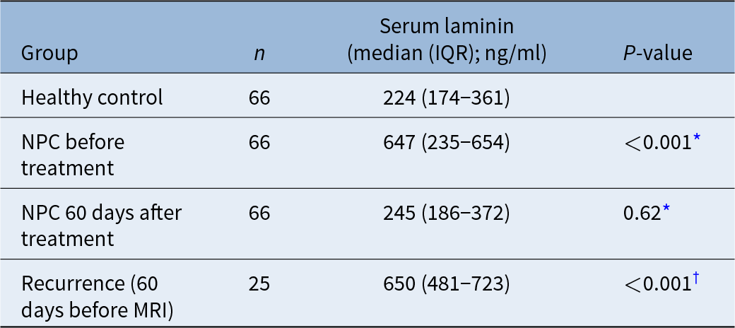
Serum laminin concentrations differed significantly between patients with NPC and healthy controls. Before treatment, NPC patients exhibited markedly elevated serum laminin levels compared with healthy individuals (median = 647 ng/ml (interquartile range = 235–654) vs 224 ng/ml (interquartile range = 174–361); p < 0.001, Mann–Whitney U test).
Following concurrent chemoradiotherapy, serum laminin concentrations measured 60 days after treatment decreased to 245 ng/ml (interquartile range = 186–372), which was not significantly different from levels observed in healthy controls (p = 0.62).
Among the 25 patients who developed recurrence, serum laminin levels increased before radiological detection of relapse. Approximately 60 days before recurrence confirmed on MRI, median serum laminin was 650 ng/ml (interquartile range = 481–723), significantly higher than the corresponding 60-day post-treatment value (p < 0.001, Wilcoxon signed rank test).
These findings suggest that circulating laminin may reflect dynamic systemic extracellular matrix turnover during tumour progression and recurrence (Table 2).
Serum laminin levels in healthy controls and NPC patients

Table 2 Long description
Serum laminin is reported as median with interquartile range in ng per ml, with group size and a P value column. Healthy control, n 66, median 224 with IQR 174 to 361, with no P value shown. NPC before treatment, n 66, median 647 with IQR 235 to 654, P value less than 0.001 with an asterisk marker. NPC 60 days after treatment, n 66, median 245 with IQR 186 to 372, P value 0.62 with an asterisk marker. Recurrence 60 days before MRI, n 25, median 650 with IQR 481 to 723, P value less than 0.001 with a dagger marker.
* Versus healthy controls (Mann–Whitney U test).
† Versus corresponding 60-day post-treatment value in the same 25 patients (Wilcoxon signed rank test). NPC = nasopharyngeal carcinoma; IQR = interquartile range; MRI = magnetic resonance imaging
Furthermore, pre-treatment serum laminin levels were significantly associated with tissue laminin expression. Patients with laminin-positive tumours exhibited significantly lower circulating laminin concentrations than those with laminin-negative tumours (median = 274 ng/ml (interquartile range = 188–648) vs 679 ng/ml (interquartile range = 641–738); p < 0.001) (Table 3).
Pre-treatment serum laminin levels by tissue laminin expression

Table 3 Long description
Tissue laminin groups are listed with sample size, serum laminin median with interquartile range in ng per ml, and a p value column. For tissue laminin, positive, n is 43 and serum laminin is 274 with an interquartile range from 188 to 648. For tissue laminin, negative, n is 23 and serum laminin is 679 with an interquartile range from 641 to 738. The p value shown is less than 0.001.
IQR = interquartile range
Spearman correlation analysis confirmed a significant negative correlation between tissue laminin expression and pre-treatment serum laminin concentrations (ρ = −0.634, p < 0.001).
Survival analysis
Kaplan–Meier survival analysis demonstrated significant differences in overall survival according to both tissue laminin expression and serum laminin levels. Patients with laminin-positive tumours exhibited significantly improved survival compared with those with laminin-negative tumours (log-rank p = 0.001) (Figure 2).
Kaplan–Meier overall survival according to tissue laminin expression. Kaplan–Meier curves showed significantly better overall survival in patients with laminin-positive tumours than in those with laminin-negative tumours. The difference between groups was significant by the log-rank test (p = 0.001). In Cox proportional hazards analysis adjusted for age and sex, tissue laminin positivity was associated with a reduced risk of mortality (hazard ratio (HR) = 0.27, 95 per cent confidence interval (CI) = 0.12–0.63). Tick marks indicate censored observations.

Similarly, patients with high serum laminin levels showed markedly worse survival outcomes compared with those with lower circulating laminin concentrations (log-rank p < 0.001) (Figure 3).
Kaplan–Meier overall survival according to serum laminin level. Patients were stratified using the receiver operating characteristic (ROC) derived pre-treatment serum laminin cut-off of 649 ng/ml. Patients with high serum laminin (>649 ng/ml; n = 18) had significantly worse overall survival than those with low serum laminin (≤649 ng/ml; n = 48). The difference between groups was significant by the log-rank test (p < 0.001). Tick marks indicate censored observations. AUC = area under the curve.

Receiver operating characteristic analysis was performed to evaluate the apparent discrimination of pre-treatment serum laminin for vital status.
The area under the curve was 0.896, indicating high apparent discrimination in this cohort. The cut-off value determined by the Youden index was 649 ng/ml, yielding a sensitivity of 69.6 per cent and specificity of 95.3 per cent for classifying vital status.
Patients were further stratified according to both tissue laminin expression and serum laminin levels. Overall survival differed among the four groups (log-rank p < 0.001) (Figure 4).
Kaplan–Meier survival curves according to combined laminin biomarkers. Patients were stratified by tissue laminin status and the receiver operating characteristic derived pre-treatment serum cut-off of 649 ng/ml: tissue-positive/low serum (n = 38), tissue-positive/high serum (n = 5), tissue-negative/low serum (n = 10) and tissue-negative/high serum (n = 13). Low and high serum laminin were defined as ≤649 and >649 ng/ml, respectively. Overall survival differed among the four groups (log-rank p < 0.001). The tissue-negative/high-serum group had the poorest survival, whereas both low-serum groups retained high survival. No pairwise comparisons were performed. Tick marks indicate censored observations. + = tissue laminin-positive; − = tissue laminin-negative.

Figure 4 Long description
The x-axis label is, Time, months. The x-axis ranges from 0 to 90. The y-axis label is, Survival probability. The y-axis ranges from 0.0 to 1.0. Four step lines are plotted. A text annotation inside the plot reads, Log-rank p less than 0.001. A legend lists four groups: Tissue plus serum less than or equal to 649 nanogram per milliliter, n equals 38; Tissue plus serum greater than 649 nanogram per milliliter, n equals 5; Tissue minus serum less than or equal to 649 nanogram per milliliter, n equals 10; Tissue minus serum greater than 649 nanogram per milliliter, n equals 13. The plotted step lines start at survival probability 1.0 near time 0 months. One line remains at 1.0 until about 50 months, then drops in steps to about 0.9 and ends near 0.9 at about 90 months. A second line remains at 1.0 until about 55 months, then drops in steps to about 0.85 and ends near 0.85 at about 90 months. A third line remains at 1.0 until about 50 months, then drops in several steps to about 0.25 by about 70 months and stays near 0.25 through about 90 months. A fourth line remains at 1.0 until about 50 months, then drops in steps to about 0.1 by about 60 months and stays near 0.1 through about 90 months.
The tissue-negative/high-serum group had the poorest survival; both low-serum groups retained high survival. No pairwise comparisons among the four groups were performed.
In order to evaluate the prognostic association of tissue laminin expression, a Cox proportional hazards regression model adjusting for age and sex was constructed. Tissue laminin positivity was associated with a reduced risk of mortality (hazard ratio = 0.27, 95 per cent CI = 0.12–0.63, p = 0.002).
Together, peri-tumoural laminin deposition and circulating laminin levels were associated with clinical outcomes in NPC, and their combined evaluation suggested potential prognostic complementarity.
Discussion
This prognostic study (level of evidence 2) found that tissue peri-tumoural laminin deposition was associated with overall survival after adjustment for age and sex, whereas longitudinal serum laminin concentrations changed after treatment and rose before radiologically confirmed recurrence (Tables 1–3, Figures 2–4). These findings extend previous nasopharyngeal carcinoma (NPC) research by combining tissue assessment with dynamic serum monitoring in one cohort and deriving a serum cut-off for vital-status classification.
• Loss or disruption of laminin-containing basement membranes is associated with tumour invasion and adverse outcomes in several epithelial malignancies
• Peri-tumoural laminin deposition and circulating laminin have not previously been evaluated together longitudinally in nasopharyngeal carcinoma (NPC)
• Circulating laminin-related analytes may reflect extracellular matrix turnover, but their longitudinal behaviour and relationship to tissue laminin in NPC remain uncertain
• In 66 patients with stage II NPC followed for five years, peri-tumoural laminin positivity was associated with improved survival
• Pre-treatment serum laminin was inversely correlated with tissue laminin status; levels decreased after chemoradiotherapy and, among patients with recurrence, rose before radiologically confirmed recurrence
• Combined tissue and serum assessment defined four groups with different overall survival, supporting potential biomarker complementarity
Peri-tumoural laminin loss and tumour progression
Our finding that loss of peri-tumoural laminin deposition was associated with poorer survival is supported by evidence from other solid tumours, where basement membrane laminin loss predicts worse outcome,Reference van der Zee, van Eijck, Hop, Biermann, Dicheva and Seynhaeve14 and is consistent with NPC literature linking matrix-degrading activity to adverse outcome. Matrix metalloproteinase-9, a protease capable of degrading basement membrane components, has also been correlated with poor prognosis in NPC.Reference Liu, Li, Yang, Luo, Li and Yang20–Reference Cheng, Kong and Li22
A comparable pattern has been reported in breast carcinoma, where continuous laminin staining is retained in benign and pre-invasive lesions but becomes disrupted in invasive disease, correlating with lymphatic invasion and nodal statusReference Zheng, Looi and Cheah23 – closely paralleling the continuity-based peri-tumoural laminin scoring used in the present study.
Loss of functional laminin anchoring also predicts outcome in other tumours: reduced expression of the glycosyltransferase LARGE, which mediates cell-surface laminin anchoring, characterises aggressive breast and brain tumours, and independently predicts poor survival in brain cancer.Reference Akhavan, Griffith, Soroceanu, Leonoudakis, Luciani-Torres and Daemen24 Low tumour expression of the laminin α3 chain (LAMA3) in ovarian cancer is likewise associated with worse five-year recurrence-free and overall survival.Reference Tang, Wang, Wang and Zhong25 A broader synthesis of the breast-cancer extracellular matrix literature similarly links higher tumoural laminin expression to better outcomes.Reference Robertson26
Some NPC studies have examined individual laminin subunits rather than organised peri-tumoural deposition. A transcriptomic study reported that tumour-cell LAMB1 promotes proliferation and metastasis in NPC,Reference Feng, Yang, Yang, Qu and Li17 seemingly opposite to our finding that peri-tumoural laminin positivity is protective. This difference may reflect distinct compartments: tumour-cell-associated LAMB1 expression versus organised extracellular deposition at the tumour–stroma interface. Similarly, laminin-332 γ2-chain reorganisation at the invasive front marks aggressive behaviour in squamous carcinomas.Reference Berndt, Gaßler and Franz9 Thus, laminin signals may differ by chain, molecular form and compartment, supporting assessment of peri-tumoural deposition as a distinct readout in NPC.
Serum laminin and recurrence
Circulating laminin-related analytes have shown disease associations in hepatocellular carcinomaReference Kiyokawa, Yasuda, Oikawa, Okuse, Matsumoto and Ikeda27 and inflammatory bowel disease.Reference Koutroubakis, Petinaki, Dimoulios, Vardas, Roussomoustakaki and Maniatis28 In colorectal cancer, pre-operative serum laminin remained an independent prognostic factor on multivariable Cox regression, with high levels predicting worse survivalReference Saito and Kameoka29 – consistent with our observed serum–vital-status association. In our cohort, serum laminin showed high apparent discrimination for vital status and rose approximately two months before radiologically confirmed recurrence. Serum laminin may provide valuable information for prognosis.
Tissue–serum inverse correlation
The inverse tissue–serum correlation is compatible with, but does not prove, proteolytic matrix remodelling with increased circulating laminin immunoreactivity. Hypoxia-driven exosomal matrix metalloproteinase-13 can degrade extracellular matrix and promote NPC metastasis,Reference Shan, You, Shi, Shi, Zhang and Zhang30 providing a possible pathway; however, neither the tissue source nor the molecular form of circulating laminin has yet been established.
Combined tissue and serum laminin assessment
This study’s originality lies in measuring laminin in two compartments and at different times. Tissue laminin, scored once at diagnosis, provides a structural snapshot of peri-tumoural deposition, whereas serum laminin was measured repeatedly and may reflect changing systemic extracellular matrix turnover. The two measures may therefore capture different temporal and biological dimensions of disease.
To our knowledge, this is the first study to combine tissue and serum laminin assessment within a single NPC cohort followed longitudinally for five years. The inverse correlation suggests potential complementarity and provides a framework for independent validation.
Limitations
Several limitations should be acknowledged. The serum cut-off and four-group analysis were derived and evaluated in the same cohort without external validation. The molecular form and tissue source of circulating laminin, and the proteases linking matrix remodelling to serum measurements, were not established. Larger multicentre cohorts with prespecified analyses are needed to validate these findings and clarify the mechanism.
Conclusion
This study identified a dual-compartment prognostic pattern in nasopharyngeal carcinoma. Preservation of peri-tumoural laminin deposition was associated with favourable outcome, whereas elevated pre-treatment circulating laminin was associated with death and levels rose before recurrence, potentially reflecting systemic extracellular matrix turnover.
Funding statement
This research was supported by the Natural Science Foundation of Fujian Province (grant numbers: 2019J01622 and 2023J011904).
Competing interests
None declared
Ethical standards
The authors assert that all procedures contributing to this work comply with the ethical standards of the relevant national and institutional guidelines on human experimentation (Institutional Ethics Review Board of Mindong Hospital (approval number: 0608-1)), and with the Helsinki Declaration of 1975, as revised in 2008.