Introduction
Vestibular schwannomas, also termed acoustic neuromas, are benign, primary, intracranial tumours. They constitute one of the extra-axial tumour types that arise from the Schwann cell sheath over the vestibular or cochlear nerve, and/or from the myelin-forming cells of the vestibulocochlear nerve (the VIIIth cranial nerve).Reference Andersen, Schrøder and Bonding1, Reference Lin, Hegarty, Fischbein and Jackler2 Vestibular schwannoma may occur sporadically or as part of von Recklinghausen neurofibromatosis.Reference McLeod, Upfold and Taylor3 Clinically diagnosed vestibular schwannomas occur in 7–10 people per 1 000 000 population; most diagnosed patients have no apparent risk factors.Reference Hansen, Clark, Gantz and Goswami4
Treatment for vestibular schwannoma includes surgical removal and radiotherapy. Approximately 25 per cent of all vestibular schwannoma cases are treated with medical management, consisting of periodic monitoring of the patient's neurological status, serial imaging studies, and the use of hearing aids when appropriate.Reference Chopra, Kondziolka, Niranjan, Lunsford and Flickinger5 Resection is indicated for patients with large tumours that have caused major neurological deficits due to brain compression. Stereotactic radiosurgery is performed for small to medium-sized tumours, with the aim of preserving neurological function and preventing further tumour growth.Reference Koerbel, Gharabaghi, Tatagiba and Samii6
Vestibular schwannoma surgery poses significant challenges with regard to definitive management. Preservation of hearing and facial nerve function is of great concern.Reference Nayak and Kumar7 Surgical removal remains the treatment of choice for tumour eradication. Various surgical approaches can be used to remove acoustic tumours. The retrosigmoid approach can be used for all acoustic tumours and provides the best wide-field visualisation of the posterior fossa.Reference Yang, Yu and Han8 The goal of the retrosigmoid approach is complete eradication of the tumour with preservation of hearing and facial nerve function.Reference Misra, Purandare, Ved, Bagdia and Mare9
Patients with vestibular schwannoma usually present late with large tumours and disabling deafness. Our study reviewed our experience over the past seven years, with an emphasis on the impact of recent refinements in microsurgical technique upon clinical outcomes.
Materials and methods
A retrospective study was conducted of patients with vestibular schwannoma treated between 2003 and 2010 at the Suez Canal University Hospital, Ismailia, Egypt. Twenty-four patients presenting with an acoustic tumour were included in our study. Surgical outcomes were assessed, along with complications, facial nerve preservation, hearing state and extent of tumour resection.
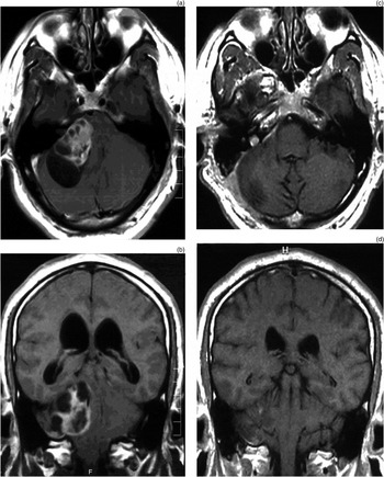
All patients underwent complete history-taking, complete ENT examination, neurological assessment, pure tone audiometry, speech audiometry, auditory brainstem response testing, contrast computed tomography, and magnetic resonance imaging with and without gadolinium contrast (Figure 1). All patients were routinely followed for at least 12 months.
(a) Axial magnetic resonance imaging (MRI) scan showing large schwannoma with cystic degeneration. (b) Coronal MRI scan showing multi-cystic degeneration within the same tumour. (c) Axial and (d) coronal MRI scans showing appearance following complete tumour removal, with some cerebellar oedema and residual intracanalicular tumour.

Tumour sizes were measured; large tumours were defined as greater than 30 × 20 mm in size, and small tumours as up to 30 × 20 mm. Tumour extent was described as follows: class T1, purely intrameatal; class T2, intra- ‘and/or’ extrameatal; class T3a, filling the cerebellopontine cistern; class T3b, reaching the brainstem; class T4a, compressing the brainstem; and class T4b, severely dislocating the brainstem and compressing the fourth ventricle.Reference Timmer and Graamans10
Facial nerve function was assessed using the House–Brackmann grading system.Reference House and Brackmann11 Hearing was classified using the Gardner–Robertson grading system, as follows: a pure tone average (PTA) of 0–30 dB was classified as grade I (i.e. good to excellent hearing); 31–50 dB (with a speech discrimination score of >50) as grade II (serviceable); 51–90 dB as grade III (non-serviceable); 91 dB or more as grade IV (poor); and no response as grade V.Reference Gardner and Robertson12
Surgical procedure
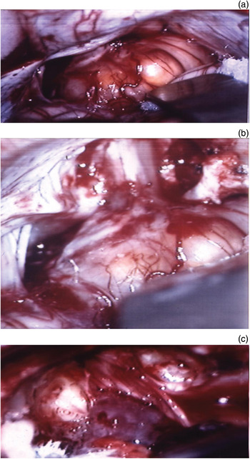
A retrosigmoid, suboccipital approach was used in all cases. Patients were placed in the supine position with the head turned towards the contralateral shoulder. Facial nerve electrodes were placed around the orbicularis oris and oculi muscles. An S-shaped incision (suboccipital craniectomy) was then made, under direct vision, to the cerebellopontine angle. Following opening of the dura, the cerebellum usually fell away with adequate exposure, and the tumour was debulked. Then, under operating microscopic vision, micro-dissection instruments were used to maintain the anterior portions of the capsule so as to avoid injury to the VIIth and/or VIIIth cranial nerves. Once the tumour had been substantially debulked, the posterior wall of the internal auditory canal could be removed using a high-speed drill, taking great care not to injure the labyrinth. Once the internal auditory canal had been exposed, the dura was opened and the tumour removed from it. The vestibular nerves were generally sacrificed, but the cochlear nerve was kept intact in order to preserve hearing (Figure 2).
Serial intra-operative photographs showing different phases of microsurgical tumour excision: (a) tumour exposure with gentle cerebellar retraction; (b) drilling the acoustic canal to expose the intracanalicular part of the tumour; and (c) peeling the tumour off the intracanalicular part of the VIIth and VIIIth nerve complex, preserving the arachnoid layer.

The root entry zone of the VIIth cranial nerve was easy identified. Facial nerve monitoring was of great help at this stage of the dissection, to facilitate preservation of VIIth nerve function. All of the lower cranial nerves and draining veins were preserved.
Good haemostasis was secured, the dura closed primarily, and the wound closed in layers. The patient was managed post-operatively on ventilation.Reference Nayak and Kumar7
Statistical analysis
Data were processed using the SPSS version 15 statistical software program (SPSS Inc, Chicago, Illinois, USA). Quantitative data were expressed as means ± standard deviations (SDs), while qualitative data were expressed as frequencies and percentages. Student's t-test was used to test the significance of differences for quantitative variables that followed a normal distribution.
Ethical considerations
Written consent was obtained from all patients, or from first-degree relatives, before the study. The stages in the operative procedure were explained to all patients. The local ethics committee approved all surgical procedures.
Results and analysis
The 24 patients with vestibular schwannoma included 2 patients with neurofibromatosis type 2. Thirteen patients were female and 11 male. The mean patient age was 39.7 years (range, 16–58 years). The tumour was on the left side in 12 patients and on the right side in 12 patients. There were no significant differences regarding sex or tumour site.
All of the patients presented with unilateral sensorineural hearing loss with marked speech discrimination impairment and tinnitus. In addition, 14 patients (58.3 per cent) had a history of vertigo attacks with unsteadiness, and 9 (37.5 per cent) reported headache.
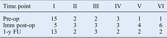
All 24 patients had pre-operative facial nerve involvement: grade I in 15 patients, grade II in 2 patients, grade III in 2 patients, grade IV in 3 patients, grade V in 1 patient and grade VI in 1 patient, according to the House–Brackmann classification of VIIth nerve function (Table I).
Viith nerve function: house–brackmann grade
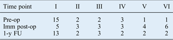
Pre-op = pre-operative; Imm post-op = immediate post-operative; 1-y FU = one-year follow up
Gardner–Robertson classification of pre-operative VIIIth nerve hearing function revealed 1 patient with grade I function, 3 patients with grade II, 12 patients with grade III, 3 patients with grade IV and 5 patients with grade V (Table II).
Viiith nerve hearing function: gardner–robertson grade

Pre-op = pre-operative; post-op = post-operative
Auditory brainstem response findings showed a prolonged I–III interval and I–V inter-peak latencies in all patients, with 85 per cent specificity for distinguishing vestibular schwannoma.
Tumours were larger than 30 × 20 mm in all patients. Three patients had tumours of class T3a size (i.e. filling the cerebellopontine cistern). Ten patients had class T3b tumours (reaching the brainstem). Six patients had class T4a tumours (compressing the brainstem), while five patients had class T4b tumours (severely dislocating the brainstem and compressing the fourth ventricle).
Four patients required ventriculoperitoneal shunting to treat hydrocephalus. Later in the study, another four patients required endoscopic third ventriculostomy.
Complete tumour removal was successful in 19 patients. In two patients, there was retained tumour attached to the internal auditory canal; this was treated with gamma knife surgery.
The mean duration of the surgical procedure was 4.9 hours (SD, 4–9 hours). The mean hospital stay was 8 days (SD, 6–16 days). None of the patients needed peri-operative blood transfusion.
Facial nerve function
Intra-operative facial nerve identification and preservation were achieved in 16 patients (66.6 per cent). The immediate post-operative House–Brackmann grading of VIIth nerve function was grade I in five patients, grade II in three patients, grade III in three patients, grade IV in three patients, grade V in four patients and grade VI in six patients (Table I).
After 1 year of follow up, 13 (54.1 per cent) patients showed complete recovery of facial nerve function (i.e. House–Brackmann grade I), while 2 patients were grade II, 3 patients were grade III, 2 patients were grade IV, 2 patients were grade V and 2 patients were grade VI (Table I).
Hearing preservation
Pre-operatively, four patients had useful hearing (i.e. Gardner–Robertson grades I or II).
Post-operatively, useful hearing was preserved in 4 patients (grade I hearing in 2 patients and grade II hearing in 2 patients), while 10 patients had grade III hearing, 6 patients had grade IV hearing and 4 patients had grade V hearing (Table II).
Postoperative complications
Post-operatively, cerebrospinal fluid (CSF) leakage was diagnosed in two patients (8.3 per cent), both of whom responded to conservative therapy.
In addition, one patient developed trigeminal nerve dysfunction, a second showed unsteadiness and a third developed hemiparesis.
Discussion
The operative mortality rate for vestibular schwannoma has dropped dramatically, from 40 per cent at the beginning of the last century to less than 1–2 per cent in the past decade.Reference Nayak and Kumar7 The introduction of microsurgical techniques and electrophysiological monitoring has improved the anatomical and functional preservation of the facial and vestibulocochlear nerves.Reference Kim and Nam13 The primary goal of vestibular schwannoma surgery has changed from preserving the patient's life to preserving their neurological function.Reference Yamakami, Uchino, Kobayashi, Yamaura and Oka14
In 1979, Cohen was one of the first to report the outcome of surgery using the retromastoid approach aimed at hearing preservation: 8 per cent facial nerve paralysis and no reported mortality.Reference Cohen15
In 1993, Glasscock and colleagues' study of 161 selected patients found that the retromastoid and middle fossa approaches resulted in a lower incidence of temporary facial nerve paresis.Reference Glasscock, Hays, Minor, Haynes and Carrasco16
That same year, Mazzoni et al. reported that, in 90 out of 300 patients operated upon using the retromastoid approach, the facial nerve was anatomically preserved in 99 per cent, with completely normal function in 78 per cent, while the cochlear nerve was anatomically preserved in 96 per cent, with functionality maintained in 44 per cent.Reference Mazzoni, Calabrese, Danesi and De Nigris17
Achievement of complete tumour excision depends on many factors, including tumour size, tumour site and extent, tumour consistency, existence of a capsule, presence of an arachnoid plane between the tumour and the brain, and anatomical variations in the cerebellopontine angle.Reference Sampath, Rini and Long18 In contrast, facial nerve outcomes usually depend solely on tumour size: if the tumour is less than 1.5 cm, a good facial nerve function can be expected (i.e. House–Brackmann grade I–II) in more than 90 per cent of patients.Reference Nayak and Kumar7
In our study, 19 tumours were completely excised, 5 of which were cystic. We found that cystic tumours were tightly adherent to the facial nerve or to other cranial nerves, making complete tumour excision more difficult and sometimes impossible without significant risk. Sampath et al. also reported this finding.Reference Sampath, Rini and Long18
Surgical treatment of vestibular schwannoma aims for complete tumour resection with ongoing reduction in morbidity, and this aim is well supported by the retromastoid approach.Reference Timmer and Graamans10 Bentivoglio et al. reported that when the retromastoid approach was used, facial and cochlear nerve function was preserved in more than 90 per cent of patients, independent of tumour size; however, in general these patients had tumours larger than 30 mm in diameter.Reference Bentivoglio, Cheesman and Symon19
In addition, hearing preservation has been reported in patients with intracanalicular tumours, with the best results reported by surgical teams experienced in such surgery.Reference Timmer and Graamans10, Reference Kondziolka, Lunsford and Flickinger20 In these studies, hearing preservation (i.e. a hearing threshold of less than 50 dB) was achieved in four of five patients with a pre-operative hearing threshold of less than 50 dB and a tumour smaller than 2.5 cm. Acoustic nerve preservation should be attempted in all cases with measurable hearing, regardless of tumour size.Reference Torrens, Maw, Coakham, Butler and Morgan21 Kurokawa et al. studied 35 cases of unilateral acoustic neurinoma in which surgery was performed using the retromastoid approach, and reported anatomical preservation of the facial nerve in all cases and anatomical preservation of the cochlear nerve in 14 of 35 cases (40 per cent).Reference Kurokawa, Uede, Ohtaki, Tanabe and Hashi22
Most of our patients with facial nerve dysfunction showed an improvement at one-year follow up. Similarly, Anderson et al. found that 80 per cent of their patients gained normal to near-normal facial nerve function.Reference Anderson, Leonetti, Wind, Cribari and Fahey23 Timmer and Graamans noted that factors affecting facial nerve preservation included tumour size, tumour extent, tumour type (especially whether cystic or not), surgical technique (i.e. revision surgery vs gamma knife surgery) and surgeon experience.Reference Timmer and Graamans10
Hearing is much more difficult to conserve in 1.5–2.0 cm diameter tumours than in small, intracanalicular tumours. The most important factors predicting hearing preservation are tumour size, tumour extent and pre-operative hearing level.Reference Timmer and Graamans10
In 2005, Betchen reviewed 142 patients after hearing preservation surgery, and found that 38 (26.7 per cent) had immediate post-operative hearing confirmed by pure tone audiometry.Reference Betchen, Walsh and Post24 Long-term follow up (over 7 years) showed that 35 patients (85.7 per cent) had long-term hearing preservation.Reference Betchen, Walsh and Post24
In our study, CSF leakage was diagnosed in two patients (8.3 per cent); these patients responded to conservative therapy with significant improvement. Suggested risk factors for CSF leakage include infection, hydrocephalus, inadequate wound closure or healing, and opened mastoid air cells.Reference Timmer and Graamans10
• Vestibular schwannoma treatment options include observation with serial imaging, stereotactic radiosurgery and microsurgical removal
• Surgery aims for complete tumour removal with hearing and facial nerve function preservation
• This retrospective series used a retromastoid, suboccipital approach
• Tumour was completely removed in 19 of 24 patients
• Anatomical facial nerve preservation was achieved in 67 per cent
• Useful hearing (where present) was preserved in 80 per cent
The major advantage of the retromastoid approach is its applicability in all acoustic tumour cases, as it provides the best wide-field visualisation of the posterior fossa. The inferior portions of the cerebellopontine angle and the posterior surface of the temporal bone anterior to the porus acousticus are clearly observed.Reference Nayak and Kumar7 The disadvantages of the retromastoid approach are: (1) it may require cerebellar retraction; and (2) manipulation of the cerebellum provides opportunities for post-operative oedema, haematoma, infarction and haemorrhage.Reference Nayak and Kumar7
Experience has a remarkable influence on the outcome of vestibular schwannoma surgery, as well as on the prevalence of surgical morbidity. Furthermore, we believe that repeated practice will never cease to enhance the surgeon's skill.
Conclusion
In patients with vestibular schwannoma, the retromastoid, suboccipital approach to the skull base can be safely and successfully utilised with minimal or no damage to neurovascular structures, using a microsurgical technique, even for large tumours. Complete removal of the tumour is possible using a minimally invasive surgical technique, with a positive effect on quality of life. It is possible to obtain near-normal to normal VIIth nerve function in most patients. Seventh cranial nerve function should be monitored for up to one year to allow a full opportunity for nerve recovery.