





Irritable bowel syndrome (IBS) is a functional gastrointestinal (GI) disorder, with a complicated pathology in which not all mechanisms underlying the aetiology and pathophysiology are understood(Reference Hojo and Nagahara1,Reference Vahedi, Ansari and Mir-Nasseri2) . As a result, no adequate treatment for patients with IBS is available yet. Nonetheless, dietary intake plays an important role in symptom onset. The majority of patients with IBS report that their symptoms are provoked by certain meals, with greasy and fried foods, high in saturated fats, being one of the most important triggers for their symptoms(Reference Zaribaf, Keshteli and Esmaillzadeh3–Reference Tomassen, Govers and Vos6). Moreover, there is evidence suggesting that patients with IBS exhibit alterations in lipid metabolism, including elevated plasma TAG levels and changes in fatty acid absorption, although the underlying mechanisms and direction of causality remain unclear(Reference Guo, Niu and Momma7–Reference Zhan, Zheng and Li10). Furthermore, genetic factors, the gut microbiota composition, stress, increased mucosal permeability and low-grade mucosal inflammation have all been reported to contribute to the pathophysiology of IBS(Reference Hojo and Nagahara1,Reference Vahedi, Ansari and Mir-Nasseri2) . An increased mucosal barrier permeability in patients with IBS can facilitate the passage of luminal antigens, leading to increased activation of the intestinal immune system(Reference Öhman, Törnblom and Simrén11). This might result in an increased release of pro-inflammatory cytokines, leading to the stimulation of the nervous system, impairments in motility, low-grade inflammation and hypersensitivity of the GI tract(Reference Öhman, Törnblom and Simrén11).
Increased activation of the immune system in patients with IBS is supported by a study of Dlugosz and colleagues(Reference Dlugosz, Nowak and D’Amato12), who have shown that IBS patients with predominant diarrhoea complaints (IBS-D) have higher serum concentrations of lipopolysaccharide (LPS) compared with controls, indicating subclinical inflammation. LPS is a cell wall component of gram-negative bacteria and naturally present on the skin and in the gut. When the gut barrier function is working effectively, only low amounts of LPS will be translocated from the gut lumen to the blood circulation(Reference Laugerette, Vors and Peretti13–Reference Fuke, Nagata and Suganuma15). Evidence from in vitro studies, animal studies and human studies shows that the ingestion of high-fat meals facilitates the translocation of LPS from the gut to the lymph and further into the circulation, potentially triggering endotoxemia associated with low-grade inflammation(Reference Laugerette, Vors and Peretti13–Reference van Dijk, Mensink and Esser18). In literature, several mechanisms have been proposed to explain this process, including transcellular transport of LPS, either via chylomicron-mediated translocation through enterocytes or via M-cells, as well as increased paracellular permeability due to disrupted tight junctions(Reference Ghoshal, Witta and Zhong14). Most likely, fat-induced LPS translocation is regulated via transcellular transport. After uptake in the enterocytes, LPS is transported to the Golgi apparatus and incorporated into chylomicrons, mainly after ingestion of a high-fat load(Reference Laugerette, Vors and Peretti13,Reference Ghoshal, Witta and Zhong14,Reference Erridge, Attina and Spickett16) .
A specific marker for circulating chylomicron particles in the blood is Apo-B48, which is formed in the small intestine(Reference Nakajima, Nagamine and Fujita19), and plays a superior role in the control of intestinal lipid transport in response to dietary lipid intake(Reference Lo and Coschigano20). Previous research indicates that there is a positive correlation between postprandial Apo-B48 and LPS levels(Reference Carnevale, Pastori and Nocella21). This reinforces the hypothesis that fat-induced LPS translocation is mediated by chylomicrons. Nevertheless, recent in vitro and animal studies indicate that also other, chylomicron-independent, mechanisms might be associated with fat-induced LPS translocation(Reference Tomassen, Govers and Vos6). In the circulation, LPS can bind to the CD14 receptor on the plasma membrane of various immune cells, which initiates the activation of these cells and induces the secretion of pro-inflammatory cytokines, such as IL-1, IL-6, TNFα and nitric oxide(Reference Laugerette, Vors and Peretti13,Reference Ciesielska, Matyjek and Kwiatkowska22) . Subsequently, this may generate inflammatory events in the vascular endothelium and peripheral tissues(Reference Michalski, Vors and Lecomte23), potentially contributing to the development of low-grade inflammation and, consequently, the pathophysiology of individuals with IBS(Reference Ng, Soh and Loke24).
There is increasing interest in medicines and food supplements to manage IBS. Curcumin and other curcuminoids, major components of turmeric and commonly used dietary supplements and colouring agents, have demonstrated anti-inflammatory and antioxidant properties and are suggested to modulate the gut microbiota(Reference Ng, Soh and Loke25–Reference Kahkhaie, Mirhosseini and Aliabadi27). Curcuminoids have been found to suppress cytokine production of macrophages, dendritic cells and T-cells and to target multiple signalling molecules such as NF-kB in humans, suggesting their potential as therapeutic agents for inflammatory and immune-related diseases(Reference Kahkhaie, Mirhosseini and Aliabadi27). A meta-analysis of three randomised controlled trials in 326 patients with IBS, with study durations ranging from 4 to 8 weeks, showed beneficial effects of turmeric supplementation on IBS symptoms(Reference Ng, Soh and Loke25). Animal and in vitro studies have shown reduced LPS levels(Reference Cai, Lu and Zou28,Reference Ghosh, Bie and Wang29) , improved intestinal barrier function(Reference Ghosh, Bie and Wang29), inhibited secretion of chylomicrons(Reference Ghosh, He and Wang26,Reference Feng, Ohlsson and Duan30,Reference Yang, Zou and Mai31) and increased adiponectin levels(Reference Den Hartogh, Gabriel and Tsiani32), upon curcumin(oids) supplementation. Therefore, we hypothesised that supplementation with a turmeric formula (TF) would result in decreased concentrations of postprandial LPS, Apo-B48 and TAG in people with IBS.
To date, no studies are available on the effect of curcuminoids on the translocation of LPS in humans. Given that IBS symptoms vary over time and are frequently meal-related, the postprandial period is clinically relevant to study mechanistic triggers. Human evidence further suggests that curcumin can modulate gut barrier-related and inflammatory markers within 3 d, further supporting the feasibility of an acute intervention approach.
Therefore, the aim of the current exploratory randomised, placebo-controlled, double-blind, crossover study was to investigate whether 3-d supplementation with a TF can diminish intestinal translocation of LPS in patients with IBS after consumption of a high-fat shake. To gain further understanding of the underlying mechanism, we also measured postprandial circulating Apo-B48 and TAG levels as markers of chylomicron metabolism. Furthermore, the effect of TF on GI complaints was determined.
Methods and materials
Ethics
This study was approved by the Medical Research Ethics Committee Utrecht, the Netherlands (NL75915.041.21, MEC approval number: 21/324), and was registered at clinicaltrials.gov (NCT05016596). The study was conducted in accordance with the principles of the Declaration of Helsinki (Fortaleza, Brazil 2013) and the requirements described in the EU Clinical Trials Directive 2001/20/EC transposed in the Revision of the Dutch Medical Research involving Human Subjects Act (WMO, effective as of 1 March 2006). Written informed consent was obtained from each subject prior to inclusion in the study.
Study participants
This study was conducted between February and March 2022 at Wageningen University & Research, the Netherlands. Subjects were recruited between November 2021 and January 2022, using the in-house database, advertisements on social media and via the website and social media of the Dutch IBS patient association. Inclusion criteria were patients with IBS meeting the Rome IV criteria including the criteria specific for the diarrhoea (IBS-D) or the mixed (IBS-M) predominant subtype, age between 18 and 70 years and BMI between 18·5 and 30 kg/m2. The main exclusion criteria were having GI or significant systemic diseases, history of intestinal surgery, smoking and the use of medication or dietary supplements that could interfere with the study outcomes, including codeine, antibiotics and prebiotics or probiotics. Eligibility was assessed during a screening visit, where a questionnaire was administered to check inclusion and exclusion criteria, and BMI was assessed. Subjects were enrolled by the study coordinator.
Study design and procedures
The study consisted of a randomised, double-blind, placebo-controlled, crossover design. The primary outcome was to determine the effect of 3-d TF supplementation on fasting and postprandial LPS concentrations, compared with placebo. Secondary outcomes included fasting and postprandial Apo-B48 concentrations, and tertiary outcomes included fasting and postprandial TAG concentrations, GI complaints, stool frequency, stool consistency and symptom severity.
Subjects visited the research facility in Wageningen twice to attend two high-fat shake challenges, with a wash-out period of at least 3 weeks in between. On the 2 d before and on the morning of each visit, subjects took either a turmeric or control placebo supplement. On the night before each visit, subjects consumed a low-fat standardised meal (nutritional composition is detailed in Table S1 supplementary data), after which they stayed fasted overnight. During each visit, subjects consumed a high-fat shake of 500 mL (consisting of 95 g fat, 25 g carbohydrate, no lactose and 8 g protein; full nutritional composition is detailed in Table S1 supplementary data) in the morning, which had to be consumed within 10 min. Blood samples were collected before and up to 5 h after consumption of the high-fat shake. Subjects reported their GI complaints (bloating, flatulence, nausea, heartburn and abdominal pain) on a VAS scale before and each hour after consumption of the high-fat shake, for 5 h in total. Three hours after consumption of the high-fat shake, subjects ate a banana, followed by a lunch after the last blood collection. On the 2 d before, during and the 2 d after each visit, subjects completed daily questionnaires via an ecological momentary assessment app (EMA, LifeData, LLC). In this app, subjects reported their stool frequency, stool consistency by using the Bristol Stool Scale (BSS), GI complaints and information about supplement compliance and medication intake. Before each test day, they completed a validated questionnaire to assess symptom severity (IBS-SSS)(Reference Francis, Morris and Whorwell33).
Dietary supplement
The TF (a dried colloidal suspension of curcuminoids as TurmipureGold®, Givaudan France Naturals) and the acacia gum control (yellow/orange dyed acacia gum with Sunset yellow dye (E110), Givaudan France Naturals, Avignon, France) were provided in capsules with the same appearance (Swedish orange capsules, Capsugel, Lonza). The 300 mg TF capsule supplement consisted of 101·2 (sd 2·3) mg curcuminoids of which 86·6 (sd 1·9) mg curcumin (mean and standard deviation of the quantification realised on ten capsules). Subjects had to consume one capsule with breakfast on the 3 d before the challenge test, of which the last capsule was consumed in the fasted state on the morning of the challenge test visit, 1 h before consuming the high-fat shake. Based on a previous human pharmacokinetic study with the same TF dosage, the time to maximal concentration of curcuminoids in blood is reached approximately 3 h after consumption of the TF capsule in fasting conditions(Reference Fança-Berthon, Tenon and Bouter-Banon34), for that reason the peak concentration of curcuminoids is expected approximately 2 h after consumption of the high-fat shake.
Lipopolysaccharide, Apo-B48 and TAG analysis
Fasting and postprandial LPS, Apo-B48 and TAG levels were measured in serum samples collected at baseline and every hour, up to 5 h, following high-fat shake consumption. Apo-B48, as a specific marker of chylomicrons, was assessed using an Apo-B48 ELISA assay (AKHB48, XCeltis GMBH), according to the manufacturer’s protocol. LPS was measured with a chromogenic Pyrochrome® assay (Associates Cap. Cod Inc.; distributed by Nodia BV). The assay was performed in a Pyros Kinetix Flex tube reader (pKFlex; Nodia BV). Pyrochrome lysate was reconstituted with 3·4 mL Pyrochrome reconstitution buffer (C1500-5; Associates Cap. Cod Inc.) and for the calibration curve, Limulus Amebocyte lysate (LAL) control standard with an LPS concentration of 0·5 µg/vial (CSEE0005-1; Associates Cap. Cod Inc.) was used. First, the serum samples were diluted with LPS-free Milli-Q water and incubated for 15 min in a water bath at 70 °C. After 1 h cooling down at 4 °C, the samples were placed at room temperature for 15 min. Subsequently, 200 µL of the pre-treated sample or control standard sample was added in a pKFlex glass tube with 50 µL Pyrochrome lysate. The mixture was stirred briefly and directly after measured on the pKFlex. The endotoxin concentration was calculated using the Pyrosexpress 21 CFR Part 11 compliant software. TAG levels were measured using the Triglycerides liquicolor mono kit (#10720P, Human Diagnostics), with minor modifications to the manufacturer’s protocol, as the assay was completely performed in a 96-well plate. To this end, the used amounts in the original protocol were reduced by a factor 4. TAG levels were determined after enzymatic hydrolysis by lipases, followed by a series of coupled reactions in which quinoneimine as an indicator was formed under the catalytic influence of peroxidase. This reaction product was measured at an absorbance of 500 nm in a Tecan Spark spectrophotometer (Tecan Life Sciences). The assay was executed in duplicate, and TAG concentrations were calculated using the manufacturer’s instructions. To align with clinical interpretations for atherosclerotic risk, a conversion factor of 200 mg/dL was applied, where 1 mg/dL corresponds to 0·0114 mmol/L.
Sample size and randomisation
No data on effect size for the intervention product tested in the current study were available. Therefore, data from an earlier study that measured postprandial LPS levels after an intervention with resveratrol and polyphenol in healthy subjects (n 10; μ = 0·07; σ = 0·05) were used to calculate the sample size(Reference Ghanim, Sia and Korzeniewski35). To allow for a smaller effect, the sample size was calculated based on a mean difference of 0·035 and an sd of 0·05. With a power of 80 % and a significance level of 0·05, nineteen subjects would be needed in a crossover design. To ensure equal treatment orders, twenty patients with IBS-D or IBS-M were included.
Subjects were stratified according to their age (two groups) and BMI (two groups) and randomly assigned to one of the two treatment orders using block randomisation by the study coordinator. Blocks of two subjects were created, and the allocation sequence within each block was generated using randomly generated numbers in Excel. An independent scientist assigned a unique code to the two study products. All researchers of the project team, except for the independent researcher, were kept blinded to the assignment of treatment, and so were the study subjects. After database lock and after signing the statistical analysis plan, the key to the treatment allocation was shared with the other investigators.
Data and statistical analyses
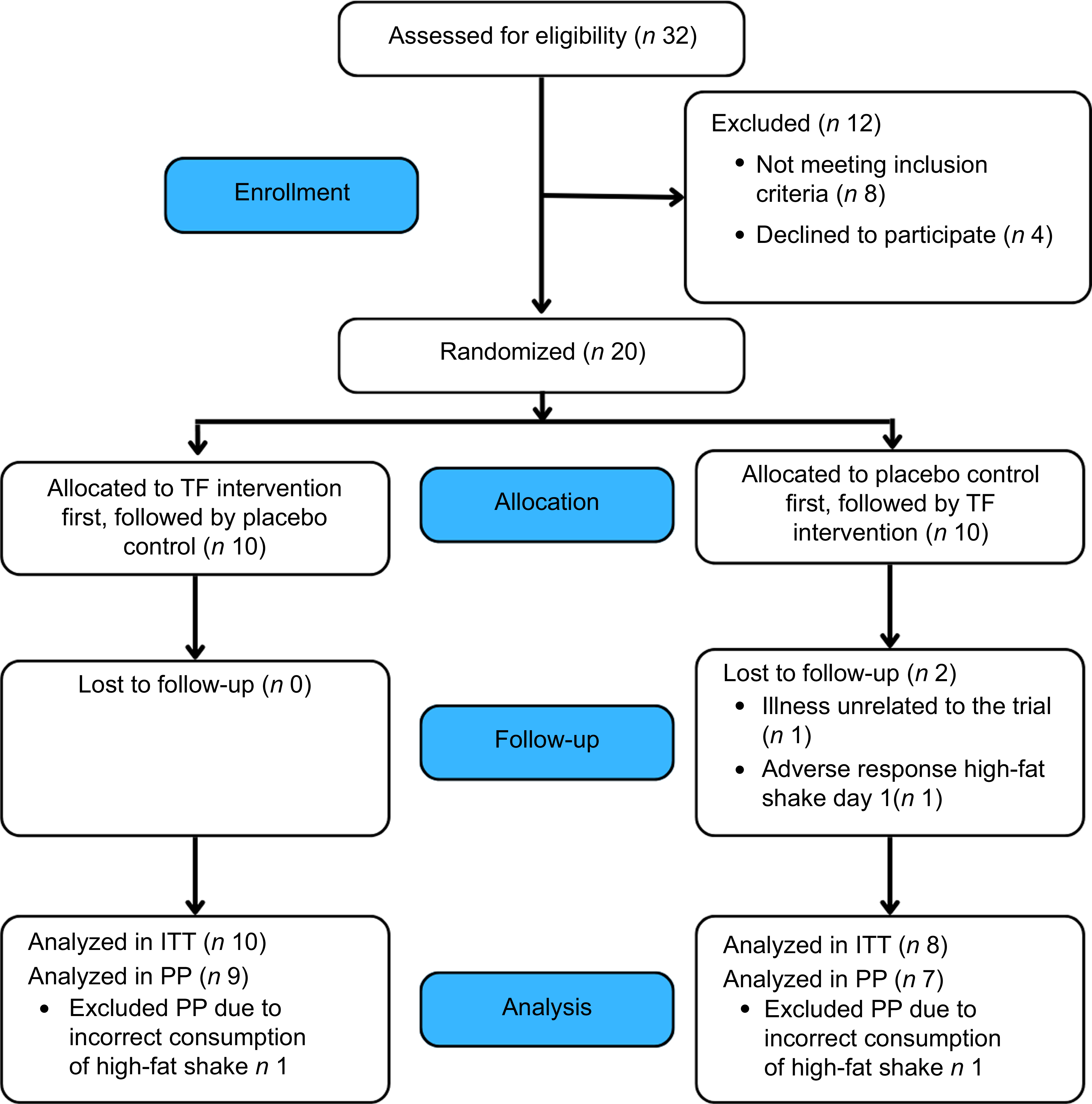
Intention-to-treat analyses were performed for all outcomes and for all randomised subjects who received any study supplement and who completed both study visits. Per protocol analyses excluded subjects with major protocol deviations. After inclusion, two subjects dropped out of the study. Therefore, eighteen subjects completed the study (Figure 1). For per protocol analyses, two subjects were excluded from the LPS and Apo-B48 analyses because of protocol deviations related to (mal-)consumption of the high-fat shake. Results show per protocol analyses for LPS and Apo-B48 and Intention-to-treat analyses for all other variables.
Flow chart diagram of the participant selection and randomisation process. ITT, intention to treat; PP, per protocol; TF, turmeric formula.

All statistical analyses were performed using R (version 4.0.2). Continuous data are presented as mean and standard deviation. Normality distributions of LPS, Apo-B48 and TAG levels were examined using the Shapiro–Wilk normality test. Since data were not normally distributed, data were transformed using log10 transformation. Statistical analyses were conducted on the log10-transformed data, and reported effect sizes and CI were back-transformed, resulting in ratios being reported.
Graphs and tables present the results in terms of absolute values. Differences in postprandial LPS, Apo-B48, TAG levels and GI complaints reported during the test days between TF and placebo were analysed using repeated-measures ANOVA.
In the repeated-measures ANOVA analysis, the influence of treatment, the kinetics of the postprandial response and the interaction of the two factors (time/treatment) were examined. Post hoc pairwise comparisons for the supplement were conducted using estimated marginal means (EMM), from the ‘emmeans’ package in R. To test for differences in fasting values (time point 0), Student’s paired t tests were performed on the log-transformed data, for LPS, Apo-b48 and TAG levels. Regression analysis was performed for Apo-B48 and LPS values, using Pearson’s correlation coefficients.
Differences in daily reports of GI complaints, stool frequency and stool consistency between TF and placebo were analysed using linear mixed model regression analyses. The model included time and supplement as factor effects, time*supplement as an interaction effect and subject as a random effect. Model comparisons were performed to assess the independent effects of time and supplement and to evaluate the interaction between time*supplement. To check whether the wash-out period of 3 weeks was sufficient, the effect of the period on daily reported GI complaints was examined using linear mixed model regression analysis, including supplement*period as an interaction effect and subject as a random effect. Questionnaire data on symptom severity, reported at baseline before each visit, was analysed using ANOVA to test for period and allocation order effects.
Results
Participants, baseline characteristics, compliance and adverse events
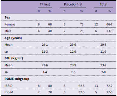
A total of eighteen participants (six males and twelve females) completed the study. Their mean age was 29·3 years, ranging from 19 to 61 years. Their mean BMI was 23·7 kg/m2, ranging from 20·5 to 28·1 kg/m2. According to the ROME-IV criteria, thirteen subjects were classified within the IBS-D subgroup and five subjects within the IBS-M subgroup (Table 1). Compliance of supplement intake was 100 %, based on reported data in the daily questionnaire. Average daily questionnaire compliance was 96·5 %. A total of six adverse events, possibly or probably related to the treatment or study procedures, were reported. All these events were mild to moderate and involved nausea, abdominal pain, dizziness and headache.
Study participant baseline characteristics (ITT population, n 18)*

Table 1. Long description
Image fully described in caption.
ITT, intention-to-treat; TF, turmeric formula; IBS-D, diarrhoea-predominant irritable bowel syndrome; IBS-M, mixed irritable bowel syndrome.
* Participants did not use any IBS-related medication or supplements; probiotic and prebiotic supplements were discontinued at least 4 weeks prior to study entry. Habitual use of vitamin supplements was permitted to continue throughout the study.
Postprandial lipids (lipopolysaccharide and Apo-B48 and TAG)
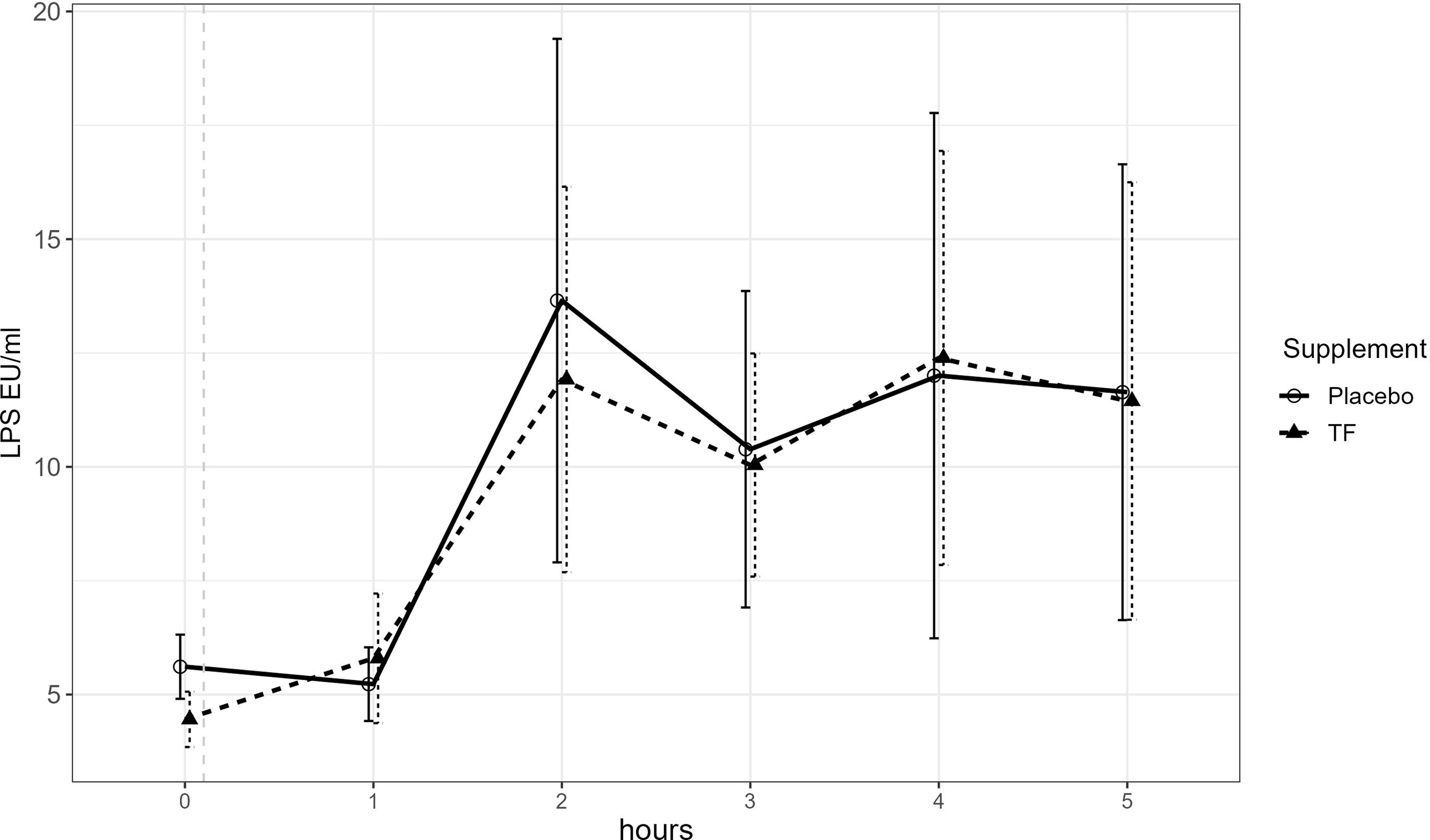
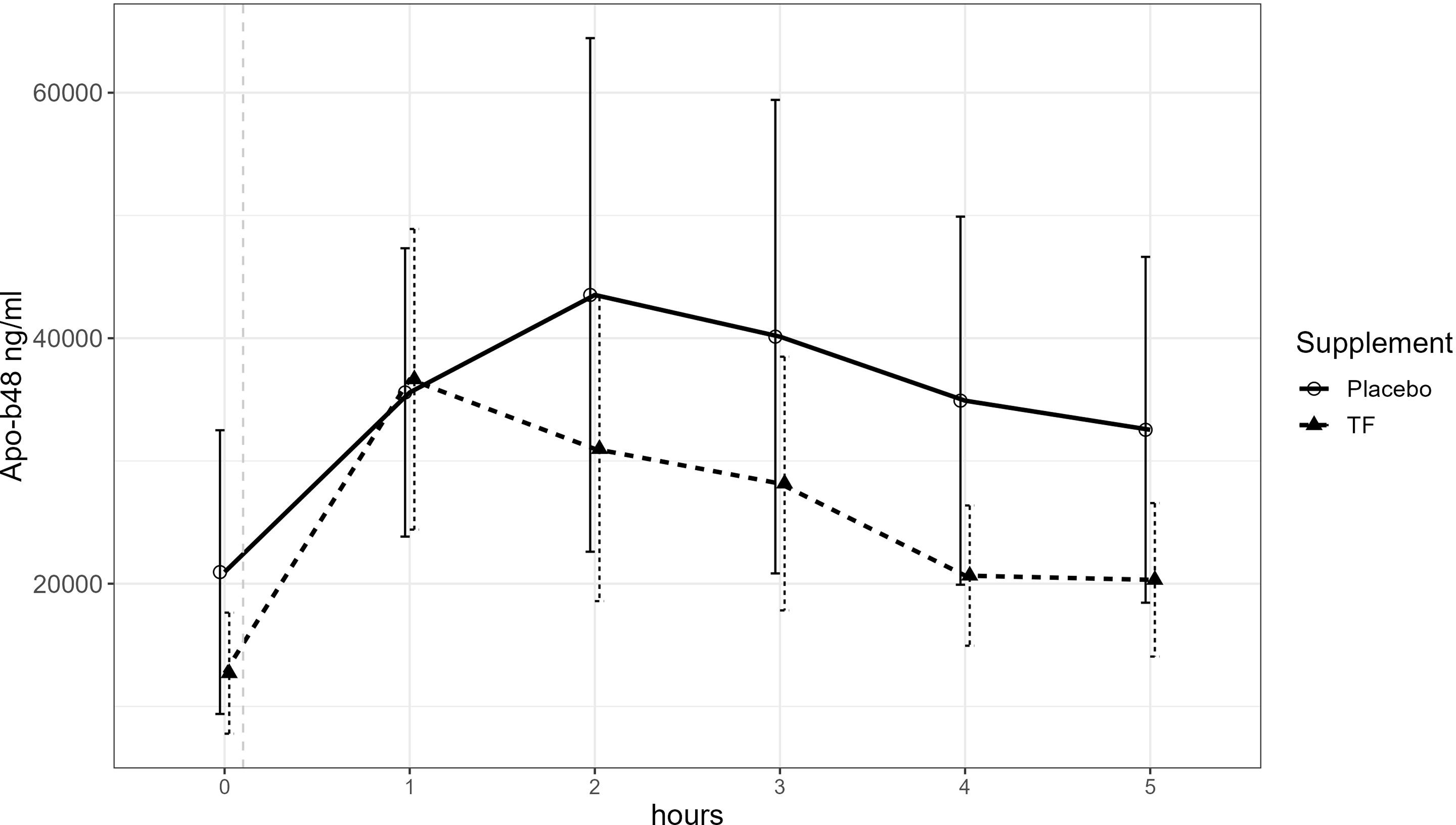
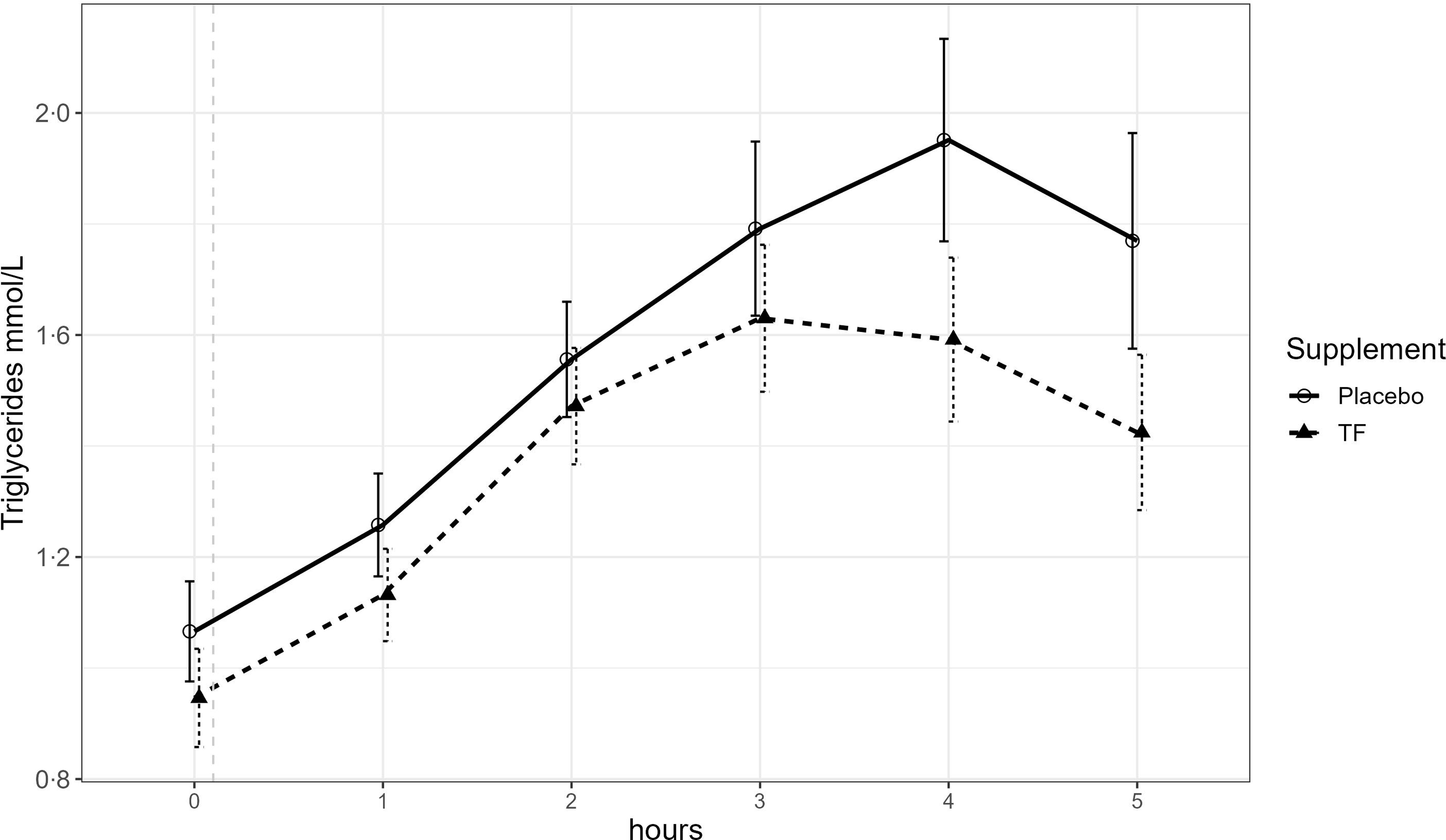
LPS in serum showed a steep increase after the high-fat challenge, as illustrated in Figure 2 and detailed in Table S2 supplementary data. The postprandial LPS response, however, was not significantly different after supplementation with either TF or placebo (mean ratio: 0·98, 95 % CI 0·85, 1·13, p = 0·98). Pre-challenge fasting LPS concentrations were slightly, but non-significantly, lower after 3-d supplementation with TF compared with placebo (mean ratio: 0·79, 95 % CI 0·61, 1·0, p = 0·06). In contrast to the results of LPS, significantly lower postprandial Apo-B48 and TAG levels were found after supplementation with TF as compared with placebo (Apo-B48: mean ratio = 0·82, 95 % CI 0·674, 0·989, p = 0·04, TAG: mean ratio = 0·87, 95 % CI 0·79, 0·97, p = 0·01, Figures 3 and 4, Table S2 supplementary data). Meaning that on average, Apo-B48 levels were 18 % lower, and TAG levels were 13 % lower, after TF supplementation compared with placebo. After 3-d supplementation with TF, fasting TAG levels were slightly, but nonsignificantly, lower (mean ratio = 0·87, 95 % CI 0·76, 1·0, p = 0·06). Fasting Apo-B48 concentrations were also not significantly different between supplements (mean ratio = 0·85, 95 % CI 0·34, 2·17, p = 0·72). When combining both treatments, LPS showed a weak but significant, positive correlation with Apo-B48 (R 0·25, 95 % CI 0·12, 0·38, p < 0·001). This indicates that higher circulating LPS levels are weakly correlated with higher circulating Apo-B48 levels.
Postprandial LPS response after an HF challenge following 3-d TF or placebo supplementation. Absolute LPS values (endotoxin units (EU)/mL) are presented before (t = 0 h) and after consumption of an HF shake, n 16. The dotted grey line indicates the time point of HF shake consumption. Based on repeated-measures ANOVA, TF did not significantly lower postprandial LPS levels compared with placebo. Fasting levels (t = 0) tend to be lower after TF supplementation compared with placebo. LPS, lipopolysaccharide; HF, high-fat; TF, turmeric formula.

Postprandial Apo-B48 response after an HF challenge following 3-d TF or placebo supplementation. Absolute Apo-B48 values (ng/ml) are presented before (t = 0 h) and after consumption of an HF shake, n 16. The dotted grey line indicates the time point of HF shake consumption. Based on repeated-measures ANOVA, TF significantly reduced postprandial Apo-B48 levels compared with placebo. Fasting levels (t = 0) did not differ after TF supplementation compared with placebo. HF, high-fat; TF, turmeric formula.

Postprandial TAG response after an HF challenge following 3-d TF or placebo supplementation. Absolute TAG values (mmol/L) are presented before (t = 0 h) and after consumption of an HF shake, n 16. The dotted grey line indicates the time point of HF shake consumption. Based on repeated-measures ANOVA, TF significantly reduced postprandial TAG levels compared with placebo. Fasting TAG levels (t = 0) tend to be lower after TF supplementation compared with placebo. TF, turmeric formula; HF, high-fat.

Gastrointestinal complaints, stool pattern and irritable bowel syndrome severity symptoms
Overall, subjects reported low to moderate GI complaints on the 2 d before, during and the 2 d after the challenge day (Figs. S1 and S2, supplementary data) with mean scores below 3 (on a ten-point scale) for all complaints, except for nausea (mean is 4). For none of the complaints, there was a significant difference in scores between supplements over time. For both supplements, nausea and bloating showed a peak 1 hour after the high-fat shake, whereas flatulence complaints decreased throughout the challenge day.
For stool frequency and stool consistency, there were no significant changes over time between supplements.
To evaluate the presence of any carry-over effect from the previous intervention period, GI complaints at day 0 (the day before the start of the 3-d supplementation) were compared between the first and second intervention periods. Scores for none of the symptoms were significantly different at the beginning of both intervention periods. Additionally, GI complaints at the start of the challenge day (t = 0 h), indicating the effect of the 3-d supplementation, showed no significant differences on GI complaints between the supplements.
Additionally, to exclude that IBS symptom severity affected the magnitude of response, IBS symptom severity was assessed at day 0 (before the start of the supplementation) of both intervention periods. Most of the subjects reported moderate IBS symptom severity, with scores between 175 and 300 (see Fig. S3, supplementary data). There was no significant difference between their symptom severity at the start of the two intervention periods (mean difference: 1·75, 95 % CI −32·3, 35·8, p = 0·91). Besides, no significant difference in baseline IBS symptom severity between the TF and placebo supplement group was observed (mean difference: 25·8, 95 % CI −8·26, 59·8, p = 0·13), regardless of allocation order.
Discussion
This randomised, double-blind, placebo-controlled crossover study shows that 3-d supplementation with a turmeric compound reduced the postprandial response of Apo-B48 and TAG after consumption of a high-fat shake, without affecting the postprandial LPS response. Stool pattern and GI complaints were not affected by TF supplementation.
Postprandial LPS, Apo-B48 and TAG responses after a high-fat shake have neither been measured before in patients with IBS nor after supplementation with turmeric. In the current study, the high-fat challenge effectively increased postprandial LPS levels in patients with IBS. Additionally, 3-d supplementation with TF led to a diminished postprandial Apo-B48 and TAG response compared with placebo, suggesting a lower level of circulating TAG and chylomicrons in blood. Previous research suggests that fat-induced chylomicron production is involved in LPS translocation(Reference Ghoshal, Witta and Zhong14,Reference Schönfeld and Wojtczak36,Reference Tomassen, Govers and Vos37) , although the exact mechanisms remain to be elucidated(Reference Vanuytsel, Tack and Farre38).
In our study, the reduction in postprandial Apo-B48 did not result in lower postprandial LPS values. Together with the weak, but significant, positive correlation between LPS and Apo-B48, this suggests that additional underlying mechanisms may be involved in the fat-induced translocation of LPS. Recent in vitro and animal models indeed show that blocking the chylomicron production almost completely abolishes Apo-B48 secretion, while LPS translocation was only reduced by 50 %(Reference Tomassen, Govers and Vos37,Reference Akiba, Maruta and Takajo39) . Akiba et al. recently reported an alternative lipid-dependent route of intestinal LPS translocation in rats, independent of chylomicrons, but via lipid rafts and CD36-mediated mechanisms(Reference Akiba, Maruta and Takajo39). Other possible mechanisms involved in LPS translocation are via the transcellular clathrin-mediated endocytosis and the goblet cell-associated pathways, or via paracellular transport across the intestinal epithelium(Reference Vanuytsel, Tack and Farre38,Reference Akiba, Maruta and Takajo39) . These pathways do not seem to be necessarily related to a high-fat diet or lipid metabolism. Paracellular transport is mainly regulated by tight-junction proteins, playing an important role in the intestinal barrier function(Reference Vanuytsel, Tack and Farre38,Reference de Punder and Pruimboom40) . Postprandial LPS, Apo-B48 and TAG responses were in line with those reported in previous intervention studies, although literature also shows a large variability in those responses(Reference Harte, Varma and Tripathi41–Reference Masuda, Sakai and Sugimoto43). Comparing absolute LPS values across studies is complicated, due to the technical difficulties involved in LPS measurements(Reference Gnauck, Lentle and Kruger44), and varying compositions of the high-fat shake between intervention studies(Reference Gnauck, Lentle and Kruger44,Reference Mani, Hollis and Gabler45) .
Previously, IBS has been associated with an increased intestinal permeability(Reference Leech, Schloss and Steel46) and increased levels of inflammatory markers(Reference Hellström47). It has been shown that an enhanced gut barrier function can effectively lower systemic LPS levels(Reference Dey48), which might result in a reduction of inflammatory events. Supplementation with TF resulted in slightly lower fasting LPS levels. This was a basal effect and did not sustain postprandially, after the high-fat challenge.
Apart from its potential role in dietary fat-mediated LPS translocation, Apo-B48 has been proposed as a surrogate marker for postprandial lipemia(Reference Martins, Pileggi and Maranhão49,Reference Su, Nzekwu and Cabezas50) . Moreover, research suggests that elevated postprandial chylomicrons and TAG levels activate leucocytes in the circulation(Reference de Vries, Klop and Alipour51–Reference Keirns, Sciarrillo and Koemel53), leading to postprandial inflammation, which is known to contribute to atherosclerosis(Reference de Vries, Klop and Alipour51,Reference Karpe54) , a risk factor for CVD(Reference de Vries, Klop and Alipour51). Therefore, accelerated clearance of TAG and Apo-B48, indicative of a reduction of postprandial chylomicrons, could be linked to a reduction of postprandial inflammation and subsequently a reduced risk of CVD.
Curcuminoids, compounds deriving from turmeric, have previously been associated with beneficial effects on IBS symptoms, in dosages ranging from 40 to 330 mg per d(Reference Ng, Soh and Loke25). The underlying mechanism is not clear yet but may be related either to the PG cascade, affecting visceral hypersensitivity(Reference De Palma, Reed and Bercik55,Reference Koeberle, Northoff and Werz56) , or to the anti-inflammatory effects of curcuminoids. The latter might also affect the translocation of LPS via both paracellular and transcellular pathways(Reference Ghosh, He and Wang26).
The effect of curcuminoids on the transcellular (fat-mediated) pathway is suggested in in vitro and animal studies, indicating that curcuminoids are able to inhibit cholesterol uptake by reducing the expression of cholesterol transporter Niemann–Pick C1-Like 1 (NPC1L1), resulting in attenuated chylomicron secretion(Reference Ghosh, He and Wang26,Reference Feng, Ohlsson and Duan30,Reference Yang, Zou and Mai31) . Our study also showed the TF-induced reduction of chylomicron secretion; however, this could not be linked to lower postprandial LPS levels. The effect of curcuminoids on the paracellular pathway is supported by research showing that curcumin(oids) can promote intestinal integrity(Reference Ghosh, He and Wang26,Reference Cai, Lu and Zou28) by increasing the expression of tight-junction proteins directly or by their anti-inflammatory potential that can improve intestinal barrier function(Reference Ghosh, He and Wang26,Reference Burge, Gunasekaran and Eckert57) . Additionally, anti-inflammatory properties of curcuminoids can promote blood clearance of LPS in rats, through enhancing the expression of low-density lipoprotein receptors in liver cells(Reference Cai, Lu and Zou28). Both enhanced intestinal integrity and increased LPS clearance may explain the reduction in fasting LPS values observed in the current study, following 3-d supplementation with TF. How turmeric is able to affect Apo-B48 and TAG levels, besides its possible effect on NPC1L1, and how this can be linked exactly to intestinal inflammation and IBS, remains largely unclear. Earlier studies in mice have shown increased adiponectin levels after curcuminoids supplementation, which might explain the reduction in postprandial TAG levels after TF supplementation(Reference Den Hartogh, Gabriel and Tsiani32). Furthermore, it is reported that TNF-α, which is elevated in patients with IBS compared with healthy controls(Reference Mitselou, Grammeniatis and Varouktsi58), induces an Apo-B48 overproduction(Reference Qin, Dawson and Anderson59). Previous research suggests that curcuminoids can block TNF-α activity(Reference Aggarwal, Gupta and Sung60), and it might thereby lower the TNF-α-induced overproduction of Apo-B48. Together this might be an explanation for the diminished postprandial Apo-B48 response after TF supplementation observed in the current study.
The timing of the reduced postprandial Apo-B48 levels after TF supplementation aligns with an earlier human pharmacokinetics study with TF(Reference Fança-Berthon, Tenon and Bouter-Banon34) that showed a peak in curcuminoids in plasma 3 h after consumption of TF. Our findings, showing a reduced Apo-B48 response 2 h after consumption of the high-fat shake (TF was taken 1 hour before shake consumption), correspond with this timing. Since the half-life of curcuminoids is approximately 5 h, this could potentially explain the sustained reduction in Apo-B48 levels up to five hours postprandial.
Besides the anti-inflammatory effects of curcuminoids that are mentioned above, curcuminoids might have beneficial effects on IBS symptoms via the ‘gut-brain’ axis, since psychosocial factors have been implicated in the aetiology of IBS(Reference Luscombe61), and positive effects of curcuminoids have been seen in a rat model of IBS(Reference Yu, Wu and Li62). A recent meta-analysis study also found indications that curcuminoids can have a beneficial impact on IBS symptoms, but statistical significance was not found(Reference Ng, Soh and Loke25). The effect of TF on IBS symptom severity, such as IBS-related pain, distension, bowel dysfunction and quality of life, was not assessed in the current study. We did assess the effect of TF supplementation on general GI symptoms (abdominal cramping, nausea, heartburn, reflux, bloating and flatulence), but no major effects were observed. There were some time effects independent of supplement intake for nausea, flatulence and bloating, which were likely caused by the high lipid load with the consumption of the shake. Since GI complaints remained low, also during and after the challenge day, we can conclude that supplements were well tolerated in general. Previous studies that showed a beneficial effect on IBS symptoms were all long-term studies of 4–8 weeks, with similar or higher doses of turmeric (40–330 mg/d)(Reference Ng, Soh and Loke25). The current study tested a dose of 300 mg TF, containing 101·2 ± 2·3 mg curcuminoids of which 86·6 ± 1·9 mg curcumin. Previous research with this specific formula, however, has shown a 24·2-fold higher bioavailability for TF compared with a standard turmeric extract (95 % curcuminoids) and demonstrated a bioequivalence of >1500 mg of the same standard turmeric extract(Reference Fança-Berthon, Tenon and Bouter-Banon34). Therefore, a dedicated long-term study with TF on IBS symptoms might be interesting to further explore additional benefits of this formula for patients with IBS.
The current study was conducted during a period of high COVID-19 prevalence in the Netherlands (January 2021–April 2021)(63). During the study, three subjects indicated COVID-19-related symptoms or tested positive. However, the true number of infections might have been higher due to the high incidence of asymptomatic cases(Reference Oran and Topol64). Patients with COVID-19 infections are associated with alterations in the mucosal barrier, elevated levels of pro-inflammatory cytokines, and increased microbial translocation, including higher LPS levels(Reference Teixeira, Dorneles and Santana Filho65,Reference Oliva, Cammisotto and Cangemi66) . This might have affected our results, contributing to the relatively high CV as observed with the levels of LPS and Apo-B48 in response to the high-fat shake. Additionally, previous studies have shown that postprandial LPS levels can vary between obese and healthy controls(Reference Harte, Varma and Tripathi41,Reference Vors, Pineau and Drai67) , suggesting the influence of BMI on these levels. The wide range of BMI (20·5–28·1) and age (19–61 years) in the current study might also partly explain the high CV. Although the effect of age on the levels of LPS and Apo-B48 has not yet been studied, it is known that age can influence fasting systemic inflammation, and it is suggested that it can also impact several postprandial inflammatory markers(Reference Emerson, Sciarrillo and Kurti68).
Another limitation is that dietary intake during the 3-d supplementation period was not assessed, which can have influenced postprandial lipid and LPS responses. To minimise dietary variability, participants were instructed to keep their habitual diet stable during the intervention period, and the dinner meal prior to the test day was standardised. Furthermore, measurement of other relevant inflammatory biomarkers such as faecal calprotectin, high-sensitivity C-reactive protein, TNF-α or IL-6 should be considered in future research. Inclusion of these markers would have allowed a more comprehensive assessment of the inflammatory response in patients with IBS and may strengthen the mechanistic interpretation of our findings.
Given the limited understanding of how a high-fat meal impacts lipid markers in patients with IBS, future studies should include a healthy control group. This enables comparison of the results against findings from previously conducted studies.
To conclude, supplementation with TF did not significantly affect dietary fat-mediated postprandial levels of LPS. However, TF treatment did significantly reduce postprandial Apo-B48 and TAG levels, compared with the control group. These findings are indicative of a reduction in postprandial chylomicrons and can thereby potentially reduce postprandial inflammation. Together with the observed trend towards a reduction in fasting LPS levels, these results suggest that TF might have beneficial properties for patients with conditions related to low-grade subclinical inflammation, such as IBS and CVD. The mechanisms by which turmeric influences Apo-B48 and TAG levels and how these are connected to intestinal inflammation and IBS remain largely unclear. Future studies should include a healthy control group to explore this further.
Supplementary material
For supplementary materials referred to in this article, please visit https:/doi.org/10.1017/S0007114526108101
Acknowledgements
The authors would like to thank Odette Paling, Els Oosterink and Henriette Fick for their part in the execution of the study. The authors would like to thank Geertje van Bergen for her help with the statistical analyses.
Data described in the manuscript will not be made available because this was not stated in the ethics application.
Maartje Van den Belt: Data curation-Lead, Formal analysis-Lead, Investigation-Lead, Methodology-Supporting, Project administration-Lead, Visualisation-Lead, Writing – original draft-Lead; Lonneke L.M. JanssenDuijghuijsen: Conceptualisation-Equal, Investigation-Lead, Methodology-Equal, Project administration-Supporting, Writing – review and editing-Equal; Monic Tomassen: Data curation-Equal, Investigation-Equal, Writing – original draft-Supporting, Writing – review and editing-Supporting; Nicole M. de Roos: Conceptualisation-Supporting, Formal analysis-Supporting, Methodology-Supporting, Supervision-Equal, Writing – review and editing-Supporting; Ben Witteman: Conceptualisation-Supporting, Investigation-Supporting, Methodology-Supporting, Supervision-Supporting, Writing – review and editing-Supporting; Julie Laval: Conceptualisation-Supporting, Writing – review and editing-Equal; Pascale Fança-Berthon: Conceptualisation-Supporting, Writing – review and editing-Equal; Nicole de Wit: Conceptualisation-Lead, Data curation-Supporting, Formal analysis-Supporting, Funding acquisition-Equal, Investigation-Supporting, Methodology-Lead, Supervision-Lead, Writing – original draft-Supporting, Writing – review and editing-Lead.
This research was supported by the Ministry of Economic Affairs and Climate Policy of the Netherlands via a Public–Private Partnership grant from the Top Consortium for Knowledge and Innovation Agri & Food, project AF16012 ‘Nutrition to improve quality of life of IBS patients’. The following companies contributed to the funding of this project: Givaudan France Naturals (Avignon, France), Nexira (Rouen, France), WeCare Probiotics (Suzhou City, China), Winclove Probiotics B.V. (Amsterdam, the Netherlands), Bioiberica (Barcelona, Spain), Roquette (Lestrem, France), Ingredia (Arras, France), Ingredion (Illinois, USA) and Darling Ingredients Nederland Holding B.V. (Son, the Netherlands).
Open access funding provided by Wageningen University & Research.
PFB and JL are employed by Givaudan France Naturals, and all other authors declare no conflict of interest. No brand names or specific products were communicated to the study participants.


 Open access

