Introduction
The genus Mycoblastus is a widely distributed group of mainly epiphytic species found in cool temperate to arctic regions of both hemispheres. Its type species, M. sanguinarius (L.) Norman, is one of the common and familiar crustose lichens of boreal conifer forests, and is circumboreal. Despite being easily recognized and often collected, the genus has never been subjected to a complete global revision. Northern Hemisphere species concepts in Mycoblastus developed gradually through the description of forms and varieties of M. sanguinarius that were later raised to species rank. More species were added to the genus as regions of the Southern Hemisphere became better explored and species previously described under Lecidea were combined into Mycoblastus (e.g., Müller-Argoviensis Reference Müller (Argoviensis)1894; Zahlbruckner Reference Zahlbruckner1926). Recent European taxonomic concepts and nomenclature were outlined by Schauer (Reference Schauer1964), who recognized two species, and were expanded by James (Reference James1971), who provided a key. Recently Kantvilas (Reference Kantvilas2009) revised cool temperate Southern Hemisphere material, recognizing eight species, which he considered to belong to two different species groups, the ‘M. sanguinarius group’ which always contains atranorin, and the ‘M. dissimulans group’, the members of which always contain perlatolic acid.
Mycoblastus in the Northern Hemisphere is currently considered to include eight species, namely M. affinis, M. alpinus, M. glabrescens (Kantvilas Reference Kantvilas2009), M. sanguinarius, M. sanguinarioides (Spribille et al. Reference Spribille, Klug and Mayrhofer2011), M. japonicus (Müller-Argoviensis Reference Müller (Argoviensis)1891), M. fucatus (James Reference James1971) and M. caesius (Tønsberg Reference Tønsberg1992). A dichotomy between atranorin- and perlatolic acid-containing species is present in the Northern Hemisphere as well, with M. caesius containing perlatolic acid and all other taxa containing atranorin and other substances. The atranorin-containing Mycoblastus species of the Northern Hemisphere have been accorded renewed attention recently with a detailed study of the M. sanguinarius group by Spribille et al. (Reference Spribille, Klug and Mayrhofer2011). Specifically, these authors inferred the phylogenetic relationships with an emphasis on testing monophyly of M. sanguinarius in a phylogeny in which all known atranorin-containing Northern Hemisphere species were represented. Mycoblastus fucatus was represented by a single specimen, and was resolved to be only distantly related to the core group of Mycoblastus.
Mycoblastus fucatus is enigmatic among the Northern Hemisphere atranorin-containing species, for at least two reasons. First, its brilliant violet hymenial pigment, termed ‘Fucatus-violet’ by Kantvilas (Reference Kantvilas2009), sets it apart from other Mycoblastus species, which contain the dull greenish to green-blue pigment ‘Cinereorufa-green’. Second, it is the common and sole host of a lichenicolous fungus, Tremella lichenicola, which does not invade any other Mycoblastus species (Coppins & James Reference Coppins and James1979; Diederich Reference Diederich1986, Reference Diederich1996). Apart from James (Reference James1971), little attention has been paid to the ascocarps of M. fucatus, in part because they are so rare; in Norway, apothecia were observed in only three of 103 specimens studied by Tønsberg (Reference Tønsberg1992). Sterile forms were described in Britain as a separate species, M. sterilis (Coppins & James Reference Coppins and James1979) until it was later realized that they were sterile forms of M. fucatus (Tønsberg Reference Tønsberg1992).
The recovery of Mycoblastus fucatus outside of the core of Mycoblastus by Spribille et al. (Reference Spribille, Klug and Mayrhofer2011) motivated us to expand our sampling in line with our previous phylogenetic work on Tephromela s. lat. (Muggia et al. Reference Muggia, Grube and Tretiach2008), a lineage which has repeatedly been found to be related to Mycoblastus (Miądlikowska et al. Reference Miądłikowska, Kauff, Hofstetter, Fraker, Grube, Hafellner, Reeb, Hodkinson, Kukwa and Lücking2006; Arup et al. Reference Arup, Ekman, Grube, Mattsson and M.2007; Ekman et al. Reference Ekman, Andersen and Wedin2008). We also wanted to explore the possible relationship of M. fucatus with the saxicolous genus Calvitimela and some of the species groups discussed by Kantvilas (Reference Kantvilas2009). Sequence motifs in M. fucatus indeed suggested affinities to Tephromela or Calvitimela rather than to Mycoblastus. At the same time, another taxon clearly related to M. fucatus was collected by the first two authors of this paper in Russia and China, providing more fresh material and further solidifying the concept of this as a recognizable species group with distinct morphological characters. Here, we present the results of molecular phylogenetic and morphological investigations on the M. fucatus group and propose for it the new genus Violella.
Materials and Methods
Taxon sampling and hypothesis testing
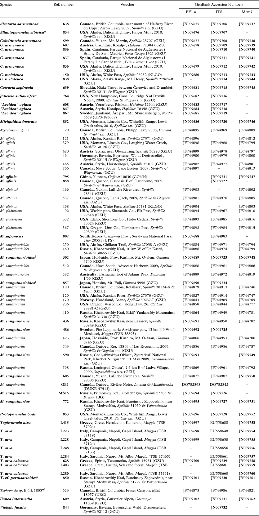
We designed our taxon sampling to include the core groups of Mycoblastus for which we could obtain fresh material, as well as representatives of major groups in the Tephromelataceae identified by Hertel & Rambold (Reference Hertel and Rambold1985), including Tephromela, Calvitimela and the “Lecidea” aglaea group, which has been treated as belonging to both Tephromela and Calvitimela in the past. We also generated sequences for several taxa of Parmeliaceae, which is a group often retrieved in BLAST searches of Mycoblastus sequences in GenBank. We included one specimen of Japewia (Lecanoraceae), hypothesized as being close to Mycoblastus by Kantvilas (Reference Kantvilas2009), and spent some sequencing effort examining the possibility of relationships to Megalaria, also proposed as a relative of Mycoblastus by Kantvilas (Reference Kantvilas2009), and Psorinia, suggested as a possible relative to Calvitimela by Hafellner & Türk (Reference Hafellner and Türk2001). We ultimately excluded Megalaria and Psorinia from our sampling because 1) morphological evidence, especially the strongly gelatinized proper exciple of Megalaria, argues against close relationships with that genus, and 2) DNA sequence data we obtained for single loci for both Megalaria and Psorinia were so different from the other taxa in our dataset as to be easily ruled out as close relatives. Heppsora indica, a species and genus described from Tamil Nadu state, India (Awasthi & Singh Reference Awasthi and Singh1977; Singh & Sinha Reference Singh and Sinha2010: photograph), exhibits clear morphological affinities to Tephromelataceae (Poelt & Grube Reference Poelt and Grube1993). Unfortunately we did not have access to any fresh material; a specimen distributed under this name in a recent exsiccate (Lumbsch & Feige, Lecanoroid Lichens #85) differs in chemistry and ascocarp pigmentation and is not H. indica. In the end, our taxon sampling (Table 1) provided a sufficient taxonomic neighbourhood to test the hypothesis of whether Mycoblastus fucatus would be recovered within Mycoblastus, in the vicinity of Tephromela, or in the vicinity of Parmeliaceae or Lecanoraceae.
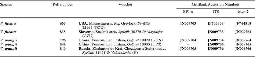
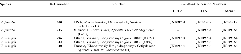
DNA vouchers and GenBank Accession Numbers of the species used in this study; bold species names and accession numbers indicate new accessions
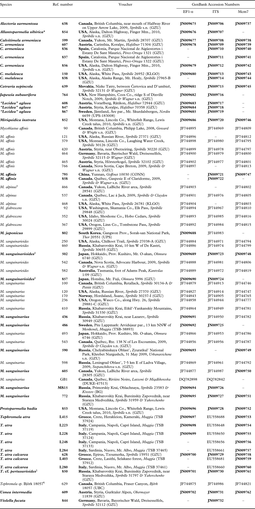
* first confirmed record for North America (TLC: α-collatolic and alectoronic acids)
† reported as M. affinis by Spribille et al. (Reference Spribille, Klug and Mayrhofer2011), this specimen actually corresponds to the alpinus morphotype
‡ first modern record for Japan
§ first record for Russia
¶ previously published as T. atra s.lat. by Spribille et al. (Reference Spribille, Klug and Mayrhofer2011), but probably an undescribed taxon
Laboratory methods
Material for DNA extraction was taken from apothecia if present, otherwise from parasite-free thallus fragments inspected in water droplets on a microscope slide under ×20 magnification. Prepared material was transferred into reaction tubes, dried and pulverized using a TissueLyserII (Retsch). DNA was extracted using the DNeasy Plant Mini Kit (Qiagen) extraction kit using the manufacturer's instructions. For Tephromela specimens already studied by Muggia et al. (Reference Muggia, Grube and Tretiach2008), existing extractions were used. Dilutions (mostly 5 × 10⁻²) of the genomic DNA extractions were used as a template for the PCR reactions. After screening potential markers (Spribille et al. Reference Spribille, Klug and Mayrhofer2011), we settled on using three loci: two protein-coding genes, namely translation elongation factor 1-α (EF1-α) and the DNA replication licensing factor mini-chromosome maintenance complex 7 (Mcm7), and the nuclear ribosomal internal transcribed spacer region (ITS). For amplification of EF1-α from Mycoblastus japonicus, we employed a Mycoblastus-specific primer pair which will be described in detail elsewhere. PCR reactions were performed with Illustra Ready-To-Go RT-PCR Beads (GE Healthcare) in a thermocycler (AlphaMetrix) using conditions detailed by Spribille et al. (Reference Spribille, Klug and Mayrhofer2011). Two µl aliquots of PCR products were viewed on 1% agarose gels stained with GelRed™ (Biotium, VWR); whole products were subsequently purified with NucleoSpin Extract II Kit (Macherey-Nagel). PCR product sequencing was outsourced to Macrogen, Inc. (Seoul, South Korea). Sequence fragments were obtained electronically from Macrogen and electropherogram ambiguities checked in BioEdit (Hall Reference Hall1999). All DNA sequences were submitted to GenBank and are retrievable under the accession numbers listed in Table 1.
Phylogenetic analyses
Alignments were performed using ClustalW (Thompson et al. Reference Thompson, Higgins and Gibson1994) and subsequently optimized by hand in BioEdit (Hall Reference Hall1999). Non-conserved regions and positions with missing data in >50% of sequences were removed using Gblocks (Talavera & Castresana Reference Talavera and Castresana2007). Candidate nucleotide substitution models were identified for each partition using the likelihood ratio test implemented in jModelTest (Posada Reference Posada2008); likelihood scores were then compared based on the Akaike Information Criterion (AIC). Individual gene alignments were analyzed using a maximum likelihood (ML) and Bayesian Markov Chain Monte Carlo (B/MCMC) approach. We tested for conflict between partitions by examining frequencies of bipartitions for the same taxon sets across all three partitions using a set of B/MCMC gene trees; a conflict was interpreted as significant if two well supported different relationships were detected for the same taxon set (Kauff & Lutzoni Reference Kauff and Lutzoni2002); we used the threshold of ≥ 95%. Maximum likelihood analyses were performed using the program PhyML 3.0 (Guindon et al. Reference Guindon, Dufavard, Lefort, Anisimova, Hordijk and Gascuel2010). Bootstrapping was carried out on 500 tree replicates. B/MCMC analyses were performed using the program MrBayes v. 3.1.2 (Huelsenbeck & Ronquist Reference Huelsenbeck and Ronquist2001) using substitution models approximated by jModeltest (see above). For each analysis, two runs with ten million generations each starting with a random tree and running four simultaneous chains was employed. Every 1000th tree was sampled and saved to a file. The first 5 000 000 generations (5000 sampled trees) were discarded as chain ‘burn-in’. Of the remaining 5001 trees a majority consensus tree with averaged branch lengths and annotated with posterior probability values at every node was calculated using the sumt command in MrBayes. The program TRACER v. 1.5 (http://tree.bio.ed.ac.uk/software/tracer/) was used to assess whether likelihood values had reached stationarity within the allocated burn-in window by plotting log likelihood against the number of generations. In addition, we examined the distributions of split frequencies using the online program AWTY (Nylander et al. Reference Nylander, Wilgenbusch, Warren and Swofford2007) to test whether runs had converged. Only clades that received bootstrap values ≥ 70% in ML and posterior probabilities ≥ 0·95 were considered significantly robust. Phylogenetic trees were visualized in TreeView (Page Reference Page1996).
Morphological and chemical analyses
To test whether our phylogenetic results could be matched by morphological traits, we sorted specimens under a Leica Wild M3Z dissecting microscope and examined anatomical sections on material mounted in water with a Zeiss Axioskop light microscope fitted with Nomarski differential interference contrast and outfitted with a ZeissAxioCam MRc5 digital camera. Some images were digitally optimized through ‘stacking’ using CombineZM open source image processing software (www.hadleyweb.pwp.blueyonder.co.uk/CZM/). Ascospore, areole, soredia and apothecia measurements are given as (smallest absolute measurement–)smallest average – largest average(–largest absolute measurement). Ascus morphology was investigated in asci with immature ascospores (following Hafellner Reference Hafellner1984). In addition, we examined specimens for chemical patterns that could corroborate phylogenetic differentiation using thin-layer chromatography (TLC), following the methods of Culberson (Reference Culberson1972) with modifications (Culberson & Johnson Reference Culberson and Johnson1982). We used silica-coated glass plates (Macherey-Nagel 821 030) run their full length in solvent systems A, B' and C. Aliphatic acids were visualized by immersing completely dried plates post-development into a tank of water for 1–2 s, quickly dripping off the plates and marking spots over the next 4 min. No attempt was made to separate roccellic and angardianic acids, which are indistinguishable in TLC (Tønsberg Reference Tønsberg1992).
Pigments were examined under the light microscope and named according to Meyer & Printzen (Reference Meyer and Printzen2000), except for Fucatus-violet, which was not treated by those authors. Fucatus-violet would key in Meyer & Printzen's key under lead 2 as N+ violaceous as it goes from its natural violet colour in H2O to a deep raspberry red. It has the following standard reactions: K+ peacock-blue, N+ raspberry-red, HCl− (slowly fading but maintaining hue), C+ grey, eventually bleaching altogether; after pretreatment with N: K greenish yellow ↔ HCl completely clear. The pigment was mentioned already by Stirton (Reference Stirton1879) as an ‘intense violaceous colour’ and has also been previously referred to as ‘gentian violet’ (James Reference James1971; James & Watson Reference James, Watson, Smith, Aptroot, Coppins, Fletcher, Gilbert, James and Wolseley2009). We adopt the name proposed by Kantvilas (Reference Kantvilas2009).
Reference material studied for morphological comparisons. Calvitimela armeniaca (DC.) Hafellner: Austria: Carinthia: Koralpe, c. 12 km NE above St. Paul in Lavanttal, 2008, Hafellner 71304 & Hafellner (GZU).
“Lecidea” aglaea Sommerf.: Austria: Styria: Koralpe, c. 15·5 km WNW of Deutschlandsberg, 2007, Hafellner 70358 (GZU); Vorarlberg, Rätikon, 2008, Hafellner 72944 (GZU).
Mycoblastus dissimulans (Nyl.) Zahlbr.: Chile: Region de los Lagos: Isla Grande de Chiloé, 2009, Pérez-Ortega 1186 & Etayo (GZU).
Mycoblastus sanguinarius (L.) Norm.: USA: Alaska: Kenai Peninsula, Russian River, 2008, Spribille 27359 & Wright (GZU).
Tephromela atra (Huds.) Hafellner: Greece: Epirus: Tzoumerka, near Kataraktis, Shrine of Profitis Ilias, 2005, Spribille 16260 (GZU).
Results of phylogenetic analysis
We obtained 91 new DNA sequences from 43 individuals, including 30 of EF1-α, 31 of ITS and 30 of Mcm7. Following exclusion of positions with missing or ambiguous data, the sequences consisted of 852, 478 and 564 characters, respectively, for a combined total of 1894 characters. Tests of nucleotide substitution models returned TIM3ef+I+G for EF1-α, GTR+I+G for ITS, and HKY+I+G for Mcm7. We ran individual B/MCMC analyses for each locus but detected no significant conflict between the loci, and thus combined them. Our partitioned B/MCMC analysis employed six, six and two substitution rate categories, respectively, for the three partitions; four rate categories, predicted in the TIM3ef model, are not possible to implement in current software. Overall rate heterogeneity was modelled using a gamma density function. ML and B/MCMC returned congruent phylogenies for the concatenated data set. Analysis of B/MCMC log likelihood outputs in Tracer indicated that convergence was reached well before our burn-in threshold; plotting of split frequencies between runs in AWTY also showed stationarity had been reached. The average standard deviation across runs for splits with a frequency of at least 0·1 was 0·003493.
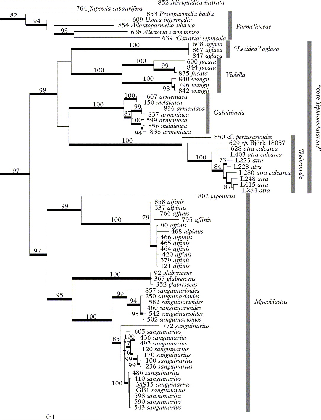
We recovered two strongly supported core groups (Fig. 1), one of which includes Tephromela, Calvitimela s.lat. and the Mycoblastus fucatus group (which we call here ‘core Tephromelataceae’), and another including Mycoblastus s.str. Both of these clades were separated from the five taxa of Parmeliaceae at the base of the tree and Japewia subaurifera, which was recovered close to Miriquidica instrata (Lecanoraceae). The combined Mycoblastus clade consists of a strongly supported monophyletic M. sanguinarius, M. sanguinarioides and M. glabrescens. Mycoblastus alpinus was recovered within a strongly supported M. affinis clade and the single individual of M. japonicus, which for the first time is represented by two markers in a molecular phylogeny, is recovered as strongly supported sister to M. affinis.
Majority rule B/MCMC consensus tree of the concatenated EF1-α , ITS and Mcm7 data set. Posterior probabilities ≥95% are shown as thick branches; bootstrap support results of maximum likelihood analysis are shown where ≥70%. Reference numbers refer to Table 1.
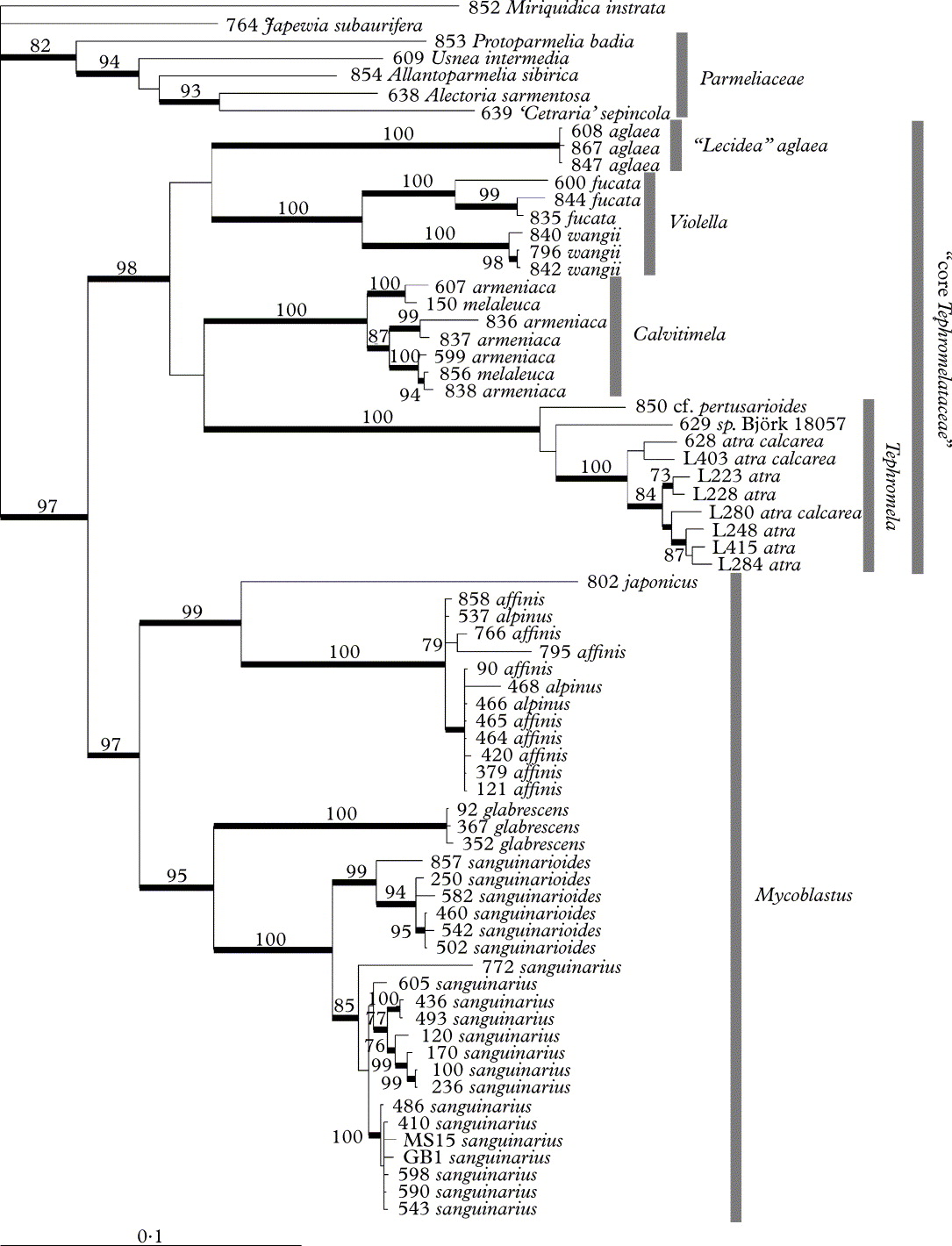
The ‘core Tephromelataceae’ clade consists of four distinct, well supported groups; the relationships to each other are, however, not supported. These groups correspond to Tephromela (T. atra, T. cf. pertusarioides and the undescribed Tephromela sp. Björk 18057), Calvitimela s.str. (C. armeniaca and C. melaleuca), the Mycoblastus fucatus group, interpreted here as the new genus Violella (see below), and “Lecidea” aglaea on its own long branch separate from the rest of Calvitimela.
Discussion
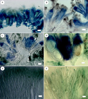
Hertel & Rambold (Reference Hertel and Rambold1985) provided an overview of species groups in what they considered Tephromela, and later Kantvilas (Reference Kantvilas2009) proposed a range of potential relatives for Mycoblastus. Our results shed new light on potential relationships and invite a reassessment of meaningful morphological characters (Table 2). In his study of Lecanoralean ascus types, Hafellner (Reference Hafellner1984) implied deep differences between Tephromela and Mycoblastus, sufficient for him to recognize them as belonging to different families, Tephromelataceae and Mycoblastaceae. Indeed, our results strongly support the distinctness of Mycoblastus s. str. from a ‘core Tephromelataceae’ (Fig. 1). This does not necessarily translate to different families, however. We did not structure the taxon sampling of our phylogenetic analysis to test family-level relationships within a broader Lecanoralean context, and cannot predict the outcome of such a study. Morphologically, however, the distinction of two families would appear to be untenable. Mycoblastus shares a similar ascus apical apparatus with members of Tephromelataceae, similar development of a peculiar thalline cushion below the apothecia (see below), similar pycnidial development, conidiophores, shared ascocarp pigments and widely overlapping thallus secondary chemistry. Morphologically, the only difference we have found may relate to the basic type of hymenial matrix formed by the paraphyses. In ‘core Tephromelataceae’, paraphyses can be branched and anastomosing, but more often than not they form long, straight, multicellular ‘beams’ that separate easily in K and are substantially thicker than the cross-bridges (Fig. 2F). In M. sanguinarius, by contrast, paraphyses almost never form straight segments even within a single paraphysis cell, the anastomosing network is intricate, with bridges often nearly as thick as the main beams (Fig. 2E), and the entire network enmeshes the asci; even in K, squashing of the hymenium results in breakage of the hymenium rather than separation of asci and paraphyses. We never found the extreme degree of branching and anastomosing without straight beams depicted by James (Reference James1971: fig. 7) for M. fucatus but instead always found the paraphysis beams to be much thicker than the bridges and easily separable in K, and thus similar to other core Tephromelataceae.
Selected asci and paraphyses. A–D, ascus variation in the Tephromelataceae, showing asci with immature ascospores; A, Calvitimela armeniaca (Hafellner 71304); B, Tephromela atra (Spribille 16260); C, “Lecidea” aglaea (Hafellner 72944); D, Violella wangii (holotype). E & F, paraphyses; E, Mycoblastus sanguinarius (Spribille 27359); F, Violella wangii (holotype). A–D in ILugol's after pretreatment with K, E & F in K. Scales: A–F = 10 µm.

Characters of genera and major groups in the Tephromelataceae and Mycoblastus
* description based on Awasthi & Singh (Reference Awasthi and Singh1977) and Poelt & Grube (Reference Poelt and Grube1993);
† outer wall considered an epispore by Stirton (Reference Stirton1879), but not dissolving in C;
‡ Fucatus-violet not seen in Chilean material but reported from Tasmania by Kantvilas (Reference Kantvilas2009);
§ ‘exciple’ of Kantvilas (Reference Kantvilas2009, p. 158: table);
¶ illustrated by Hertel & Rambold (Reference Hertel and Rambold1985);
** in T. siphuloides (Poelt & Grube Reference Poelt and Grube1993).
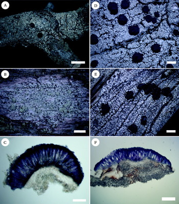
Another enigmatic structure linking Tephromelataceae and Mycoblastus is the so-called thalline exciple, especially evident in Tephromela. Hertel & Rambold (Reference Hertel and Rambold1985) and Kantvilas (Reference Kantvilas2009) have interpreted the ‘thalline exciple’ of Tephromela to be homologous, or at least worthy of providing in the same table category, to the proper exciple in other genera. We have, however, found apparently homologous thalline tissue, in addition to the presence of a rudimentary proper exciple, in all genera of Tephromelataceae and Mycoblastus. We hesitate to refer to this as an amphithecium or thalline exciple because it lacks an algal layer and consists of differentiated, dense, prosoplectenchymatous tissue not normally found in the thallus. Instead we will refer to it as a ‘thalline cushion’. The thalline cushion occasionally emerges to outer view as a thin or thick white line in M. sanguinarioides (T. Spribille, unpublished data), is visible in section in the M. fucatus group (Fig. 3C & 3F), and in Tephromela it forms a ‘thalline rim’. However, it is even present in Calvitimela, where it forms a dense layer between the subhymenium and the thallus medulla.
Violella species, habit. A–C, V. fucata; A, fertile specimen (Tønsberg 19004); B, sterile specimen on wood (Spribille 32161); C, section of apothecium, in water (Tønsberg 19004). D–F, V. wangii; D, (holotype); E, sorediate morph (Spribille 31621); F, section of apothecium, in water (Goffinet 10033). Scales: A, D & E = 2 mm; B = 1 mm; C = 100 µm; F = 200 µm.

Our phylogenetic results re-open a discussion on the generic boundaries in Tephromelataceae, begun by Hertel & Rambold (Reference Hertel and Rambold1985) and continued by Hafellner & Türk (Reference Hafellner and Türk2001), with the description of Calvitimela. Tephromela possesses Biatora-type asci with a sometimes bulbous masse axiale (Fig. 2B). Hafellner & Türk (Reference Hafellner and Türk2001) separated out Calvitimela in part based on its Lecanora-type ascus, though even in describing their new genus they already anticipated that the “Lecidea” aglaea group, with its Biatora-type asci (Fig. 2C), might not be closely related to the type species C. armeniaca (Fig. 2A). Even so, they transferred it to Calvitimela. Our results confirm that the two are not closely related and we thus maintain this taxon in the genus Lecidea in the broad sense until its generic disposition can be resolved. To this medley can now be added the M. fucatus group with its Biatora-type asci (Fig. 2D). Mycoblastus fucatus has long been recognized for its unusual hymenium pigmentation, a character absent from Mycoblastus s. str. Furthermore, M. fucatus, and in particular material from Asia that will be described here as a new taxon, possesses a character not known from any of the other associated genera studied here, namely the tendency of the internal ascospore wall to turn brown. This character was already noted by Leighton (Reference Leighton1879, see also below). These characters also do not reconcile with those of Tephromela and Calvitimela, which differ in hymenium pigmentation, ascus type and, in part, secondary chemistry (Table 2). We accordingly propose recognizing M. fucatus and this new taxon as constituting the new genus Violella. The alternative generic solution would require all taxa from Tephromela s. str. through Calvitimela, Violella and the “Lecidea” aglaea group to be referred to Tephromela s. ampl. but in our opinion this would defeat the purpose of genera to circumscribe like species groups, and make Tephromela, which even in its narrow definition has more than 20 described species, unnecessarily large and unwieldy. We expect “Lecidea” aglaea will eventually be placed in its own genus, possibly together with C. perlata (Haugan & Timdal) R. Sant., which has Bacidia-type asci, and some of the various entities currently treated as chemotypes of “Lecidea” aglaea (Haugan & Timdal Reference Haugan and Timdal1994). Already Andreev (Reference Andreev2004) has postulated that these two taxa are closely related, though he retained them in Calvitimela. We leave these problems unresolved until a better sampling of Calvitimela s. lat. has been achieved, perhaps including Southern Hemisphere taxa (Fryday Reference Fryday2011) and Heppsora, a south Asian genus (Awasthi & Singh Reference Awasthi and Singh1977), for which DNA could not be obtained for the current study.
Taxonomy
Violella T. Sprib. gen. nov
MycoBank No: MB 519831
Genus novum ad Tephromelataceas pertinet. Generi Calvitimela simile sed differt pigmentis hymenialibus violaceis (haud viridibus), ascis ut in Biatora constructis (haud ut in Lecanora), ascosporis primum hyalinis, demum strato interno fuscescenti (haud persistenter hyalinis) et substanciis chimicis aliis (atranorinum vice acidi alectorialici).
Typus: Violella fucata (Stirt.) T. Sprib.
Thallus crustose, areolate to rimose; photobiont chlorococcoid algae. Thallus chemistry includes the depside atranorin, a depsidone and a fatty acid.
Apothecia apparently biatorine, macroscopically black, formed on a rudimentary thalline cushion, this prosoplectenchymatous, tawny with brown streaks; proper exciple reduced; epihymenium not differentiated as a distinct layer, epipsamma lacking; hymenium inspersed with violet granules (‘Fucatus-violet’) that react N+ raspberry red, K+ peacock green; paraphyses straight or slightly curved with thinner cross-bridges; asci Biatora-type; ascospores simple, in the known species two per ascus (reported as occasionally 1 or 3: Stirton Reference Stirton1879; Leighton Reference Leighton1879; James Reference James1971), initially with a single wall, eventually a differentiated internal wall turning brown.
Pycnidia apparently rare, colourless or with light brown pigment around ostiole, sunken in thallus areoles; conidiophores Parmelia-type; conidia bacilliform.
Etymology. Diminutive of Viola, a reference to the characteristic pigment in the hymenium of both known species (Fig. 3).
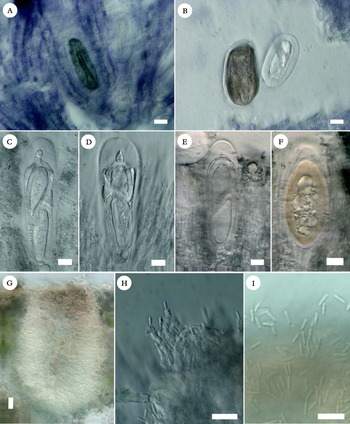
Comments. Species of Violella are distinguished from related genera first and foremost by their abundant Fucatus-violet pigment and the tendency of the inner ascospore walls to become brown. The latter character appears to have been recorded only once previously in the literature, by Leighton (Reference Leighton1879: 545), who noted the tendency of the ‘protoplasm’ of the ascospores of V. fucata to turn a ‘nigro-fulvaceous colour’ in K. However, this colour, apparently produced in the internal ascospore wall, is present even without treatment with K and seems to occur in all mature ascospores in both species of the genus (Figs. 2D, 3C & F, 4A, B & F). It appears that many ascospores with a brown inner wall are collapsing internally and are possibly abortive (Fig. 4A & B). However, healthy ascospores with brown internal walls were also observed (Fig. 4F) and no mature ascospores were observed in either species that had not turned brown internally.
Microscopic characteristics of Violella apothecia and pycnidia. A, V. fucata ascospore in water (Tønsberg 19004). B–I, V. wangii internal anatomy; B, mature and immature ascospore, in water; C–F, asci at different stages of development, ending in nearly mature ascospore, using differential interference contrast following pretreatment with C; G, section of pycnidium in water (holotype); H, conidiophores (in water); I, conidia (in water). B–F from Goffinet 10033, G–I from holotype. All scales = 10 µm.

Kantvilas (Reference Kantvilas2009) speculated that the perlatolic acid-containing species of Mycoblastus, the so-called M. dissimulans group, may constitute a distinct genus. We were not able to sample that group for our phylogeny, but we note that Violella differs from M. dissimulans in 1) its paraphyses linked over small bridges to other paraphyses, as opposed to the dense network paraphyses similar to those in Mycoblastus s. str. formed in M. dissimulans; 2) its ascospores, in which the internal ascospore wall frequently becomes brown or olivaceous (remaining hyaline in M. dissimulans); and 3) its secondary thallus chemistry. We suspect that M. dissimulans will ultimately be found to cluster more closely with Mycoblastus s. str., which also has brittle, anastomosing paraphysis networks. We are not aware of any existing generic name in this group that would need to be considered before describing Violella.
Violella fucata (Stirt.) T. Sprib. comb. nov
MycoBank No.: MB 519832
Basionym: Lecidea fucata Stirton, Scottish Naturalist 5: 16 (1879); type: Great Britain, Scotland, Mid Perth, Tyndrum, on wood, July 1878, Stirton s.n. (BM!—holotype).— Megalospora fucata (Stirt.) H. Olivier, Bull. de Géogr. Bot. 21: 187 (1911; on p. 207 Olivier incorrectly attributes the combination to Leighton Reference Leighton1879).—Mycoblastus fucatus (Stirt.) Zahlbruckner, Cat. Lich. Univ. 4: 3 (1926).
The first species of the genus to be described was Violella fucata (Stirton Reference Stirton1879, as Lecidea fucata), but this taxon rarely produces apothecia. A detailed description is provided by James (Reference James1971). Violella fucata is widely reported from western Europe (e.g., Tønsberg Reference Tønsberg1992; James & Watson Reference James, Watson, Smith, Aptroot, Coppins, Fletcher, Gilbert, James and Wolseley2009), the Pacific Coast of North America (British Columbia and Washington: Tønsberg Reference Tønsberg1993; Alaska: Spribille et al. Reference Spribille, Pérez-Ortega, Tønsberg and Schirokauer2010) and eastern North America (Massachusetts: Spribille et al. Reference Spribille, Klug and Mayrhofer2011 and below; Newfoundland: Tønsberg Reference Tønsberg1993; New York: Schmull et al. Reference Schmull, Hauck, Vann, Johnson and Runge2002; Harris Reference Harris2004). A distribution map of its obligate parasite Tremella lichenicola (Diederich Reference Diederich1996: 102) includes many European and some western North American records.
Selected specimens examined. Norway: Hordaland: Fjell, Sotra, Tælavåg, W of Midttveit, UTM 32V, KM 766874, map 115 IV, alt. 10 m, [corticolous] on maritime Calluna vulgaris, 1993, T. Tønsberg 19004 (BG, c.fr.); Sogn og Fjordane, Askvoll, W of Fure, S of Djupevika, hill 48, UTM 32V, KP 8601, Map 1117 IV, alt. 20–48 m, [corticolous] on Calluna vulgaris, 1989, T. Tønsberg 11779 (BG, c.fr.).—Great Britain: Scotland: V.C. 105, West Ross, Dundonnell, Allt a' Chàirn ravine, on lignum of fallen Betula trunk, alt. 160 m, 1999, A. M. & B. J. Coppins 18794 (E, c.fr.); V.C. 107, East Sutherland, N side of Dornoch Firth, Spinningdale, Ledmore Wood, on lignum of fallen, decorticate Quercus trunk, alt. 10–30 m, 2001, B. J. & A. M. Coppins 20015 (E, c.fr.); V.C. 97, Westerness, N side Loch Sunart, Coel na mara, on lignum of fallen trunk of Quercus, alt. c. 40 m, 2004, B. J. Coppins 21427 & H. L. Andersen (E, c.fr.).—USA: Massachusetts: Berkshire Co., Mount Greylock, 42°38·231′N, 73°10·208′W, 1034 m alt., lignicolous on snags, 2009, T. Spribille 32161 & V. Wagner (FH, GZU, NY).
Violella wangii T. Sprib. & Goffinet sp. nov
MycoBank No.: MB 519833
A Violella fucata areolis maioribus bullatisque, apotheciis maioribus et substanciis chimicis aliis (atranorinum et acidum roccellicum/angardianum vice atranorini et acidi fumarprotocetrarici) differt. Habitat in montibus altis Asiae extratropicae.
Typus: China, Yunnan, Lijiang Prefecture, Lijiang Co. S of Lijiang, Jinhue village, Laojunshan Mountain, at the border with Jianchuan Co., 26°38·538–37·936′N, 99°43·509–45·992′E, 3510–3900 m, montane forest dominated by Abies and further up by Rhododendron, along trail from parking lot to peak, epiphytic, 16 July 2010, B. Goffinet 10029, with L. Wang, S. L. Guo and S.Y. Huang (KUN—holotypus; CONN, GZU—isotypi); same locality, same date, B. Goffinet 10033, with L. Wang, S.L. Guo and S.Y. Huang (TNS, UPS).
Thallus crustose, covering patches as much as 8 cm diam., consisting of discrete areoles (0·15–)0·2–0·6(–1·5) mm diam., these sometimes confluent forming a rimose thallus; colour white to ashen grey, surficial thallus granules corticate, corticate surface finely pruinose; cortex in esorediate thalli prosoplectenchymatous, 30–55 µm thick; algal layer c. 50 µm thick, grading into medulla that is variably thin to as much as 200 µm thick, to 300 µm thick under apothecia; soredia when present borne in soralia at tips of areoles, rarely areoles dissolving into soredia, internal and external soredia white; soredia roundish, (40–)64–88(–110) µm diam., sometimes forming consoredia; hypothallus not observed; photobiont chlorococcoid, cells rounded to irregularly angular, (7–)8·4–11·1(–17) µm diam.
Apothecia always present, rounded, single or clustered in groups of 2–3 and becoming confluent, (0·7–)1·3–2·6(–4·1) mm diam., base broadly adnate, disc ± flat to weakly convex, jet black and shiny; margin indistinct, visible from above only in the youngest apothecia, concolorous with the disc; ‘thalline cushion’ present, rarely visible from above and forming a thin white line, in section prosoplectenchymatous, variable in thickness, 25–230 µm thick, typically tawny brown with streaks of darker brown pigment, clearly differentiated from subhymenium above and medulla below; proper exciple similar in structure to the hymenium, hyphae radiate, similar to paraphyses, when well developed in young apothecia to 170 µm wide laterally, filled with Fucatus-violet granules and often suffused with Cinereorufa-green pigment; differentiated hypothecium absent; subhymenium consisting of a thin layer of ascogenous hyphae, c. 20–50 µm tall, filled like the hymenium with Fucatus-violet granules but sometimes also infused with Cinereorufa-green; hymenium highly variable in thickness even within one and the same apothecium, (80–)100–300(–350) µm tall, strongly infused with Fucatus-violet granules and collectively forming a deep violet impression in section, but hymenial gel itself hyaline in thin section; epithecium not differentiated, epipsamma lacking; paraphyses mostly simple, arranged vertically and linked to each other in their lower halves by thin bridges, the main beams stouter than the bridges and not readily breaking when squashed in K; paraphysis tips not or scarcely expanded, 4–6 µm wide including gel sheaths, lumina to 1·5 µm wide, paraphyses completely coated on the outside by Fucatus-violet granules; asci clavate, 85–110 × 25–33 µm when mature, inner and outer walls staining blue, tholus strongly ILugol+ blue, pierced by a broad, conical non-amyloid structure, thus similar to the Biatora-type; ascospores 2 per ascus, beginning colourless and apparently with a single wall, eventually developing a secondary inner wall, which quickly turns brown while still in the ascus; outer wall thick, to 5 µm in some cases, the inner brown wall thin, often collapsing (spore aborting?), live, healthy ascospores also with brown endospore, (35–)41·7–54·2(–65) × (15–)20·8–30·8(–35) µm in water.
Pycnidia apparently rare, barely visible externally, in small colourless bumps on the thallus, to 60 µm diam.; wall 10–20 µm thick, pigmented a pale rufous brown or hyaline; conidiophores of Parmelia-type (type VI of Vobis Reference Vobis1980), with zig-zag cells sprouting conidia in upper part of each cell; conidia bacilliform, c. 4–5 × 1 µm.
Chemistry. Atranorin and roccellic/angardianic acid detected by TLC.
Etymology. The species is named in honour of Dr. Wang Li-Song, for his ongoing efforts to describe the lichen diversity in western China.
Habitat and distribution. Found on bark of Rhododendron sp. in China (Hengduan Shan, Yunnan) and on wood of Pinus pumila in the Russian Far East (Bureinskiy Khrebet, north-western Khabarovskiy Krai). Substratum was not recorded for the Indian and Bhutanese material. Collections came from elevations of 3500 to 4000 m in the southern area and c. 1000 m in the northernmost collection. In two of the collections it was associated with Mycoblastus affinis; one of these specimens is included in our phylogeny.
Comments. Violella wangii is a distinct species that seems to be widespread, if rarely collected, in the mountains of high Asia. It occurs in two intergrading morphs, one esorediate with granular, corticate areoles that can become heaped and almost phyllocladioid, and another in which these areoles remain small and erupt in apical soralia, in one specimen even disintegrating completely into soredia in parts of the thallus. The two morphs exhibit no other consistent differences however and several specimens are intermediate. The apparently fluid gradient between esorediate and sorediate morphs recalls the case of Mycoblastus sanguinarius (Tønsberg Reference Tønsberg1992), in which fully leprose morphs have not been found to be genetically distinct from esorediate morphs (T. Spribille, unpublished data).
Violella wangii differs from the only other species in the genus, V. fucata, in possessing much larger thalli (frequently covering patches 4–8 cm in diam. (rarely >3 cm diam. in V. fucata), robust areoles 0·2–0·6 mm across (to 0·3 mm in V. fucata), external soredia, if present, which remain white (often turning bluish grey in V. fucata), and chemistry (roccellic/angardianic instead of fumarprotocetraric acid). Ascospores average larger in V. wangii than in V. fucata; though based on a limited number of apothecia available and paucity of ascospores, our measurements in V. fucata (38·5 ± 6·7 × 18·5 ± 3·3 µm, n = 24) fall exactly within the ranges given by Stirton (Reference Stirton1879) and James & Watson (Reference James, Watson, Smith, Aptroot, Coppins, Fletcher, Gilbert, James and Wolseley2009). The apothecia of V. wangii are larger than anything we have measured in V. fucata but this may not be a reliable character given that apothecia are rare and often poorly developed in V. fucata, a primarily sterile species.
Specimens examined (V. wangii). Bhutan: Tongsa District: Black Mountains NW of Nubji, 27°12′N, 90°22′E, 4040 m elev., Rhododendron thicket with Abies densa at treeline on ridges, on Rhododendron, 2000, G. & S. Miehe 00-13-07/06 (GZU).—India: Darjeeling: Phalut-Dentam, 11 v 1960, Togashi et al. s.n. (TNS). Sikkim: Jongri, elev. 4000 m, 20 v 1960, Togashi et al. s.n. (TNS).—Russia: Khabarovskiy Krai: Chegdomyn-Sofiysk road, high pass, watershed between Niman and Umal'ta Rivers, c. 7·1 km S of the bridge over the Niman River, 26 km (air line) SW of Sofiysk, 52°05·866′N, 133°42·433′E, Pinus pumila-Rhododendron aureum woodland under Larix gmelinii, on hard wood of P. pumila, 1016 m, 2009, T. Spribille 31621 & L. Yakovchenko (H).
Vězda (Reference Vězda1993) issued an exsiccate of a specimen from China under the name Mycoblastus fucatus, but as Kantvilas (Reference Kantvilas2009) has pointed out, it is distinct from that taxon. It was collected near the type locality of V. wangii but is distinct from that species in its chemistry (fumarprotocetraric instead of roccellic/angardianic acid, in this respect recalling V. fucata) and thallus morphology (larger, flatter areoles). It is also distinct from the chemically concordant V. fucata in, amongst other characters, developing larger thalli, large, flattened areoles and large apothecia, and apparently lacking soredia. We regard this as probably another species distinct from V. wangii and V. fucata based on thallus chemistry and morphology. However, we were unable to obtain fresh material of this species and hesitate to describe it without getting a better overview of its variability. We have seen three specimens conforming to this morphology and chemistry, all from China.
Specimens examined (unnamed fumarprotocetraric acid-containing form): China: Prov. Yunnan: montes Yulong Shan, 30 km ad septentriones ab oppido Likiang, alt. 4000 m s.m., 25 vii 1990, Soják s.n. (Vězda, Lich. Rar. Exs. 66, GZU). Prov. Sichuan: Hengduan Shan, Daxue Shan, 57 km S of Kangding, Gongga Shan, Hailougou glacier and forest park, 29°34′35″N, 101°59′56″E, 2940–3130 m, on Betula utilis, 2000, W. Obermayer 08686 (GZU); ibid., northern Qionglai Shan, Barkam, 31°57′N, 102°39′E, 4050 m, 1995, G. & S. Miehe 94-502-2/14 & U. Wündisch (GZU).
Status of Mycoblastus indicus
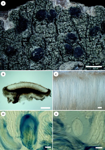
A candidate name for our new taxon that required examination was Mycoblastus indicus (Awasthi & Agarwal Reference Awasthi and Agarwal1968, as “indicum”), described from Darjeeling district, India, near to where Violella wangii has also been collected. We did not receive a response to repeated requests for type material from Lucknow (LWU), but we did find a specimen of M. indicus at UPS, collected and identified by Awasthi and Agarwal only days before they collected the type specimen. The specimen fits the description provided by Awasthi & Agarwal (Reference Awasthi and Agarwal1968) and in habit resembles the photograph of the holotype provided by Singh & Sinha (Reference Singh and Sinha2010), though the latter appears to have more mature apothecia. Mycoblastus indicus is clearly not a member of Mycoblastus or Tephromelataceae. Instead, detailed study of the UPS specimen (Fig. 5) revealed brown epihymenial and hypothecial pigments, a strongly developed proper exciple, mostly simple, loose paraphyses, and asci with a dark apical amyloid cylinder. We obtained an unknown phenolic substance from the thallus, with Rf values similar to confluentic acid in TLC. We concur with Awasthi & Agarwal's original statement that the species appears similar to the group of tropical species around Lecidea granifera Vain., for which the genus Malmidea has been erected by Kalb et al. (Reference Kalb, Rivas-Plata, Lücking and Lumbsch2011). We accordingly combine the species into that genus, where it appears similar to M. coralliformis Kalb. We note that it has larger ascospores than any of the members of the genus discussed by Kalb et al. (Reference Kalb, Rivas-Plata, Lücking and Lumbsch2011).
Malmidea indica (Awasthi & Agarwal 67.224, UPS). A, habit; B, section of apothecium; C, section through hymenium showing paraphyses; D, ascus, squash preparation stained in ILugol's after pretreatment with K; E, ascospore, in ILugol's. Scales: A = 2 mm; B = 200 µm; C–E = 10 µm.

Malmidea indica (Awasthi & Agarwal) Hafellner & T. Sprib. comb. nov
Basionym: Mycoblastus indicum Awasthi & Agarwal, Current Science 37: 84 (1968); type: India, Darjeeling District, Pashkok Road, 19 March 1967, D. D. Awasthi & M. R. Agarwal 67.78 (LWU—holotype, n.v.).
Specimen examined (Malmidea indica). India: Darjeeling District: Rangit river valley, near Lebong, alt. 1520 m, on bark of trees, 1967, D. D. Awasthi & M. R. Agarwal 67.224 (UPS).
We thank J. Hafellner (Graz) for helpful ascus and ascospore observations and for translating the diagnoses into Latin. S. Pérez-Ortega, Y. Ohmura, Göran Thor and Irina Urbanavichene provided fresh material for DNA analysis and S. Clayden and L. Yakovchenko facilitated sampling in Atlantic Canada and the Russian Far East, respectively. The director of the Bureinskiy Nature Reserve is thanked for permission to collect in that reserve in 2009. Field work by BG was funded by grant DEB-0919284 from the American National Science Foundation and made possible by Drs Wang Li-Song (Kunming Institute of Botany) and Guo Shuiliang (Shanghai Normal University), for which BG is grateful. We thank the curators of the herbaria BG, BM, E, TNS and UPS for loans of specimens. This project was supported by the Austrian Science Foundation (project P21052-B16, Circumboreal Lichen Diversification).