INTRODUCTION
Coral reefs are amongst the most productive biological zones on Earth and are home to an extraordinary biodiversity which provides food for nearly 500 million people (Wilkinson, Reference Wilkinson2008). These ecosystems are certainly the most vulnerable marine environments and have declined dramatically over the last 30 years owing to the combined effect of global warming and increasing anthropogenic pressure (Hoegh-Guldberg, Reference Hoegh-Guldberg2010). The most visible and widespread manifestation of this decline is coral bleaching and to a lesser extent the emergence of other microbial pathologies (yellow blotch, black band disease, white band disease, white plague, etc) which now affect coral reefs throughout the world (Normile, Reference Normile2010). However, despite efforts made by the scientific community to protect this biological heritage, the ecological and physiological processes that lead to these coral diseases are still unproven and often challenged.
The most common explanation for bleaching is related to the expulsion of endosymbiotic zooxanthellae (Symbiodinium spp.) by the coral polyps as a result of prolonged increase in temperature or other environmental changes (Rosenberg et al., Reference Rosenberg, Koren, Reshe, Efrony and Zilber-Rosenberg2007). An alternative theory, put forward during the past ten years by an increasing number of microbiologists, is that bleaching could be the result of a sudden structural change in the bacterial and archaeal communities in the mucus in response to environmental disturbance (increase in temperature, in particular, but also changes in nutrients, acidification, etc) (Bourne et al., Reference Bourne, Lida, Uthicke and Smith-Keune2008; Rosenberg et al., Reference Rosenberg, Kushmaro, Kramarsky-Winter, Banin and Yossi2009).
Viruses are surely the least studied of the biological entities that form the coral holobiont. Their role remains uncertain and little is known about the possible pathogenic or mutualistic relationships that they may have with the host animal (Davy et al., Reference Davy, Burchett, Dale, Davies, Davy, Muncke, Hoegh-Guldberg and Wilson2006; van Oppen et al., Reference van Oppen, Leong and Gates2009). A few recent studies have shown that the surface microlayer of mucus contains a large number of viruses that are morphologically and genetically very diverse and differ from the viral communities in the surrounding water (Davy et al., 2007; Marhaver et al., Reference Marhaver, Edwards and Rohwer2008), suggesting specific interactions between these viruses and the other members of the holobiont. The high levels of viral abundance found in healthy corals also suggest that their activity may stabilize or even improve the coral biological balance rather than disturbing it. For example, Rohwer et al. (Reference Rohwer, Seguritan, Azam and Knwolton2002) and van Oppen et al. (Reference van Oppen, Leong and Gates2009) suggested that viruses could be determinant in preventing the emergence of pathogens. Viruses were also thought to improve coral viability by encoding for genes that provide benefits to the coral host (Patten et al., Reference Patten, Harrison and Mitchell2008).
Viruses have also been found in thin sections of zooxanthellae from thermally-stressed corals (Wilson et al., Reference Wilson, Francis, Ryan and Davy2001, Reference Wilson, Dale, Davy and Davy2005; Lohr et al., Reference Lohr, Munn and Wilson2007), implying that a fraction of these dinoflagellate symbionts could harbour latent viruses. Unfortunately, such observations were only made in corals that had been cultured under artificial conditions (aquarium) and not in a natural environment. Therefore, the hypothesis that viral infection of zooxanthellae is a mechanism involved in temperature-induced coral bleaching clearly lacks supporting evidence. Furthermore, observations of virally-infected coral tissues are rare and no scleractinian diseases have yet been firmly attributed to viral origin. Viruses were first reported in Cnidaria cells of the plumose anemone Metridium senile (Wilson & Chapman, Reference Wilson and Chapman2001) and the scleractinian Pavona danai (Wilson et al., Reference Wilson, Dale, Davy and Davy2005). Although these two species were also grown artificially in aquaria, a more recent study conducted in the Great Barrier Reef (Australia) revealed the presence of intracellular viruses in epidermal and gastrodermal cells of the coral Acropora muricata (Patten et al., Reference Patten, Harrison and Mitchell2008). Viruses were found in both healthy and diseased colonies (white syndrome), suggesting that they might not be directly involved in coral morbidity or mortality. Different sizes and morphologies of viruses were found in the tissues of P. danai, which led the authors (Patten et al., Reference Patten, Harrison and Mitchell2008) to conclude that ‘viruses are infecting different organisms within the holobiont, or that one host is susceptible to infection from more than one type of virus’. Although there is conclusive evidence that viruses are a component of the coral holobiont, little is known about whether or not they contribute to the worldwide decline of coral reef environments. This study showed that bleaching colonies of the scleractinian Acropora cytherea harboured intracellular viruses in their epidermal cells.
MATERIALS AND METHODS
Our study used a single sample of those taken in the Hon Mun Marine Protected Area (MPA) in Vietnam (12°09′53″N 109°18′38″E) on 11 November 2010. Coral colonies were sampled at depths between 2 and 4 m in a zone where coral tissues were subject to partial bleaching. The superficial microlayer of A. cyntherea was removed gently from the coral surface using 10 ml syringe (Davy & Patten, Reference Davy and Patten2007). It was then fixed in a solution of 2.5% glutaraldehyde in 0.1M cacodylate buffer, pH 7.4 and stored overnight at 4°C. Samples were then washed three times in 0.1M cacodylate buffer for 15 minutes and post-fixed in 1% osmium tetroxide for 1 hour, at room temperature. Samples were then rinsed twice in 0.1M cacodylate buffer (10 minutes), dehydrated in an ethanol series (50%, 70%, 90% and 100%) and propylene oxide (10 minutes/each), embedded in Epon 812. Thin 60–80 nm sections were obtained by ultra-microtome (Leica, UC6), using collodion coated copper grids (300 meshes). The sections were double-stained for 5 minutes with 5% uranyl acetate and 10 minutes with lead citrate, and examined at 80 kV by transmission electron microscopy (TEM:JEOL 1010).
RESULTS AND DISCUSSION
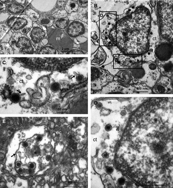
In the single sample that was analysed, only two out of more than 20 epidermal cells showed obvious signs of viral infection. The virions observed by TEM display different stages of maturation, suggesting that the viruses effectively replicate in the coral host. Virus-like particles (VLPs) were all found in the cytoplasm, were pentagonal or hexagonal in section, indicating icosahedral symmetry (Figure 1B). These VLPs had an electron-dense core surrounded by a thin double layer. The diameter of these VLPs ranged from 90 to 140 nm. The VLPs did not have an outer envelope. Although it was difficult to assign them to a viral family from their morphology, their characteristics resembled those previously published for another subspecies of Acropora (i.e. muricata) in the Great Barrier Reef (Patten et al., Reference Patten, Harrison and Mitchell2008), which were thought to belong to the Phycodnaviridae and Iridoviridae viral families. Recent metagenomic analyses also revealed the presence of abundant viruses of plant, algae, prokaryotes and also, more surprisingly, of viruses with herpes-like sequences within the tissues of Diploria strigosa (Marhaver et al., Reference Marhaver, Edwards and Rohwer2008) and Porites compressa (Thurber et al., Reference Thurber, Willner-Hall, Rodriguez-Mueller, Desnues, Edwards, Angly, Dinsdale, Kelly and Rohwer2009). However, unlike herpes viruses, those observed in A. cytherea had no envelope. Some even had tail-like structure similar to that of phages of the order Caudovirales. Although eukaryotic viruses are typically tailless, the protein of the virion coat is sometimes produced in excess and extends as a tubular tail on one side of the virus particle that becomes partially filamentous. For example, some viruses of the Hepadnaviridae family such as hepatitis B virions are known to vary between tubular and spherical forms (Kaito et al., Reference Kaito, Ohba, Chiba, Kohara, Tanaka, Fujita, Gabazza, Watanabe, Konishi and Adachi2006).
Virus-like particles (VLPs) within epidermis cells of Acropora cytherea coral colonies from the Hon Mun Marine Protected Area (Vietnam). (A) Uninfected polymorphic epidermal cells (ec) and mesoglea (m); (B) overall view of epidermal cells with numerous VLPs within the cytoplasm (ct); nucleus (n); (C) detail of (B). Spherical VLPs with a capsid diameter of 90–140 nm, showing an electron-dense core surrounded by an electron-lucent layer. Note the presence of a tail-like structure (arrow) in two of the three VLPs; (D) second detail of (B). Here, the VLPs lack tails and are hexagonal and spherical in shape with capsid diameter of 80–130 nm; (E) an inclusion of untailed VLPs, 130–140 nm in diameter within a membrane bound vacuole (arrowed) in epidermal tissue.

Herein, A. cytherea was likely infected by more than one type of virus. Indeed, the epidermal cells were also infected by tailless viruses whose size of the virions varied from 80–130 nm and 130–140 nm (see insets B, E Figure 1). The disparity between the virions could not simply be explained by the fact they were sectioned in different planes. In their recent study, Patten et al. (Reference Patten, Harrison and Mitchell2008) also showed that coral tissues can harbour viruses differing in size and/or morphology.
However, no clear causal link between their presence and the current bleaching event can be established. Pathogenicity is in question as viruses have also been found in healthy (and diseased) Acropora muricata (Patten et al., Reference Patten, Harrison and Mitchell2008) and Diploria strigosa (Marhaver et al., Reference Marhaver, Edwards and Rohwer2008). This raises several questions: (i) is there a threshold beyond which the abundance of intracellular viruses becomes lethal to the host? This is not unrealistic as previous experiments showed that herpes-like viral sequences were abundant in most ultraviolet/nutrient/temperature-stressed Porites compressa but were rarely detected in healthy individuals (Thurber et al., Reference Thurber, Willner-Hall, Rodriguez-Mueller, Desnues, Edwards, Angly, Dinsdale, Kelly and Rohwer2009). It was, therefore, suspected that stress could trigger the activation of latent viruses in coral tissues. However, further investigation is needed to determine the potential pathogenicity of these viruses; (ii) are corals simply subjected to chronic or asymptomatic infection? In the first case, viruses are still able to persist and viral replication continues despite the coral immune response (Reed et al., Reference Reed, Muller and van Woesik2010), the balance of the immune response thus being in favour of the virus. In the case of asymptomatic infection, infected corals still produce numerous viruses without developing disease; and finally (iii) can viruses manipulate corals? In other words, are viruses a source of functions that are beneficial for the corals by providing additional genes that may optimize their performance in nutrition, reproduction, resistance to pathogens, etc? (Rohwer & Thurber, Reference Rohwer and Thurber2009). It is also possible that, as suggested for zooxanthellae (Wilson & Chapman, Reference Wilson and Chapman2001), the presumed latent infection of coral tissues may confer immunity against other viral infections and thus ensure the stability of the polyps. These are all valid hypotheses and they are not mutually exclusive. Clearly, the successful isolation and characterization of these holobiont viruses is imperative if we are to understand their ecological role.
ACKNOWLDEGEMENTS
The research reported here was financially supported by the EC2CO project ‘CORINE’ and the Total Foundation. We gratefully acknowledge advice, discussion, encouragement and assistance provided by Drs Vo Si Tuan, Bui Hong Long, Nguyen Van Long, Chu Van Thuoc, Jean-Pascal Torréton and Aurore Trottet. We also sincerely thank the staff of the Electron Microscopy Laboratory at the National Institute of Hygiene and Epidemiology of Hanoï for their help with the sample preparation for TEM analyses.