Background
Diaphragm dysfunction has recently been diagnosed using sonographic evaluation of diaphragmatic excursion.Reference Hamadah, Kabbani and Elbarbary 1 This method has several advantages compared with fluoroscopy, such as being simple, rapid, and eliminating the need for radiation and movement for fluoroscopy. However, positive pressure respiratory support must be interrupted during the sonographic examination. Several reports have described the diaphragm thickening in adults during mechanical support, which has been used to evaluate respiratory function and predict extubation.Reference Goligher, Fan and Herridge 2 – Reference Matamis, Soilemezi and Tsagourias 4 As this technique has not been evaluated extensively in children,Reference Lee, Hsia and Hsiao 5 we report a case in which we used the diaphragm thickness fraction to diagnose diaphragm paralysis in a neonate during positive pressure ventilation support.
Case report
A male neonate (2465 g at 40 weeks of gestation) was born with a type A interrupted aortic arch and ventricular septal defect. At the age of 10 days, he underwent aortic arch repair with modified end-to-end anastomosis combined with subclavian flap aortoplasty and main pulmonary artery banding via thoracotomy. However, on postoperative day 5, he developed tachypnoea, and plain radiography revealed that his left diaphragm was elevated during synchronised intermittent mandatory ventilation through tracheal intubation. We suspected diaphragm paralysis and performed ultrasonography to test for diaphragm dysfunction. Before the test, the ventilator setting was changed to continuous positive airway pressure with pressure support (6 cm of H2O).
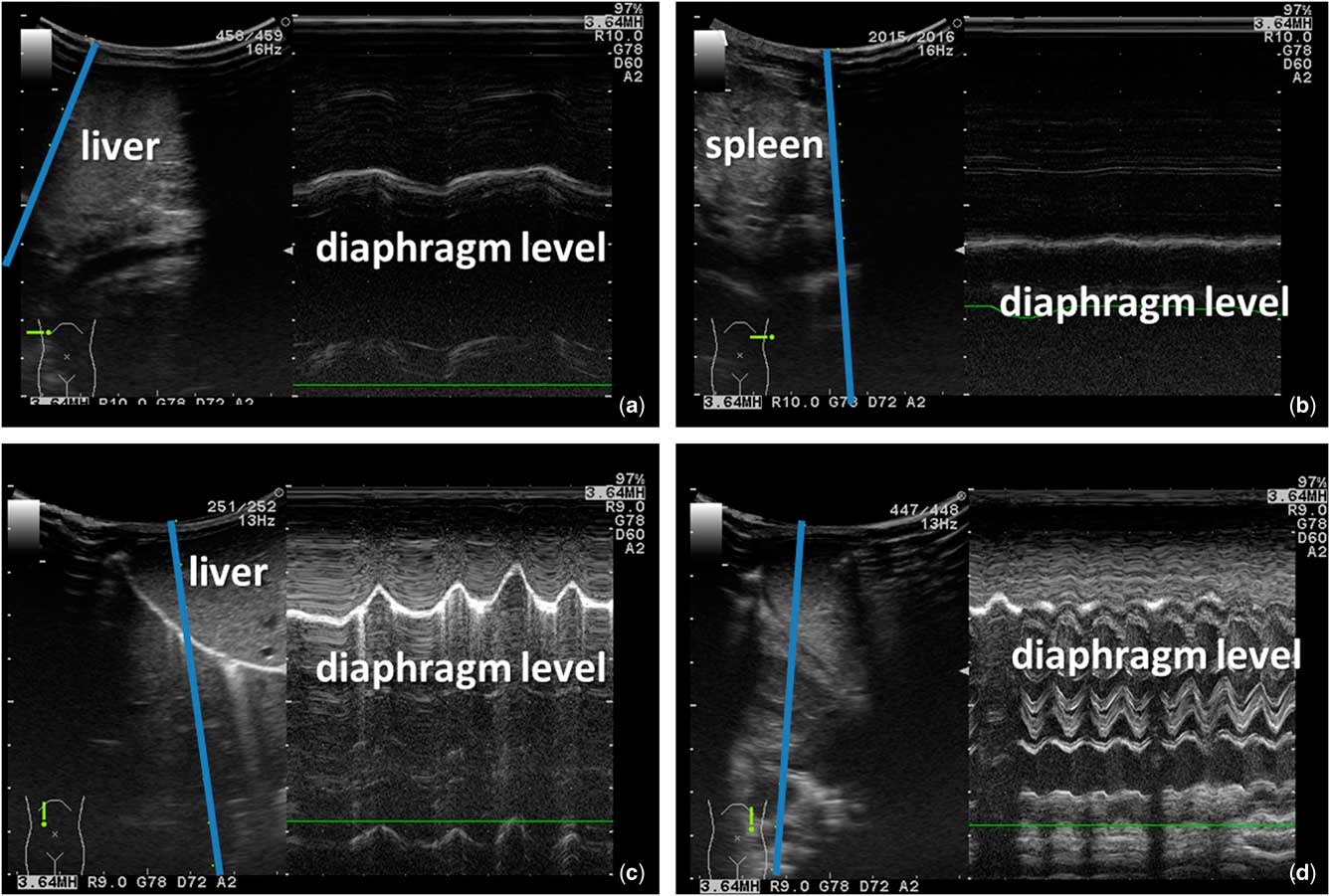
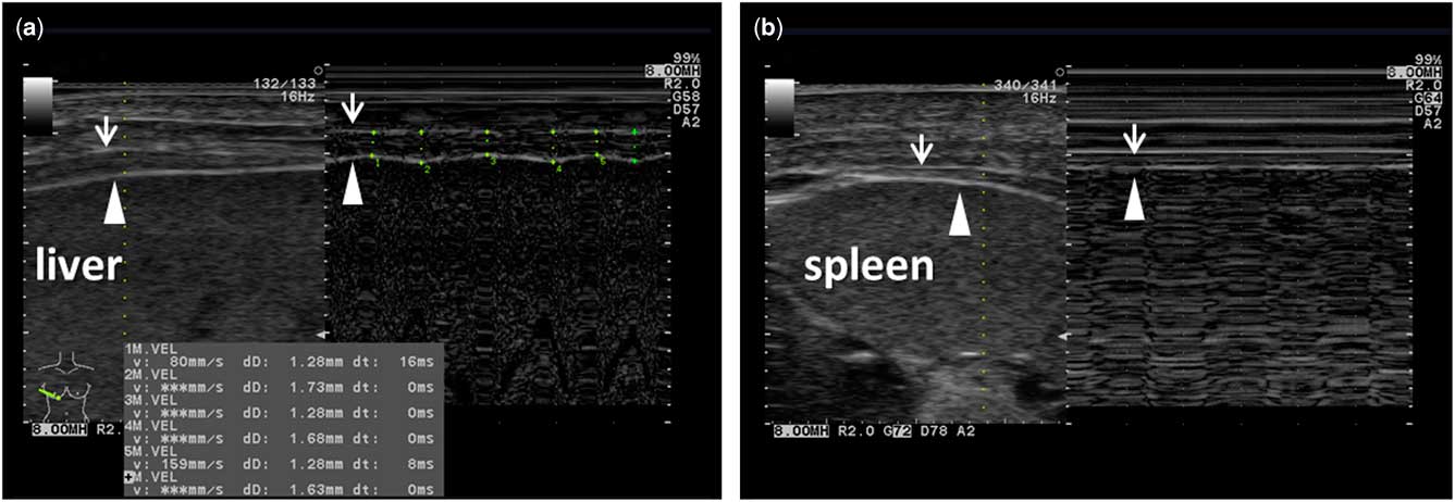
The ultrasonography was performed using a Prosound Alpha 6 device (Hitachi Aloka Medical, Mitaka, Japan) with a convex probe (3–5 MHz) and an 8-MHz linear probe. The initial examination attempted to identify diaphragm excursion at the abdomen, although it was difficult to obtain clear images of the left diaphragm through the sub-xiphoid and spleen echo windows, because of bowel gases (Fig 1a and b). Thus, we evaluated the diaphragm’s thickness at the zone of apposition, which was located on the mid-axillary line between the 8th and 10th ribs. During inhalation, the right diaphragm’s thickness increased, although no change was observed in the left diaphragm (Fig 2). A previous reportReference Lee, Hsia and Hsiao 5 calculated the per cent change in diaphragm thickness as ([end-inspiration thickness−end-expiration thickness]/[expiration thickness])×100, with diaphragm dysfunction being identified in patients with a value of <17%. During three respiratory cycles, our patient had average diaphragm thickness fractions of 31% (right) and 0.2% (left), which indicated left diaphragm paralysis. We confirmed paralysis of the left diaphragm using the excursion method with continuous positive airway pressure mode but no pressure support at 14 days after surgery, which clearly identified the left diaphragm (Fig 1c and d).
Echography of the neonate’s diaphragm using the excursion method. The blue lines indicate the M-mode reference line. The initial evaluation revealed normal right diaphragm movement ( a ), although the left side of the diaphragm was blocked by the bowel gases ( b ). At 14 days after the surgery, diaphragm paralysis was identified by comparing the right side ( c ) and left side ( d ) of the diaphragm.

Measurement of diaphragm thicknesses using M-mode ultrasonography. The distance between the inner pleura (arrow) and peritoneum (arrow head) was increased on the right side ( a ) but not on the left side ( b ) during inhalation.

The patient did not undergo diaphragm plication, based on the possibility of the paralysis resolving, as well as sustained heart failure caused by the ventricular septal defect after the pulmonary banding. However, monitoring of the diaphragm thickness fraction was performed during the mechanical ventilation (Supplementary Table S1 and Figure S1). On postoperative day 33, an increased diaphragm thickness fraction was identified but the excursion was not identified. We confirmed that the paralysis had resolved using both the diaphragm thickness fraction and the excursion method after closure of the ventricular septal defect at 53 days after surgery.
Discussion
We evaluated the diaphragm thickness fraction in a neonate who was receiving mechanical ventilation after cardiac surgery, which revealed diaphragm paralysis. The diaphragm thickness fraction method had two advantages compared with the excursion method. First, this approach facilitated the diagnosis without needing to interrupt the mechanical ventilation, which is less stressful for these patients. Second, the excursion method can be affected by bowel gases in rare cases,Reference Epelman, Navarro, Daneman and Miller 6 although the diaphragm thickness fraction method is unaffected by bowel gases. Nevertheless, the diaphragm thickness fraction method requires greater experience and a longer examination time, 10 minutes at least, compared with the excursion method, 5 minutes. The echographic diagnosis of diaphragm dysfunction can be enhanced by evaluating the diaphragm thickness fraction in patients who cannot be accurately evaluated using the excursion method, which can prevent patients from undergoing unnecessary irradiation or movement for fluoroscopy.
Conclusion
The diaphragm thickness fraction can be evaluated in children during mechanical ventilation, and can complement the echographic diagnosis of diaphragm dysfunction using the excursion method.
Acknowledgements
The authors thank Editage for English language editing.
Financial Support
This research was not supported by specific grants from any funding, commercial, or not-for-profit organisations.
Conflicts of Interest
None.
Ethical Standards
The authors assert that all procedures contributing to this work comply with ethical standards of the relevant Japanese guidelines on human experimentation and with Helsinki Declaration of 1975, as revised in 2008, and has approved by the Ethics committee of the Tsukuba University Hospital.
Supplementary material
To view supplementary material for this article, please visit https://doi.org/10.1017/S1047951118000197