Introduction
Gastrointestinal nematodes have long been identified as parasites infecting elephants, and are often associated with protein-losing gastroenteropathy that can cause hypoalbuminaemia, anaemia, enteritis and even death in younger elephants (Fowler, Reference Fowler, Fowler and Mikota2006). Among the gastrointestinal nematodes, those in the genera of Murshidia and Quilonia, belonging to the subfamily Cyathostominae of the family Strongylidae, are specific to elephants and rhinoceroses (Lane, Reference Lane1914). Five species of each Murshidia and Quilonia have been reported to infect Asian elephants (Elephas maximus) (Lane, Reference Lane1914; Ware, Reference Ware1924; Witenberg, Reference Witenberg1925; Baylis, Reference Baylis and Sewell1936; Van der Westhuysen, Reference Van der Westhuysen1938; Gupta and Jaiswal, Reference Gupta and Jaiswal1984; Gupta and Trivedi, Reference Gupta and Trivedi1984; Zhang and Xie, Reference Zhang and Xie1992), whereas 17 Murshidia and eight Quilonia species have been recorded from African elephants (Loxodonta africana or Loxodonta cyclotis) (Khalil, Reference Khalil1922; Van der Westhuysen, Reference Van der Westhuysen1938; Fowler, Reference Fowler, Fowler and Mikota2006).
Recently, optical light photomicrographs and the findings of genetic analyses of mitochondrial cytochrome c oxidase subunit I (COI) gene sequences have been presented for three Murshidia species (Murshidia falcifera, Murshidia indica and Murshidia neveulemairei) and two Quilonia species (Quilonia renniei and Quilonia travancra) infecting Asian elephants in Myanmar (Chel et al., Reference Chel, Iwaki, Hmoon, Thaw, Soe, Win, Bawm, Htun, Win, Oo, Masum, Ichii, Nakao, Nonaka and Katakura2020a). In terms of species identification, although the number of coronal leaflets is one of the definitive characteristics used in taxonomic keys for Quilonia species in elephants (Van der Westhuysen, Reference Van der Westhuysen1938), establishing the number of leaflets based on light microscopy (LM) is typically difficult. Moreover, the numbers of these leaflets in the different Quilonia parasites infecting Asian elephants tend to be quite similar: 18, 20, 24 and 20‒22 for Q. renniei, Quilonia edentata, Quilonia guptai and Quilonia simhai, respectively (Van der Westhuysen, Reference Van der Westhuysen1938; Gupta and Jaiswal, Reference Gupta and Jaiswal1984; Gupta and Trivedi, Reference Gupta and Trivedi1984; Zhang and Xie, Reference Zhang and Xie1992).
In the current study, in order to resolve issues relating to coronal leaflet number, scanning electron microscopy (SEM) was conducted to determine the exact number of coronal leaflets in Q. renniei specimens that were collected from Asian elephants in Myanmar, and confirmed taxon identity based on COI gene sequencing.
Materials and methods
Parasites
The parasite specimens examined in this study were adult worms collected in 2018 from the feces of Asian elephants maintained in two elephant camps, Hmaw Yaw Gyi (H) and Taung Kya (T), in Myanmar, after antihelminth administration. The locations of these two elephant camps are separated by a distance of more than 150 km, and elephants in the camps are cared for under semi-captive conditions, in which they are able to freely consume plants growing in the surrounding forests. The specimens were previously examined morphologically for the anterior and posterior parts, extracted genomic DNAs from the middle part of the same worms and identified parasite species based on their morphological characteristics and COI gene sequencing (Chel et al., Reference Chel, Iwaki, Hmoon, Thaw, Soe, Win, Bawm, Htun, Win, Oo, Masum, Ichii, Nakao, Nonaka and Katakura2020a). In this study, a total of 19 Q. renniei specimens (11 and 8 from camps H and T, respectively) were used (Table 1).
Variation in the number of coronal leaflets in Quilonia renniei specimens that were isolated from Asian elephants in Myanmar and identified based on COI gene sequencinga

M, male; F, female; UD, undetermined; NS, not significant.
a Data, except for the number of coronal leaflets based on SEM, were adapted from Chel et al. (Reference Chel, Iwaki, Hmoon, Thaw, Soe, Win, Bawm, Htun, Win, Oo, Masum, Ichii, Nakao, Nonaka and Katakura2020a).
*Significance in males between LM and SEM; **significance in females between LM and SEM; ***significance in SEM between males and females.
Scanning electron microscopy
For the purpose of SEM examination, formalin-fixed parasites were further fixed with 2.5% glutaraldehyde for 3 h, followed by treatment with tannic acid for 6 h. Thereafter, the specimens were subjected to five 10-min washes with 0.1 m phosphate buffer (PB) and then post-fixed with osmium tetroxide for 1 h, followed by a further five 10-min washes with 0.1 m PB. The specimens were subsequently dehydrated using an ascending series of alcohol, transferred to 3-methyl butyl acetate and finally dried using an HCP-2 critical point dryer (Hitachi, Tokyo, Japan). The dried samples were placed on a mounting stub and coated for 1 min with gold ions using a Hitachi E-1030 ion sputter coater (Hitachi, Tokyo, Japan). The worms were examined using an S-4100 scanning electron microscope (Hitachi, Tokyo, Japan) with an accelerating voltage of 4 kV.
Statistical analysis
The number of coronal leaflets was calculated from the LM (Chel et al., Reference Chel, Iwaki, Hmoon, Thaw, Soe, Win, Bawm, Htun, Win, Oo, Masum, Ichii, Nakao, Nonaka and Katakura2020a) and the SEM photographs in this study, and expressed as the mean ± standard error (s.e.). Student's t-test was performed using Microsoft Excel 2019 to assess differences between the number of coronal leaflets counted for males and females based on LM and SEM. Statistical significance was set at P < 0.05.
Phylogenetic and haplotype analysis
A total of 19 COI gene sequences (469 nucleotides) previously obtained from Q. renniei specimens (Chel et al., Reference Chel, Iwaki, Hmoon, Thaw, Soe, Win, Bawm, Htun, Win, Oo, Masum, Ichii, Nakao, Nonaka and Katakura2020a) were aligned using MUSCLE option within MEGA 7 (Kumar et al., Reference Kumar, Stecher and Tamura2016), and a corresponding maximum likelihood (ML) tree was generated. The Hasegawa–Kishino–Yano (HKY) + G model was adapted for analysis, as this showed the lowest Bayesian information criterion scores to describe the substitution pattern the best. Bootstrap values were determined based on 1000 replicates, with a total of 469 positions in the final dataset. The COI gene sequence of Quilonia africana (JN252511) from an African elephant was included as an outgroup. Nucleotide diversity of the COI genes was calculated using MEGA 7. A median-joining (MJ) network was created for a total of 16 COI haplotypes from the 19 Q. renniei specimens using PopART 1.7 (Leigh and Bryant, Reference Leigh and Bryant2015). ARLEQUIN ver. 3.5 software (Excoffier and Lischer, Reference Excoffier and Lischer2010) was used for calculating Tajima's D and Fu's Fs for the mismatch distribution analysis and the neutrality tests. Analysis of molecular variation (AMOVA) was performed to determine the genetic structure of population and their differentiation (F ST) using ARLEQUIN ver. 3.5.
Results
Scanning electron microscopic analysis
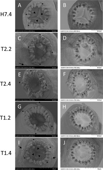
Scanning electron micrographs of the anterior end of adult worms (males and females) showed the exact number of coronal leaflets for each specimen (Table 1). In males, the numbers detected using SEM ranged from 16 to 19 (average 18.1 ± 0.4, n = 8), which was significantly higher (P < 0.05) than those determined based on the LM (15–18, average 16.8 ± 0.4, n = 6) (Table 1). Similarly, in females, leaflet number ranged 19–20 (average 19.7 ± 0.2, n = 9) based on the SEM, which was significantly higher (P < 0.001) than that estimated using the LM (16–18, average 17.4 ± 0.3, n = 7). Furthermore, a comparison of the number of coronal leaflets determined for males (18.1 ± 0.4, n = 8) and females (19.7 ± 0.2, n = 9) based on the SEM observation revealed a significantly higher number (P < 0.005) in females. Regardless of parasite sex, specimens bearing 19 coronal leaflets were most numerous (n = 7), followed by those with 20 (n = 6) and 18 (n = 2) leaflets based on the SEM. Updated information, including the number of coronal leaflets, for all five Quilonia species associated with Asian elephants, is summarized in Table S1.
Typical scanning electron micrographs of parasite specimens bearing 16, 17, 18, 19 or 20 coronal leaflets are shown in Fig. 1. The numbers of coronal leaflets were established using both the original SEM photomicrographs and the corresponding photomicrographs with inverted tone (Fig. 1A–J). Four papillae, two amphids and three mouth lobes were clearly observed in some specimens. In addition, scanning electron micrographs of the remaining 12 Q. renniei specimens are shown in Fig. S1.
Typical SEMs of the anterior end of Quilonia renniei specimens bearing different numbers of coronal leaflets. Specimens of H7.4 (A and B), T2.2 (C and D), T2.4 (E and F), T1.2 (G and H) and T1.4 (I and J) show 16, 17, 18, 19 and 20 coronal leaflets in the original photographs (A, C, E, G and I) and the corresponding images with inverted tones (B, D, F, H and J), respectively. In some specimens, four papillae (asterisks), two amphids (arrows) and three mouth lobes (arrowheads) are clearly visible.

Phylogenetic and haplotype analysis
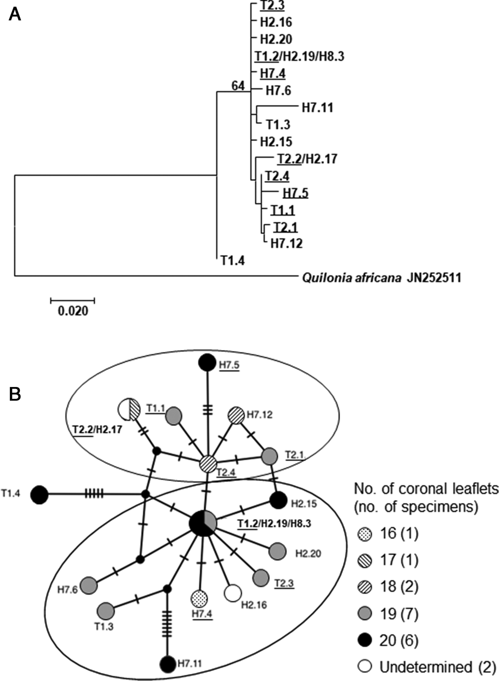
Among COI gene sequences (469 nucleotides) obtained from the 19 Q. renniei specimens, three (T1.2, H2.19 and H8.3) and two (T2.2 and H2.17) specimens were characterized by the same haplotype, respectively, and thus in total, 16 unique haplotypes were identified in this study. All nucleotide changes detected were, however, non-synonymous substitutions, thereby giving rise to the same amino acid sequences (data not shown). ML tree analysis revealed that the COI sequence of specimen T1.4 was different from other 18 sequences (Fig. 2A). The nucleotide diversity of the 19 sequences (16 haplotypes) was as low as 0.0087 ± 0.0019 (average ± standard deviation).
ML (A) and MJ network (B) analyses of the 16 haplotypes of 19 Q. renniei COI genes derived from parasites infecting Asian elephants in Myanmar. The ML tree is drawn to scale, with branch lengths measured in the number of substitutions per site, and the percentage of trees in which associated taxa clustered together is shown next to the branches (A). Parasite specimens with different numbers of coronal leaflets (16–20) are indicated by circles of different patterns. Short bars on nodes in the MJ network indicate the number of nucleotide substitutions (B). Male parasite specimens are indicated by underlines of the parasite IDs (A and B).

MJ network analysis displayed two subgroups (two circles in Fig. 2B). Mismatch distribution analysis yielded a τ value of 1.98 (0.88–6.09). Significant negative values of Tajima's D (−1.85, P = 0.02) and Fu's Fs (−11.15, P = 0.00) were obtained. AMOVA revealed that the genetic variation within groups (63.72%) was higher than that among populations (36.28%), and the overall genetic differentiation (F ST = 0.3628) between two groups was observed. However, there appeared to be no clear association between two COI groups and the number of parasite coronal leaflets, nor between COI groups and parasite sex (male or female) (Fig. 2B).
Discussion
In the current study, the exact number of coronal leaflets of Q. renniei specimens collected from Asian elephants in Myanmar was determined based on SEM observations. It is noteworthy that variable numbers of these leaflets were detected in both males (16–19, average 18.1) and females (19 or 20, average 19.7), and that the number was significantly higher in females. Gupta and Trivedi (Reference Gupta and Trivedi1984) have similarly reported variability in the number of coronal leaflets (referred to as leaf crown elements by these authors) in Q. simhai from 20 to 22, following their proposal of this parasite as a new species, although they did not describe the exact number of coronal leaflets for each male and female specimen. To the best of our knowledge, there have been no studies that have attempted determine differences in the cuticular structures of male and female nematodes. However, it is unknown the reasons why female parasites possess more leaflets than males. Furthermore, this study established that the number of coronal leaflets is not associated with the COI haplotypes of these parasites, and at present, little is known regarding functions of coronal leaflet and the genes or factors that contribute to regulating the numbers of cuticular structures in the mouths of nematode parasites.
The number of coronal leaflets or coronal rays is one of the major characteristics used in taxonomic keys for Quilonia species (Lane, Reference Lane1914; Van der Westhuysen, Reference Van der Westhuysen1938). However, the numbers of these features have been found to be very similar among related Quilonia species: 18 for Q. renniei from India (Lane, Reference Lane1914), 20 for Q. edentata from China (Zhang and Xie, Reference Zhang and Xie1992), 20−22 for Q. simhai from India (Gupta and Trivedi, Reference Gupta and Trivedi1984) and 24 for Q. guptai from India (Gupta and Jaiswal, Reference Gupta and Jaiswal1984). To determine the precise number of coronal leaflets for these parasites, further investigations of the mouth structure of adult worms by SEM or advanced methods are required. In this regard, the consistency of ten coronal leaflets based on LM and SEM observations has been reported for Q. travancra derived from Asian elephants in Sumatra, Indonesia (Prahardani et al., Reference Prahardani, Firdausy, Yanuartono and Nurcahyo2019).
The finding of negative values of Tajima's D and Fu's Fs in the COI haplotype analysis in the current study suggested the occurrence of population expansion of Q. renniei parasites in elephants in Myanmar, as significant negative both values indicate recent selective sweep or population expansion (Theint et al., Reference Theint, Thidalay Thwe, Zaw, Shimada, Bawm, Kobayashi, Saing, Katakura, Arai and Suzuki2021; https://en.wikipedia.org/wiki/Tajima%27s_D). A similar result of parasite expansion has been reported for Fasciola gigantica flukes from domestic ruminants in Bangladesh (Mohanta et al., Reference Mohanta, Ichikawa-Seki, Shoriki, Katakura and Itagaki2014). In addition, an F ST value of 0.3628 observed between two COI groups suggested the genetic variation among Q. renniei parasite populations in Myanmar, as F ST values >0.25 indicate strong genetic differentiation (Guernaoui et al., Reference Guernaoui, Hamarsheh, Garcia, Fontenille and Sereno2020). Since the sample size tested was low in this study, further studies using additional samples will clarify the genetic variation among the parasite populations.
Given the extinction of most proboscidean species, African and Asian elephants are the only proboscidean species that have survived into the current epoch (Roca et al., Reference Roca, Ishida, Brandt, Benjamin, Zhao and Georgiadis2015). Currently, populations of Asian elephants survive in a number of South and Southeast Asian countries, although they are listed as an endangered species (IUCN, 2020; http://www.iucnredlist.org/details/7140/0). The current population of Asian elephants is estimated to range between 40 000 and 50 000, with Myanmar representing a species stronghold (4000–5000 animals) (Leimgruber et al., Reference Leimgruber, Oo, Aung, Kelly, Wemmer, Senior and Songer2011). Analysis of the mitochondrial DNA (mtDNA) haplotypes of Asian elephants has revealed unique haplotypes with α and β clades (Vidya et al., Reference Vidya, Sukumar and Melnick2009). The mtDNA analysis of Myanmar elephants has indicated that both haplotype clades were distributed in Myanmar but there was the lack of evidence of a signature of either past expansion or bottlenecks of the elephants (Vidya et al., Reference Vidya, Sukumar and Melnick2009; Kusza et al., Reference Kusza, Suchentrunk, Pucher, Mar and Zachos2018; Sharma et al., Reference Sharma, Goossens, Heller, Rasteiro, Othman, Bruford and Chikhi2018). Although mtDNA haplotype analysis of elephants was not performed in this study, it is conceivable that the indicated expansion of the Q. renniei parasites is unrelated to elephant mtDNA, given the occurrence of two mtDNA haplotype clades, and detection of high levels of mtDNA diversity in both clades in Myanmar elephants (Kusza et al., Reference Kusza, Suchentrunk, Pucher, Mar and Zachos2018). Since there have been no reports on the genetic analysis of Q. edentata, Q. guptai or Q. simhai, it remains to be determined whether Q. renniei and other strongyloid nematodes in other countries show genetic variations and differences in the number of coronal leaflets. Further genetic analyses are accordingly warranted to elucidate the phylogenetic relationships among these Quilonia species in Asian elephants.
Finally, it is noteworthy that there are differences between Asian and African elephants with respect to parasite species within the same genus (Fowler, Reference Fowler, Fowler and Mikota2006), and consequently, comparative genetic studies on the parasites derived from Asian and African elephants might provide new insights into the evolution, variation, speciation and separation of elephant parasites, as shown for nematodes of Murshidia and Quilonia species and Cobboldia species of stomach bot fly (Chel et al., Reference Chel, Iwaki, Hmoon, Thaw, Soe, Win, Bawm, Htun, Win, Oo, Masum, Ichii, Nakao, Nonaka and Katakura2020a, Reference Chel, Nakao, Ohsawa, Oo, Nonaka and Katakura2020b).
In conclusion, it is evident that coronal leaflet number in Q. renniei specimens shows intra-specific variation and overlaps with that of other species of Quilonia derived from Asian elephants. As the number of coronal leaflets is one of the definitive characters of the diagnostic keys used to identity Quilonia species, further morphological and genetic studies using additional specimens are needed to gain a better understanding of the evolution, variation and distribution of Quilonia parasites in Asian elephants.
Supplementary material
The supplementary material for this article can be found at https://doi.org/10.1017/S0031182021002110
Data
Data are available from the corresponding author upon reasonable request.
Acknowledgements
We thank Prof. Hitoshi Suzuki of Hokkaido University for the MJ network analysis and Editage (www.editage.com) for English language editing.
Author contributions
KK and SB conceived and designed the study. HMC, MAM and OI conducted data collection. LLH and SB performed statistical analyses. KK, NN and RN wrote the manuscript.
Financial support
This study was supported in part by KAKENHI (grant number 17H04638) from MEXT (the Japanese Ministry of Education, Culture, Sports, Science and Technology), and the Bilateral Open Partnership Joint Research Project from JSPS (the Japan Society for Promotion of Science), Japan.
Conflict of interest
The authors declare there are no conflicts of interest.