Endoscopy is an invasive procedure that can contribute to the transmission of microorganisms responsible for infections, and the incidence of pathogen transmission attributed to gastrointestinal (GI) endoscopic procedures has been estimated at 1 to 1.8 million procedures.Reference Kimmery, Burnett and Carr-Locke 1 , Reference Epstein, Hunter and Arwady 2 To prevent transmission of infection, flexible GI endoscopes are completely cleaned and subjected to high-level disinfection. Multiple society guidelines have been developed to detail the reprocessing sequence of appropriate flexible GI endoscopes.Reference Petersen, Chennat and Cohen 3 Endoscope drying has been described as one of the most important steps in limiting bacterial proliferation and the risk of contamination during endoscope storage.Reference Muscarella 4 Storage cabinets for heat-sensitive endoscopes (SCHEs) were developed to maintain the microbiological quality of reprocessed endoscopes. The aim of the SCHE is to store GI endoscopes hanging vertically in a clean, dry, and well-ventilated cupboard. SCHEs are validated by manufacturers,Reference Pineau, Villard, Duc and Marchetti 5 but very few studies have evaluated the performance of channel-purge storage cabinets for GI endoscopes that have been used on patients.Reference Grandval, Hautefeuille, Marchetti, Pineau and Laugier 6 The objective of this study was to evaluate the benefits of SCHEs by analyzing microbiological testing of GI endoscopes in a French teaching hospital.
METHODS
We compared the results of microbiological surveillance testing of 29 GI endoscopes before and after the installation of an SCHE in the GI endoscopy unit of Brest teaching hospital. During the first part of the study (from January 1, 2010 to July 31, 2013), these GI endoscopes were stored in a clean, dry, dedicated cupboard between uses. They were subjected to high-level disinfection after 12 h of storage as recommended in France. 7 In the second part of the study (from August 1, 2013 to February 28, 2015), these 29 GI endoscopes were stored in an AS300 Hysis SCHE (Hysis Medical, La Ciotat, France). All channels of the endoscopes were purged with medical air for 1 hour. The maximal storage time without cleaning before use was 72 hours, as recommended by health authorities in France. Beyond this delay, GI endoscopes were cleaned and disinfected before use. We reviewed all of the results of programmed microbiological testing of endoscopes performed for quality assurance during this period. We did not include tests done after a first improper control or tests performed before the use of endoscopes returned from the manufacturer after maintenance.
Endoscope Reprocessing Cycle
All endoscopes stored in the SCHE received manual cleaning and were then treated using an automatic endoscope washer-disinfector (AFER, POKA-YOKE AER, Getinge), including a dry cycle at the end of reprocessing.
Endoscope Sampling Method
The hospital microbiology laboratory uses an endoscope sampling protocol consistent with national recommendations. Endoscope sampling was undertaken by the same well-trained technicians during the study period. Endoscopes were sampled by injecting neutralizing pharmacopoeia diluent (NPD) buffer with sodium thiosulfate (AES Laboratoire, Combourg, France) into each channel using sterile connectors. The elevator–guide-wire channel in each duodenoscope was not unsealed, but the sample from this channel was collected using sterile connectors. A 2-way irrigation pathway was made systematically in each channel to improve sampling efficiency. The total sample (120 mL) was recovered at the distal end of the endoscope and was incubated at 30°C on plate count agar after 100 mL was filtered through a 0.45-µm filter. After 5 days of incubation, results were expressed as the number of colony-forming units (cfu) per endoscope. Microorganisms were identified, and the results of endoscope sampling were interpreted according to the National Technical Committee on Nosocomial Infection guideline (Table 1). 8 The sample collected represented organisms from all channels and it was not possible to differentiate the channel from which the organisms originated.
Interpretation Criteria of Microbiological Testing for GI Endoscopes

note. Cfu, colony-forming units.
a Main indicator microorganisms: Staphylococus aureus, Enterobacteriaceae, Pseudomonas aeruginosa and other Pseudomonas spp., Stenotrophomonas maltophilia, Acinetobacter spp., and Candida spp.
When microbiological testing was not performed (alert or action level), the endoscope was subject to double manual reprocessing. If a new sample revealed microbial contamination, the endoscope was sent to the manufacturer for maintenance.
Statistical analysis was performed using R software (version 2.12.1). Percentages were compared using the χ2 test. All tests performed were 2-sided, and P<.05 was considered significant.
RESULTS
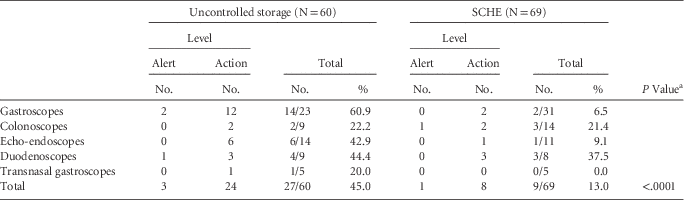
A total of 29 GI endoscopes (12 gastroscopes, 8 colonoscopes, 4 echo-endoscopes, 3 duodenoscopes, and 2 transnasal gastroscopes) were included in the study (Table 2). Over the study period, 129 programmed microbiological tests of these endoscopes were performed. During the first part of the study (n=60), 45.0% of the microbiological tests showed a level of contamination higher than the target. The results differed depending on the type of endoscope: 60.9% of gastroscopes reached the alert or action level, while only 1 transnasal gastroscope was contaminated. In the second part of the study (n=69), only 13.0% of the GI endoscopes stored in the SCHE had microbiological tests revealing contamination. Improved results were significant; the rate of achieving the target level rose from 55.0% to 87.0% with the use of an SCHE (odd ratio [OR], 5.37; 95% confidence interval [CI], 2.15–14.63; P<.0001).
Rate of Endoscope Contamination According to the Storage Conditions

NOTE. SCHE, storage cabinets for heat-sensitive endoscopes.
a χ2 test.
The main indicator microorganisms recovered during the study were Gram-negative bacteria identified as Pseudomonas aeruginosa and other Pseudomonas spp. (n=6), Stenotrophomonas maltophilia (n=4), Acinetobacter pitii (n=2), Citrobacter freundii (n=1), Klebsiella oxytoca (n=2), K. pneumonia (n=5), and Escherichia coli (n=1). Staphylococus aureus was found on 1 sample, and Candida spp. were found on 4 samples.
DISCUSSION
Storage is an important factor in the maintenance of bacterium-free endoscopes. The use of SCHE in the GI endoscopic unit has significantly improved the microbiological quality of endoscopes. Rates of endoscopes contamination were usually high, even when they were programmed. Nearly 50% of endoscope tests identified microbiological contamination, and this rate was particularly high for gastroscopes (60.9%). After the SCHE was implemented, only 13.0% of microbiological tests reached the alert or action level. This difference was especially marked for gastroscopes, for which the contamination rate is now 6.5%.
We can reasonably attribute this difference to the use of the SCHE insofar as the personnel of the endoscopy teams and microbiology laboratory did not change. Similarly, the endoscope treatment protocols and the automated flexible endoscope reprocessors used were the same throughout the study. Rates of microbiological contamination described in our study are similar to those described in other French studies.Reference Saliou, Garlantézec and Baron 9 However, the endoscope contamination rates we observed were very high compared to those of other countries such as Australia, where contamination rates are on the order of 1 contamination per 250 samples.Reference Gillespie, Kotsanas and Stuart 10 The observed difference could be due to the variation in endoscope sampling techniques between countries. Sample collection using 120 mL NDP buffer with sodium thiosulfate to flush all channels may be a more sensitive sample method than flushing each channel with 10 mL sterile water. Moreover, interpretation criteria of microbiological testing for GI endoscopes are more stringent, which may explain the observed contamination rate in France. These results show that although rate of infected patients has remained low, endoscope decontamination is not always effective and contamination may lead to outbreaks of healthcare-associated infection. Such outbreaks after endoscopy have been described, and cross contamination due to carbapenem-resistant enterobacteriaceae have already been associated with the lack of a drying procedure.Reference Carbonne, Thiolet and Fournier 11 The use of an SCHE did not improve contamination rates for duodenoscopes. Cleaning the elevator–guide-wire channel in the duodenoscope is difficult. Debris can remain under the elevator piece and biofilm can develop, which may explain the results of microbiological testing.
Improving the quality of microbiological decontamination of endoscopes might reduce the number of control samples performed and the number of endoscopes returned to the manufacturer for maintenance. Next, it will be appropriate to conduct a cost-effectiveness analysis of the implementation of the SCHE in endocopy units.
ACKNOWLEDGMENTS
The authors are grateful to Raymond Kerouanton and Monique Magnin for their contribution to the microbiological testing.
Financial support: No financial support was provided relevant to this article.
Potential conflicts of interest: All authors report no conflicts of interest relevant to this article.