INTRODUCTION
Although most parasite studies focus on systems involving a single parasite species and a single host species, in natural populations ‘concomitant’ or ‘mixed’ infections by more than one parasite species or genotype are common (Petney and Andrews, Reference Petney and Andrews1998; Cox, Reference Cox2001; Read and Taylor, Reference Read and Taylor2001). Consequently, interactions between different parasite genotypes or species frequently occur. These interactions may be synergistic or antagonistic (Cox, Reference Cox2001), with potential fitness implications for both the host (morbidity and/or mortality) and parasite (transmission potential). In turn, these effects at the scale of the individual host may influence host-parasite dynamics at the population level (Graham et al. Reference Graham, Cattadori, Lloyd-Smith, Ferrari and Bjornstad2007) and evolutionary processes in both the host and parasite (May and Nowak, Reference May and Nowak1994; Brown and Grenfell, Reference Brown and Grenfell2001; Read and Taylor, Reference Read and Taylor2001; Brown, Hochberg and Grenfell, Reference Brown, Hochberg and Grenfell2002). Understanding interactions at the level of the individual and the population, and evolutionary consequences, may have important implications for predictions of disease emergence, disease control programmes and bio-control initiatives (Thomas, Watson and Valverde-Garcia, Reference Thomas, Watson and Valverde-Garcia2003; Lello et al. Reference Lello, Boag, Fenton, Stevenson and Hudson2004).
Various mechanisms can cause interactions between parasite species or genotypes within an individual host (Petney and Andrews, Reference Petney and Andrews1998; Cox, Reference Cox2001). Parasites that infect the same site within a host can directly interact through interference competition, whilst indirect interactions may be mediated by competition for resources or via the host immune system. Immune-mediated interactions may result from a number of processes. Whereas cross-effective immune responses between antigenically similar parasite species or genotypes may cause antagonistic interactions (harmful to at least one of the parasites), parasite-induced immunosuppression may result in synergistic interactions (beneficial to at least one of the parasites). Additionally, interactions may be a consequence of differences between parasite species in the immune effector mechanisms that are stimulated and to cross-regulation among different components of the immune system, e.g. the modification of responses induced in either type 1 or type 2 T helper cells (Th1 and Th2, respectively) following helminth infection due to the effects of regulatory T cells (Treg) that act to reduce inflammatory responses (Maizels and Yazdanbakhsh, Reference Maizels and Yazdanbakhsh2003). As there have been several reviews of potential mechanisms published, here we focus on the impact that interactions between co-infecting parasite species have on host and parasite ecology.
Ecologists, in particular, are interested in co-infection in natural populations for two main classes of reason. First, the effects of parasites, especially microparasites, on the condition of individuals in natural populations, and on the dynamics of those populations, are largely unknown (Hudson, Dobson and Newborn, Reference Hudson, Dobson and Newborn1998; Telfer et al. Reference Telfer, Bennett, Bown, Cavanagh, Crespin, Hazel, Jones and Begon2002; Tompkins et al. Reference Tompkins, Dobson, Arneberg, Begon, Cattadori, Greenman, Heesterbeek, Hudson, Newborn, Pugliese, Rizzoli, Rosa, Rosso, Wilson, Hudson, Rizzoli, Grenfell, Heesterbeek and Dobson2002). To address these questions, it is essential to know whether infection with one parasite species tends to be associated (either positively or negatively) with infection by other parasite species. This is because, for example, host survival (and ultimately abundance) may be only negligibly affected in singly-infected hosts but severely affected by multiple, coincident infections. Second, the relative importance of the forces that determine the structure of parasite communities (e.g. resource- or immunity-mediated competition), provides an interesting comparison with community ecology studies of non-parasite systems (Behnke et al. Reference Behnke, Bajer, Sinski and Wakelin2001; Telfer et al. Reference Telfer, Clough, Birtles, Bennett, Carslake, Helyar and Begon2007b).
Empirical studies of co-infection have predominantly involved laboratory model systems, where controlled experiments facilitate the detection of effects and allow mechanistic insight to be gained (e.g. Graham et al. Reference Graham, Lamb, Read and Allen2005). Although there are an increasing number of studies in natural populations of humans and other animals, to a large extent these have been cross-sectional, focusing on population-level patterns. Such data offer limited opportunities to explore either the underlying mechanisms at the individual level (Pedersen and Fenton, Reference Pedersen and Fenton2007) or ultimate effects on host fitness. Moreover, there is mounting evidence from experimental studies that the outcome of interactions during co-infections (for either the host or the parasite) is context dependent, potentially varying with different host or parasite genotypes (McClelland, Granger and Potts, Reference McClelland, Granger and Potts2003; Jackson et al. Reference Jackson, Pleass, Cable, Bradley and Tinsley2006) or environmental conditions (Malakar et al. Reference Malakar, Elkinton, Hajek and Burand1999; Thomas et al. Reference Thomas, Watson and Valverde-Garcia2003; Hodgson et al. Reference Hodgson, Hitchman, Vanbergen, Hails, Possee and Cory2004). Perhaps most critically, outcome can depend on the timing and sequence of infections (Jacobson et al. Reference Jacobson, Jenkins, Springer, Munoz, Shah, Phair, Zhang and Armenian2000; Graham, Reference Graham2002; Thomas et al. Reference Thomas, Watson and Valverde-Garcia2003; Jackson et al. Reference Jackson, Pleass, Cable, Bradley and Tinsley2006), something on which cross-sectional data can provide little information. Therefore, in natural populations, the relative importance of interactions between parasites compared to other factors for determining variation in individual fitness, population dynamics and community structure remains largely unexplored.
Longitudinal data, where the infection states of known individuals are followed through time, can provide the detailed information necessary to understand context-dependent effects. Moreover, longitudinal data are necessary to fully understand the impact of co-infection on host-parasite ecology. For example, does infection with one parasite species influence (1) host susceptibility to infection by a second parasite species, (2) aspects of the infection biology of a second parasite species (e.g. length of infection or intensity of infection) and/or (3) the impact that infection by a second parasite has on host fitness (pathology)? Such potential effects have direct bearing on the population level consequences of co-infection for both parasites and hosts. Although there are increasing numbers of longitudinal studies of human populations (e.g. Jacobson et al. Reference Jacobson, Jenkins, Springer, Munoz, Shah, Phair, Zhang and Armenian2000; Harcourt et al. Reference Harcourt, Donfield, Gomperts, Daar, Goulder, Phillips and Klenerman2005; Raimondo et al. Reference Raimondo, Brunetto, Pontisso, Smedile, Maina, Saitta, Squadrito and Tono2006; Yirrell et al. Reference Yirrell, Wright, Shafer, Campbell, Van der Paall, Kaleebu, Grosskurth and Whitworth2007), there have been no longitudinal co-infection studies in natural non-human animal populations.
The remainder of this paper is divided into two parts. First, we briefly review some of the evidence for effects of co-infection on susceptibility, infection biology and pathology, showing where possible insights gained from longitudinal studies, and the importance of infection sequence. Second, we illustrate the potential of longitudinal data for investigating the impact of co-infection on susceptibility and infection biology using results obtained from monitoring the parasite community infecting field vole (Microtus agrestis) populations. In contrast to most co-infection studies, this dataset focuses primarily on a microparasite community.
EFFECTS OF CO-INFECTION ON HOST-PARASITE ECOLOGY
Host susceptibility
Susceptibility is a property of an individual host at a given time and is the relative probability that a host becomes infected, given exposure to an infective dose. At the population level, changes in the proportion of susceptible hosts present or available could have profound consequences for parasite transmission rates and, therefore, host-parasite population dynamics (Graham et al. Reference Graham, Cattadori, Lloyd-Smith, Ferrari and Bjornstad2007).
In many cases the ability of a parasite to establish an infection successfully will depend on the initial immune response of the exposed host. On entry into the host, a parasite will experience an ‘immuno-environment’ potentially determined by both previous and current infections, as well as intrinsic factors such as sex, age, nutritional status and genotype. The immediate immuno-effectors in a naïve host will be dominated by the cells and molecules that comprise the innate immune response, and thus the efficiency of this arm of host immunity at reducing and clearing an infection will be influential in determining susceptibility. However, few co-infection studies have focused on innate immunity. The efficiency of an adaptive immune response may also affect susceptibility, especially for parasites that induce strong immunological memory. In such cases, secondary exposure of a host to the same parasite species or a parasite species that elicits a cross-effective immune response may rapidly initiate the production of specific antibodies and other effectors, potentially causing the clearance or neutralisation of infection. In contrast, other immune-mediated mechanisms may increase susceptibility. For example, in a longitudinal study of malaria infection that used molecular genotyping to distinguish between new infections and the recrudescence of existing infections, it was shown that the risk of new malaria infection was six times higher in adults infected with the immunosuppressive human immunodeficiency virus (HIV) than in HIV-uninfected adults (Kamya et al. Reference Kamya, Gasasira, Yeka, Bakyaita, Nsobya, Francis, Rosenthal, Dorsey and Havlir2006).
The continuous monitoring of hosts is not practical and, consequently, in practice it may be difficult to determine whether any observed impact of co-infection is associated with changes in host susceptibility or infection length. For example, a laboratory experiment demonstrated that whilst mice exposed only to Trichuris muris infection showed significant worm burdens at 21 days, mice infected with Schistosoma mansoni prior to T. muris exposure appeared to have become resistant, with T. muris worm burdens of zero at 21 days (Curry et al. Reference Curry, Else, Jones, Bancroft, Grencis and Dunne1995). However, in a further experiment that reversed the order of exposure, it was found that this ‘resistance’ probably resulted from a rapid expulsion of T. muris worms induced by the Th2 response stimulated by S. mansoni egg production; thus, infection length (an aspect of infection biology), rather than susceptibility per se had been affected). In this case, whether host susceptibility or infection length was affected is immaterial with respect to parasite fitness: T. muris take 30 days to reach reproductive maturity, expulsion by day 21 will have effectively reduced T. muris fitness to zero.
Infection biology
Infection biology describes the typical manifestation of an infection, including the within-host location, length and intensity of the infection. Infection by other parasites may alter infection biology and, consequently, parasite transmission success. At the population level such effects could again impact on parasite dynamics by changing the number or infectiousness of infected individuals (Graham et al. Reference Graham, Cattadori, Lloyd-Smith, Ferrari and Bjornstad2007).
Such interactions may be mediated by a range of mechanisms, including resource competition. For example, when smaller tea tortix (Adoxophyes honmai) larvae are co-infected with two viruses (A. honmai nucleopolyhedrovirus (AdhoNPV) and A. honmai entomopoxvirus (AdhoEPV)) the virus that succeeds in replicating faster experiences a significant advantage in terms of transmission potential, acquiring host resources pre-emptively and significantly reducing the viral occlusion bodies produced by its competitor. In simultaneous infections, the intrinsically faster replicating AdhoNPV wins the resource race, whilst if AdhoEPV infects first the results are reversed (Ishii et al. Reference Ishii, Takatsuka, Nakai and Kunimi2002).
Alternatively, effects on infection biology may be mediated by immune mechanisms. HIV infection, through depletion of CD4+ T cell populations, has a significant impact on a number of other co-infections in humans, including Plasmodium (increased parasitaemia; Whitworth et al. Reference Whitworth, Morgen, Quigley, Smith, Mayanja, Eotu, Omoding, Okongo, Malamba and Ojwiya2000) and Hepatitis B virus (increased viral replication; Koblin et al. Reference Koblin, Taylor, Rubinstein and Stevens1992; Kellerman et al. Reference Kellerman, Hanson, McNaghten and Fleming2003). In turn, some of these parasites appear to affect HIV infections with, for example, increases in viraemia (malaria; Hoffman et al. Reference Hoffman, Jere, Taylor, Munthali, Dyer, Wirima, Rogerson, Kumwenda, Eron, Fiscus, Chakraborty, Taha, Cohen and Molyneux1999). However, immune-mediated effects are not solely induced by parasites that have tropisms for components of the host immune system. More ‘remote’ effects occur between parasites infecting different systems or tissues of the host. For example, sheep co-infected with Trichostrongylus colubriformis (intestine) and larvae of the fly Oestrus ovis (nasal cavity) have reduced T. colubriformis worm burdens and egg production compared with sheep only infected with T. colubriformis (Yacob et al. Reference Yacob, Dorchies, Jacquiet, Bleuart, Prevot, Grisez, Bergeaud and Hoste2004). Interestingly, this effect occurs irrespective of the infection sequence, although the reductions are greater if the fly infection precedes the helminth infection (Yacob et al. Reference Yacob, Duranton-Grisez, Prevot, Bergeaud, Bleuart, Jacquiet, Dorchies and Hoste2002).
Longitudinal data can help resolve ambiguous data on effects of co-infection. Cross-sectional studies have indicated conflicting results of interactions between Hepatitis B (HBV) and Hepatitis C (HCV) viruses in humans, with some claiming that HCV inhibits HBV replication (Sato et al. Reference Sato, Fujiyama, Tanaka, Yamasaki, Kuramoto, Kawano, Sato, Mizuno and Nonaka1994; Chu, Yeh and Liaw, Reference Chu, Yeh and Liaw1998), whilst others claimed the reverse (Crespo et al. Reference Crespo, Lozano, Delacruz, Rodrigo, Rodriguez, Sanmiguel, Artinano and Ponsromero1994; Ohkawa et al. Reference Ohkawa, Hayashi, Yuki, Hagiwara, Kato, Yamamoto, Eguchi, Fusamoto, Masuzawa and Kamada1994). A longitudinal study that quantified viral replication rate found that, in many co-infected patients, patterns of decline and recrudescence in each of the viral infections were similar to those found in singly infected patients (Raimondo et al. Reference Raimondo, Brunetto, Pontisso, Smedile, Maina, Saitta, Squadrito and Tono2006), indicating that interactions between HBV and HCV may be relatively unimportant for disease progression and explaining the lack of concurrence between previous cross-sectional studies.
Pathology
Interactions between parasites may also impact on the pathology of an infection (i.e. the nature and degree of the effect on the host), which may, in turn, lead to effects on host fitness parameters such as survival or reproductive rates.
The pathology of an infection may relate directly to changes in infection biology. Again there are several examples of co-infection effects on pathology involving HIV. A longitudinal study of HIV and human herpesvirus type 8 (HHV-8) showed that men that were already HIV-positive and immunocompromised when infected with HHV-8 were more likely than those with the reverse infection sequence to develop Kaposi's sarcoma (Jacobson et al. Reference Jacobson, Jenkins, Springer, Munoz, Shah, Phair, Zhang and Armenian2000). Interestingly, there are also some parasites that appear to improve prognosis in HIV co-infected individuals. Compared with individuals only infected with HIV, HIV patients chronically infected with GB virus-C (Hepatitis G) show a comparatively slower decline in CD4+ T cells and increased survival time (Yirrell et al. Reference Yirrell, Wright, Shafer, Campbell, Van der Paall, Kaleebu, Grosskurth and Whitworth2007). The mechanism for this effect remains unresolved, but the interaction could be mediated by competition over CD4+ T cells or by immune responses.
Attempts by the immune system to simultaneously counter the multiple parasite species involved in a co-infection can lead to immunopathological disease and pathology that are more than the simple additive pathogenic effects of the different parasite species (Graham, Reference Graham2002). An experimental examination of Plasmodium-helminth co-infection demonstrated that mice infected with Litomosoides sigmodontis and P. chabaudi chabaudi lost more body mass and red blood cell density than mice infected with plasmodium alone (Graham et al. Reference Graham, Lamb, Read and Allen2005). Critically, for a given parasitaemia level, co-infected animals exhibited more severe disease symptoms, with a 25% reduction in red blood cell number compared with animals only infected with P. chabaudi chabaudi.
The above review highlights that longitudinal studies and experiments that explicitly consider the sequence of infection can investigate the impact of co-infection on host susceptibility, parasite infection biology and host fitness, and allow detailed investigation of the mechanisms involved. Data, especially longitudinal data, from natural populations of non-human animals may be better for addressing questions regarding co-infection than data from humans or the laboratory. While a number of confounding factors may also affect individuals in natural populations (such as density and season), once these are accounted for statistically, the results that emerge are more likely themselves to be ‘natural’ than caused by laboratory artefacts or influenced by medical interventions. Thus, the challenge now for ecologists is to export this approach to studies of co-infection in natural populations. For the remainder of this paper we will explore this challenge using data from field voles.
INTERACTIONS BETWEEN MICROPARASITES IN WILD FIELD VOLE POPULATIONS
Details of the trapping and diagnostic methods used in the study are given in Box 1. Below we attempt to generate predictions regarding potential interactions between the parasites, based on both the ecology of the parasites and the expected immunological response of hosts, as advocated in recent reviews (Graham et al. Reference Graham, Cattadori, Lloyd-Smith, Ferrari and Bjornstad2007; Pedersen and Fenton, Reference Pedersen and Fenton2007). We then describe two preliminary analyses, investigating the impact of co-infection on host susceptibility and on parasite infection biology respectively.
Box 1 Between May 2001 and March 2007, field vole populations were trapped at four grassy clear-cut sites within Kielder Forest, a man-made spruce forest situated on the English-Scottish border (55°13′ N, 2°33′ W). Each site was at least 3·5 km from the nearest neighbouring site. Using a 0·3 ha live-trapping grid, populations were trapped every 28 days from March to November, and every 56 days from November to March. Individual animals were identified using subcutaneous microchip transponders. Each time an animal was trapped, data were collected on mass, reproductive condition, body condition (based on estimating the degree of fat cover over the vertebral column and dorsal pelvic bones; Burthe et al. Reference Burthe, Telfer, Lambin, Bennett, Carslake, Smith and Begon2006), number of ticks, number of fleas (recorded on an ordinal scale with low=1–2 fleas, medium=3–5 fleas and high=>5 fleas) and the presence of external signs of Mycobacterium microti infection (Burthe et al. Reference Burthe, Bennet, Kipar, Lambin, Smith, Telfer and Begon2008a). In addition, a 20–30 μl blood sample was taken from the tail tip. For more information on the study sites see (Burthe et al. Reference Burthe, Telfer, Lambin, Bennett, Carslake, Smith and Begon2006).
Antibody to cowpox virus was detected in sera by immunofluorescence (IF) assay (Crouch et al. Reference Crouch, Baxby, McCracken, Gaskell and Bennett1995). DNA extracts were prepared from red blood cell pellets by alkaline digestion (Bown et al. Reference Bown, Begon, Bennett, Woldehiwet and Ogden2003). Polymerase chain reaction (PCR) assays were used to detect infection by Anaplasma phagocytophilum (Courtney et al. Reference Courtney, Kostelnik, Zeidner and Massung2004), Babesia microti (R. Birtles, unpublished data) and Bartonella spp. (R. Birtles, unpublished data). For three of the sites, samples positive by the genus-specific PCR assay for Bartonella spp. were tested by species-specific PCR assays to identify B. taylorii, B. grahamii and B. doshiae (R. Birtles, unpublished data). Here we use data from June to November 2006, primarily focusing on the presence-absence data available for the microparasite community.
Interactions between the microparasites: formulating predictions
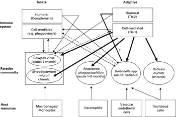
To generate hypotheses regarding interactions within a parasite community, Pedersen and Fenton (Reference Pedersen and Fenton2007) suggested constructing a diagram of the within-host parasite network, that explicitly incorporates the three trophic levels of host resources, parasite community and host immune system (Fig. 1). One of our motivations behind using this approach was to see how readily it can be used on a real system. The five microparasites considered here represent a diverse taxonomic range, with different life cycles, ecologies and interactions with the host immune system. Below, we describe these, first considering the use of host resources by the parasites, followed by a discussion of host immune responses.
A subset of the community of parasites of the field vole, Microtus agrestis, and their interaction network within a host. The basal level represents specific host resources. Arrows correspond to the flux of ‘energy’ from host resource to parasite, which are represented in the middle level of the network. The top level represents the immune system of the host. For two of the parasites, cowpox virus and Bartonella spp., we attempted to fully populate the top level of the network based on current immunological knowledge. Arrows represent the aspects of the immune system that target each parasite, with the thickness of the arrow corresponding to the relative importance of the immune response. Dashed arrows are used to signify if a particular parasite is thought to affect a particular immune response through immunomodulation. The importance of adaptive humoral responses for clearance of bartonellae infections is unclear, shown by ?. We did not attempt to repeat this exercise fully for the other parasites (see text). However, a predominantly Th1 response to all the parasites is expected (as shown by the single arrows).

Life cycle and exploitation of host resources
In rodents, the directly transmitted cowpox virus replicates in the respiratory tract and lymphoid tissues, mainly in macrophages and monocytes (Baxby and Bennet, Reference Baxby, Bennet, Webster and Granoff1999). Infection in otherwise healthy hosts is acute, lasting approximately 4 weeks and causes no obvious clinical signs (Bennett et al. Reference Bennett, Crouch, Begon, Duffy, Feore, Gaskell, Kelly, McCracken, Vicary and Baxby1997). Members of the genus Babesia are tick-borne protozoa that parasitize erythrocytes and which can cause haemolytic anaemia in infected hosts (Irwin, Reference Irwin, Shaw and Day2005). It is thought that Babesia spp. become sequestered within the capillary networks of the spleen and liver, with periodic release into circulation. Field voles are susceptible to Babesia microti and infections are usually subclinical and persistent. Bartonella spp. are facultatively intracellular bacteria transmitted by arthropod vectors, often fleas (Birtles, Reference Birtles, Shaw and Day2005a). Following infection, Bartonella spp. migrate to, and associate with, the vascular endothelium. From this niche bacteria are subsequently seeded into the bloodstream where they encounter and invade erythrocytes, thereafter replicating and persisting within these cells (Birtles, Reference Birtles, Oteo, Hechemy, Raoult, Silverman and Blanco2005b). As with Babesia, these infections do not usually result in clinical disease. Up to five Bartonella species circulate concurrently in UK woodland rodent communities (Birtles et al. Reference Birtles, Hazel, Bennett, Bown, Raoult and Begon2001; Telfer et al. Reference Telfer, Begon, Bennett, Bown, Burthe, Lambin, Telford and Birtles2007a, Reference Telfer, Clough, Birtles, Bennett, Carslake, Helyar and Begonb). Anaplasma phagocytophilum is a tick-borne bacterium that specifically parasitizes granulocytes, primarily neutrophils (Harrus et al. Reference Harrus, Waner, Bjoersdorff, Shaw, Shaw and Day2005). Although primary infections are associated with transient neutropenia, some hosts (e.g. sheep) become chronically infected without subsequent clinical signs. However, whether persistent infection occurs in field voles is uncertain. Mycobacterium microti is a member of the Mycobacterium tuberculosis complex, which includes M. tuberculosis and M. bovis (van Soolingen et al. Reference van Soolingen, van der Zanden, de Haas, Noordhoek, Kiers, Foudraine, Portaels, Kolk, Kremer and van Embden1998). Mycobacterial infection in voles is insidious, progressing from primary infection of, probably, the lungs, to the development of disseminated lesions throughout the body, and ultimately resulting in dramatic external lesions (Wells, Reference Wells1946; Cavanagh et al. Reference Cavanagh, Lambin, Ergon, Bennett, Graham, van Soolingen and Begon2004; Burthe et al. Reference Burthe, Bennet, Kipar, Lambin, Smith, Telfer and Begon2008a).
Immune responses
An understanding of immune responses to the different parasites is essential for predicting the occurrence and outcome of immune-mediated interactions during co-infections. Although previous studies do provide some information on the interactions of these microparasites with the immune system, the following two examples demonstrate the patchiness of our knowledge. Cowpox virus is known to have many gene products involved in immunomodulation of innate and acquired immunity at sites of viral replication (Alcami et al. Reference Alcami, Symons, Khanna and Smith1998). Infection results in long-term antibody production (Chantrey et al. Reference Chantrey, Meyer, Baxby, Begon, Bown, Feore, Jones, Montgomery and Bennett1999). Additionally, cell mediated immunity is also thought to play an important role in protection against infection and disease (Baxby and Bennet, Reference Baxby, Bennet, Webster and Granoff1999). Bartonellae also immunomodulate the host, subverting multiple cellular functions of vascular endothelial cells and resulting in proinflammatory activation and cell proliferation (Dehio, Reference Dehio2004). Results from experiments aimed at exploring the nature of host immune responses to bartonellae are inconsistent and different reservoir host species may respond in different ways. For example, antibodies are produced in response to natural infections in cats and humans, but apparently not in rodents (Kosoy et al. Reference Kosoy, Regnery, Tzianabos, Marston, Jones, Green, Maupin, Olson and Childs1997; Brouqui et al. Reference Brouqui, Stein, Dupont, Gallian, Badiaga, Rolain, Mege, La Scola, Berbis and Raoult2005; Kabeya et al. Reference Kabeya, Sase, Yamashita and Maruyama2006; Pearce et al. Reference Pearce, Radecki, Brewer and Lappin2006). Experimentally at least, the role of antibodies in clearing infection has been demonstrated (Koesling et al. Reference Koesling, Aebischer, Falch, Schulein and Dehio2001). Cell-mediated immune responses have also been induced in experimental models (Arvand et al. Reference Arvand, Ignatius, Regnath, Hahn and Mielke2001; Kabeya et al. Reference Kabeya, Yamasaki, Ikariya, Negishi, Chomel and Maruyama2007).
The examples above illustrate the current shortfall in our knowledge of the immune responses that occur in natural infections, and which parts of this response affect and control infections. Consequently, generating predictions about interactions between parasites within a co-infected host, while desirable, is in practice problematic. This is likely to be especially true for wild animal-parasite systems. In our system, all the parasites are likely to invoke a predominantly Th1-biased response, and 4 out 5 exploit cells that are integral to the host immune system. Consequently, we predict that immune-mediated interactions are very likely (see Fig. 1). Indeed, as A. phagocytophilum causes a transient neutropenia (Harrus et al. Reference Harrus, Waner, Bjoersdorff, Shaw, Shaw and Day2005), depleting populations of one of the key effector cells in the innate response, we might predict that infection with this parasite is especially likely to alter host susceptibility to other invaders.
Although a detailed web of predictions is clearly not possible, the approach advocated by Pedersen and Fenton (Reference Pedersen and Fenton2007) can, in a sense, be reversed (but still be equally useful): relationships apparent in the data may be used to deduce possible underlying interactions not previously predictable from studies of the pathogens concerned. Below we look at patterns of co-infection in our microparasite community of field voles and examine some of these potential interactions. Our principal aim is explore how such longitudinal data from natural populations can be used to investigate co-infection. To do this we have focused on two objectives. First, to address the question of the effects of co-infection on host susceptibility, we investigated whether infection with A. phagocytophilum influences vole susceptibility to B. microti and/or whether B. microti influences vole susceptibility to A. phagocytophilum. Secondly, we examined infection kinetics, considering whether co-infection influences infection length for Bartonella taylorii.
Data analysis
Susceptibility: host susceptibility to A. phagocytophilum and B. microti
In each of these two analyses we wished to compare animals that became infected with animals that could have become infected but did not. For animals that showed evidence of infection by a particular parasite during their capture history we incorporated knowledge of both host and parasite ecology to determine the trapping session when initial infection occurred. We also ensured that the negative captures included in the dataset provided valid comparisons.
As B. microti induces a chronic infection, the first positive capture of an animal was taken as the time of initial infection and later positive captures were excluded. Data from March to May 2006 were also available, allowing us to determine whether an animal had previous positive records. In addition, only captures from animals first caught when weighing less than 19 g were used (mostly animals first caught when <10 weeks old, unpublished data). These captures represent recently acquired infections and were compared with negative captures of animals that were first caught when weighing less than 19 g and had never shown any signs of B. microti infection.
Individuals are only rarely positive for A. phagocytophilum in more than one trap session (for example in this data set only 5 individuals out of 99 infected). Consequently, we assumed an animal's first positive result reflected a recently acquired infection. There was no evidence of re-infection in the dataset and therefore all captures after a positive result were excluded (animals not available for infection). These captures were compared with negative captures of animals that had never shown any signs of A. phagocytophilum infection.
For each parasite, generalised linear mixed models (GLMM) were used with a logit link, binomial errors and fitted using the Laplace approximation to restricted maximum likelihood estimation (lmer procedure in the R software package available under the GNU license at http://www.r-project.org). As animals caught on the same site in the same trap session experienced the same environmental conditions, site∗trap session was included as a random effect. In longitudinal studies, a random effect to account for the correlation amongst different observations of the same individual should also be included. However, in the datasets considered here the average number of captures per animal was very low (range 1·8–2·0 captures per individual) resulting in problems of estimating the random effect with many models. Where possible, individual was added as a random effect to the restricted model to verify that this did not alter the results.
As with cross-sectional data, analyses of longitudinal data need to consider whether any observed associations may be due to correlations in exposure risk. For each parasite we first considered extrinsic and intrinsic factors that may have influenced infection probability: site; season (2 seasons: June–August; September–November); sex; weight (as a crude proxy for age); maturity status (2 categories: immature vs currently or previously reproductively active) and recapture status (whether or not this animal had been caught previously). Two-way interactions were included and a polynomial relationship with weight was also considered. We used the Akaike Information Criterion index (AIC; Akaike, Reference Akaike, Petran and Csaki1973) to select the most parsimonious base model. Models with a difference in AIC (ΔAIC) of less than two may be considered similar in their ability to account for the data (Johnson and Omland, Reference Johnson and Omland2004).
After selection of the best base model for each parasite, we first considered whether infection by the other parasite at the same trapping session influenced infection probability. When B. microti infections were used as the explanatory variable we also investigated whether there was any difference in the effect of chronic and newly acquired B. microti infections. The impact of macroparasites was also considered, using the number of ticks as a continuous variable and flea infestation as a categorical score from 0 to 3.
To investigate the importance of infection sequence, for animals caught in the preceding month we investigated whether infection status in the preceding month influenced the probability of becoming infected.
Infection biology: length of Bartonella taylorii infections
To investigate whether concomitant and/or recently cleared infections influence the biology of Bartonella spp. infections, we examined whether infection by other parasites influenced the length of B. taylorii infections. Animals were only included that (1) had the start of an infection identified (i.e. were caught negative for B. taylorii in one month but were positive in the next month) and (2) were caught in a third consecutive month. The length of each infection was scored as (i) positive in only one month or (ii) positive in more than one month.
Generalised Linear Models were used. This time, in the first stage of analysis aimed at accounting for potentially confounding factors, only sex, weight and season were considered. We then investigated whether infection by other parasites (1) at the same trap session as the first positive B. taylorii record (i.e. capture 2 in the run of 3 consecutive captures), (2) prior to initial infection (capture 1) or (3) after initial infection (capture 3) influenced infection length. The effect of co-infection at each of these time points was investigated separately and then variables from the best models were combined. Again, AIC was used for model selection.
Infections by the following parasites were considered as explanatory variables: A. phagocytophilum, B.microti, cowpox, other Bartonella spp. (infection by either B. doshiae or B. grahamii) and fleas. The probability of current infection with cowpox can be calculated based on the serological history and, for young animals, the weight of an individual at first capture (Telfer et al. Reference Telfer, Bennett, Bown, Cavanagh, Crespin, Hazel, Jones and Begon2002). However, sequential probabilities are highly correlated. We therefore estimated the probability of infection with cowpox early during the B. taylorii infection based on an average of probabilities in capture 1 and capture 2; and a probability of infection late in the B. taylorii infection based on an average of probabilities in capture 2 and capture 3.
Results
In the 6 trap sessions between June and November 2006, there were 1643 captures of 775 individual animals. The number of captures per individual ranged from 1 to 6, with an average of 2·12. Of the 742 animals for which there were data on cowpox antibodies, 86% of animals were seropositive on at least one capture. Of the 753 animals for which there were data on the parasites detected by PCR, 59% became infected with Bartonella spp., 44% with B. microti and 12% with A. phagocytophilum. Only 10 animals showed evidence of mycobacterial infection based on external lesions.
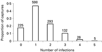
In the 3 sites that had the Bartonella spp. identified, 36% of captures (n=1279) were of animals co-infected with more than one microparasite species (Fig. 2), with some animals being co-infected with up to 5 microparasites at the same time.
Proportion of captures that represented individual field voles infected with different numbers of co-infecting microparasites. The data included information of infection status for 7 microparasites. Numbers represent sample size.

Susceptibility
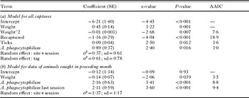
The B. microti dataset included 686 captures from 388 individuals, with 135 positive records for B. microti and 47 positive records for A. phagocytophilum. The probability of becoming infected with B. microti showed a polynomial relationship with weight (Table1), with a peak in infection risk at approximately 20 g. Recaptured animals were less likely to become infected (Table1), whilst risk increased with the number of ticks on an animal. Animals with a concomitant infection with A. phagocytophilum were more than twice as likely to become infected as animals not infected with A. phagocytophilum (odds ratio=2·44; 95% CI 1·17–5·05).
Factors influencing the probability of an individual field vole becoming infected with B. microti showing the best model for (a) all captures conforming to inclusion criteria described in text and (b) all such captures where the animal was also caught in the preceding month. ΔAIC=the change in the AIC if the single term is dropped. σ2=the variance attributable to a random effect. sd=standard deviation of σ2

When animals caught only in the preceding month were examined, there were 291 captures of 159 individuals, with 38 positive records for B. microti and 18 positive records for A. phagocytophilum. Larger animals were less likely to become infected (Table 1). Both infection with A. phagocytophilum in the current month (odds ratio=8·67; 95% CI=2·51–29·67) and infection in the preceding month (odds ratio=8·25; 95% CI 2·59–26·31) increased the likelihood of becoming infected with B. microti.
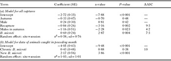
The A. phagocytophilum dataset included 1429 observations from 711 individuals, with 87 records positive for A. phagocytophilum and 633 records positive for B. microti. Recaptured animals were again less likely to be become infected than new animals, whilst in autumn males were less at risk than females (Table 2). In addition, animals with a concomitant infection with B. microti were nearly twice as likely to become infected (odds ratio=1·99; 95% CI 1·25–3·19). Although there was some evidence that animals with new B. microti infections were more likely to become infected than animals with chronic B. microti infections (new odds ratio=2·39; 95% CI 1·41–4·05; chronic odds ratio=1·28; 95% CI 0·56–2·92), distinguishing between new and chronic infections did not improve the fit of the model.
Factors influencing the probability of an individual field vole becoming infected with A. phagocytophilum showing the best model for (a) all captures conforming to inclusion criteria described in text and (b) all such captures where the animal was also caught in the preceding month. ΔAIC=the change in the AIC if the single term is dropped. σ2=the variance attributable to a random effect. sd=standard deviation of σ2

When animals caught only in the preceding month were examined, there were 771 captures of 396 individuals, with 29 positive records for A. phagocytophilum and 371 positive records for B. microti. None of the intrinsic or extrinsic variables examined showed a significant relationship with risk of infection (Table 2). However, animals infected with B. microti in the current month still showed a positive relationship with infection risk. Moreover, the model that distinguished between new and chronic B. microti infections had a lower AIC, with newly infected animals more likely to become infected with A. phagocytophilum than both uninfected animals and animals with a chronic infection (new odds ratio=8·76; 95% CI 2·92–26·25). There was no relationship with B. microti infection in the preceding month.
Infection biology
There were 49 animals infected with B. taylorii that conformed to the requirements for inclusion in the infection length analysis. Of these, 28 were only infected at one capture occasion and 21 were infected for 2 or more capture occasions.
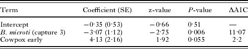
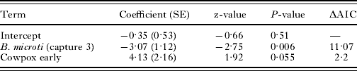
There was no effect of sex, weight or season on infection length. B. microti infection decreased the probability of an animal remaining infected with B. taylorii for a second month, irrespective of whether B. microti infection status at capture number 1, 2 or 3 was considered (capture 1: odds ratio=0·10, 95% CI 0·01–0·93 capture 2: odds ratio=0·13, 95% CI 0·03–0·64; capture 3: odds ratio=0·05, 95% CI 0·005–0·47). Infection with cowpox early in the B. taylorii infection increased the probability that an animal would remain infected (odds ratio=62·18; 95% CI 0·92–4204·9), whilst there was no evidence of such an effect of cowpox later on in the infection.
The final model included an effect of both early infection with cowpox and B. microti infection at capture 3 (Table 3). However, it is worth noting that the inclusion of infection status with other Bartonella spp. at capture 3 reduced the model AIC by 1·95. Therefore there is also some evidence that animals infected with other Bartonella species in the third capture are more likely to still be infected with B. taylorii (odds ratio=5·70, 95% CI 0·89–36·67). There was no suggestion of any effect at capture 1 or 2.
Factors influencing the probability of an individual field vole remaining infected with B. taylorii for a second consecutive capture. ΔAIC=the change in the AIC if the single term is dropped
Discussion
Even though this study has only considered a subset of the parasite community, co-infection by more than one parasite was common. Other studies and unpublished data have confirmed that these field vole populations are also infected by Trypanosoma microti (Smith et al. Reference Smith, Telfer, Burthe, Bennett and Begon2005) and Hepatozoon microti, as well as a variety of tick, flea, mite and helminth species. Thus, in these natural populations it seems probable that at any one time the majority of animals may be co-infected. Consequently, interactions between parasites within hosts have the potential to be of great significance for host-parasite dynamics. Indeed, the preliminary analyses presented in this paper indicate apparent associations between co-infection and susceptibility and co-infection and infection length. We first discuss possible explanations for the observed associations, before considering the potential and the challenges provided by longitudinal studies.
Susceptibility
The observed positive impact of A. phagocytophilum on B. microti infection risk and vice versa may be attributable to a number of factors. By including both fixed and random effects, the statistical approach adopted aimed to account for factors that may have reflected correlations in exposure risk between the two parasite species. That is, it sought to avoid inferring the existence of a biological interaction between two parasites from a positive distributional association between them, when in reality this was a reflection of both, independently, being associated with some further factor with which risk of infection was correlated. For both microparasites, several of the variables examined were indeed related to infection risk, probably reflecting, for example, behavioural changes with age and seasonal patterns in tick abundance. However, even when care has been taken to include such factors, it is impossible to rule out further correlations in exposure risk, especially as both B. microti and A. phagocytophilum are transmitted by ticks. The number of ticks on an animal was included as a potential explanatory variable (and was significant for the analysis looking at B. microti susceptibility), but the short time of tick attachment relative to the interval between trapping sessions means that many tick attachments will be missed. Moreover, the positive impact of B. microti on A. phagocytophilum susceptibility appears primarily driven by new concomitant B. microti infections, which themselves indicate a recent tick attachment. Consequently, correlations in exposure risk cannot be ruled out as a partial explanation for the positive associations between these two microparasites.
It is interesting to note that susceptibility to B. microti is associated with both concomitant and previous A. phagocytophilum infections, whereas susceptibility to A. phagocytophilum is associated with only concomitant, and mostly with new B. microti infections. Thus, it seems possible that A. phagocytophilum may increase susceptibility to B. microti infections more than vice versa. Previous studies of both humans and animals have indicated that A. phagocytophilum can influence the pathology of a range of co-infecting parasites (Reid et al. Reference Reid, Buxton, Pow, Brodie, Holmes and Urquhart1986; Gokce and Woldehiwet, Reference Gokce and Woldehiwet1999; Krause et al. Reference Krause, McKay, Thompson, Sikand, Lentz, Lepore, Closter, Christianson, Telford, Persing, Radolf and Spielman2002), which is not unexpected given that it infects neutrophils. On the other hand, if an A. phagocytophilum infection is detectable by PCR earlier than a B. microti infection, our result could still reflect simultaneous exposure during a tick attachment.
Studies elsewhere have commonly found questing ticks that are searching for hosts to be carrying more than one pathogen (Swanson et al. Reference Swanson, Neitzel, Reed and Belongia2006). Due to the much higher prevalence of B. microti infection in field voles and its chronic nature, a tick carrying A. phagocytophilum must have a high probability of also carrying B. microti. Of animals that become infected with A. phagocytophilum, 72% are infected with B. microti at some time. This compares with an overall B. microti prevalence of 44%. Moreover, it is striking that 63% of these animals (n=63) are first infected with both microparasites at the same trapping session. Irrespective of whether or not each microparasite impacts on susceptibility to the other, this highlights that many animals infected with A. phagocytophilum have to fight both infections concurrently. This is likely to have fitness costs, especially if co-infection influences pathology.
Infection biology
Sampling occurred at discrete time points and, therefore, an animal infected with a specific parasite at one trap session but negative subsequently may still have been infected for some or all of the intervening period. Despite our consequently crude estimates of infection length, B. taylorii infections were shorter in animals co-infected with B. microti and longer in animals that were co-infected with cowpox.
The negative interaction between B. microti and B. taylorii could result from competition over resources, as both species invade and replicate within red blood cells. Although haemolytic anaemia is a potential outcome of babesiosis in a range of host species (Krause et al. Reference Krause, Telford, Spielman, Sikand, Ryan, Christianson, Burke, Brassard, Pollack, Peck and Persing1996; Hofmann-Lehmann et al. Reference Hofmann-Lehmann, Meli, Dreher, Gonczi, Deplazes, Braun, Engels, Schupbach, Jorger, Thoma, Griot, Stark, Willi, Schmidt, Kocan and Lutz2004; Coleman et al. Reference Coleman, LeVine, Thill, Kuhlow and Benach2005), a large number of uninfected erythrocytes still remain. Alternatively, up-regulation of the Th1 immune response during chronic B. microti infections may speed up the clearance of bartonellae infections. Although bartonellae are to some extent protected from a host immune response when sequestered within erythrocytes, the vascular endothelial bed does not share such immunological privilege (Birtles, Reference Birtles, Oteo, Hechemy, Raoult, Silverman and Blanco2005b). An immune response that effectively cleared infection from endothelial cells would therefore ultimately control an infection. As B. microti infections are chronic, infection status at successive time points is correlated and, unsurprisingly, the effect of B. microti was apparent throughout B. taylorii infections. Previous studies of co-infections involving B. microti have provided conflicting results, with some studies suggesting enhanced Borrelia burgdorferi pathology (Krause et al. Reference Krause, Telford, Spielman, Sikand, Ryan, Christianson, Burke, Brassard, Pollack, Peck and Persing1996; Moro et al. Reference Moro, Zegarra-Moro, Bjornsson, Hofmeister, Bruinsma, Germer and Persing2002), whilst others suggest no effect (Wang et al. Reference Wang, Liang, Sangha, Phillips, Lew, Wright, Berardi, Fossel and Shadick2000; Coleman et al. Reference Coleman, LeVine, Thill, Kuhlow and Benach2005).
The positive interaction between cowpox and B. taylorii is likely to be mediated by the immune system, since there seems to be little or no overlap in the resources used by the two pathogens. Although immunomodulation by cowpox is considered localised to sites of viral replication (Bennett, Smith and Baxby, Reference Bennett, Smith, Baxby, Mahy and van Regenmortel2008), the virus is disseminated in lymphoid organs throughout the body of rodents, and thus may decrease host ability to present antigens or clear infected cells. As cowpox exploits macrophages, infection may also decrease host ability to present antigens. The effect of cowpox appeared primarily driven by an individual's infection status early in the B. taylorii infection, perhaps suggesting that, whatever the mechanism, greater persistence is achieved through effects on initial establishment and replication. Positive effects on bartonellae infection length have also been shown for other viruses in humans (Signorini et al. Reference Signorini, Simeone, Tonegatti, Colombini, Cadeo, Cristini, Matteeli and Viale2002; Resto-Ruiz, Burgess and Anderson, Reference Resto-Ruiz, Burgess and Anderson2003). Interestingly, in this population of field voles, cowpox has been shown to have a negative effect on host survival (Burthe et al. Reference Burthe, Telfer, Begon, Bennet, Smith and Lambin2008b). If cowpox infection generally has a positive effect on other parasitic infections, this result may reflect cumulative costs of infection rather than simply an effect of cowpox virus in its own right.
There was also a suggestion of an impact of infection with other Bartonella spp. Previous studies in cats have provided evidence of some cross-effective immune responses between Bartonella species and genotypes (Yamamoto et al. Reference Yamamoto, Chomel, Kasten, Hew, Weber, Lee, Koehler and Pedersen2003). By contrast, in experimental infections of the cotton rat, no such cross-protection was observed (Kosoy et al. Reference Kosoy, Regnery, Kosaya and Childs1999). In our study, the suggested effect was positive and only apparent at the end of the B. taylorii infection (i.e. animals were more likely to be infected for a second month if they acquired a new infection by a different Bartonella species). The immunomodulatory effects produced by Bartonella spp. during associations with endothelial cells will influence inflammatory responses and the recruitment of monocytes. Consequently, a new infection by a second Bartonella spp. may prolong an existing infection. An alternative explanation for the pattern observed is that these animals suffered a high exposure risk to bartonellae and the apparent persistent B. taylorii infection may have actually represented a new infection.
Analysis of longitudinal data
Whilst longitudinal data can provide a wealth of information for co-infection studies, analysis of such data requires care. To ensure only data relevant to the questions at hand were included, and to reduce noise (e.g. uncertainty regarding infection length), we employed specific criteria for data selection. In the susceptibility analyses it was also important that the data for animals that did not become infected provided a relevant comparison. The importance of accounting for confounding factors that may reflect correlations in exposure risk has been noted before (Behnke et al. Reference Behnke, Gilbert, Abu-Madi and Lewis2005; Booth, Reference Booth2006) and is also true for longitudinal studies. In addition, analyses of longitudinal data should try to account for non-independence of samples from the same individual. However, as shown here, estimation of the appropriate random effects can prove problematic when there are relatively few records per individual, and this may be a particular issue for studies of wild animal populations. One solution would be to randomly select one observation per individual. Although this would result in a drop in statistical power, it may be a useful approach for verifying results from the larger dataset.
In addition to correlations in exposure risk, associations between parasites may be due to correlations in susceptibility. Such correlations may occur due to environmental or genetic factors. For example, an individual's body condition or genotype may influence susceptibility to a number of parasites. Joint modelling of random effects could be used to investigate the possibility of correlations in susceptibility. In ecology, this approach has been used to investigate correlation between survival and reproductive success (Cam et al. Reference Cam, Link, Cooch, Monnat and Danchin2002; Wintrebert et al. Reference Wintrebert, Zwinderman, Cam, Pradel and van Houwelingen2005).
CONCLUSIONS
In accordance with previous studies of humans, the preliminary analyses presented here demonstrate that longitudinal data from natural populations can be used to address a range of questions related to co-infection, including effects on susceptibility, infection biology and pathology. Unlike cross-sectional studies, longitudinal studies can determine the importance of infection sequence. For example, cowpox infection status at the point of primary infection by bartonellae appears to have a large impact on infection length, whereas there is no evidence that contracting cowpox subsequent to infection by bartonellae has the same effect. Such data are also required for investigations of the ultimate consequences of co-infection on host fitness parameters such as survival.
Despite the relatively small datasets used in the present analyses, the results suggest that interactions within this community of microparasites are important and, given the prevalence of co-infections in wild populations, again emphasise the potential implications of interactions for both host and parasite fitness. However, due to a lack of detailed knowledge about immune responses and even, in some cases, the host niche exploited, generating a priori predictions regarding interactions proved difficult. Indeed, demonstrations of such interactions in naturally infected wild populations may address questions concerning immune responses to specific parasites, rather than the other way round. An alternative approach to focusing on a limited number of parasite species to investigate interactions between, and interactive effects of, combinations of parasites would be to examine cytokine profiles (Graham et al. Reference Graham, Cattadori, Lloyd-Smith, Ferrari and Bjornstad2007). This approach is now being tried in the Kielder field vole system.
ACKNOWLEDGEMENTS
The field vole study was funded by Natural Environmental Research Council studentships and the Wellcome Trust (070675/Z/03/Z; 075202/Z/04/Z) and was licensed under Home Office project license PPL40/1813. The Forestry Commission provided access to sites. Gill Telford, Pablo Beldomenico, Stephanie Gebert, Lukasz Lukomski, Roz Anderson, Gemma Chaloner, Jenny Rogers, Dave Jones, Gill Hutchison, Cathy Glover, and Anna Goodsall provided assistance in the field and laboratory.