Infants with hypoplastic left heart syndrome have either severe mitral stenosis or mitral atresia. The majority have a large defect in the atrial septum, thus permitting the pulmonary venous return to the left atrium to shunt into the right atrium. Around one-twentieth of cases,Reference Rychik, Rome, Collins, DeCampli and Spray1 however, have an intact atrial septum, this lesion causing severe acidosis and cyanosis at birth due to obstruction to pulmonary venous return. This finding places the infants into a group with an extremely high rate of mortality.Reference Rychik, Rome, Collins, DeCampli and Spray1
Case report
During an otherwise uneventful pregnancy, hypoplasia of the left heart was noted in our patient at routine antenatal ultrasonic scanning at 24 weeks of gestation. Further fetal echocardiography at 25 weeks revealed an intact atrial septum, with no evidence of a decompressive venous pathway. After full discussion of all the available options, the parents opted for surgery immediately subsequent to the delivery. They were aware of the high risk of subsequent death. At 39 weeks gestation, the mother was transferred to a centre offering services for both paediatric cardiac surgery and the needs of maternity.
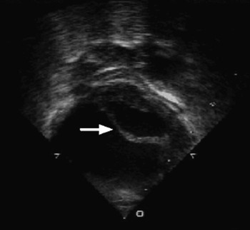
The male infant was delivered in the cardiac theatre by prearranged Caesarian section. Prior to transection of the umbilical cord, an echocardiogram was performed, which confirmed hypoplasia of the left heart, with mitral atresia and a thick and intact atrial septum (Fig. 1). After confirmation of the intracardiac malformations, the cord was transected, and the infant transferred to an adjacent operating theatre. An umbilical venous catheter was sited and used to induce anaesthesia, and an infusion of prostaglandin E1 was started. As expected ventilation was extremely difficult, due to very poorly compliant lungs, saturations were no more than 40 to 50% in 100% oxygen, and the first blood gas at 8 minutes of age revealed a partial pressure for oxygen of 3.9 kilopascals, an elevated level of carbon dioxide at 12 kilopascals, and a severe metabolic acidosis, with a base excess of −14.6. Both mother and father saw their child, and he was baptized prior to surgery.
The transthoracic echocardiogram shows an intact atrial septum (arrow), mitral atresia, and dilated pulmonary veins.

Within 30 minutes of birth the infant was established on cardiopulmonary bypass, with the venous cannula in the right atrium, and the arterial cannula in the ascending aorta. The child was cooled to 24 degrees centigrade, and circulatory arrest induced with St Thomas’ cardioplegic solution. A total atrial septectomy was performed via the right atrium, and bilateral bands applied to the pulmonary arteries. The time needed for circulatory arrest was 8 minutes.
Post-operatively, the infant was weaned from cardiopulmonary bypass in sinus rhythm, without inotropic support. As expected, his pulmonary vascular resistance was raised. Saturations of oxygen were initially measured at 80 to 90% in 100% oxygen. Over the next 2 hours, he became more hypoxic and required treatment with nitric oxide for 24 hours, and inotropic support for 6 days. He was maintained on an infusion of prostaglandin until the seventh day, when the hybrid palliation was completed in the cardiac catheterization laboratory. The sternotomy was reopened, a 6 French sheath was placed in the proximal pulmonary trunk, and a premounted Genesis stent measuring 8 millimetres by 24 millimetres was deployed in the arterial duct. He was weaned from ventilatory support four days later. At 6 weeks of age, he is at home and thriving.
Discussion
In order for infants with hypoplasia of the left heart and an intact atrial septum to survive extra uterine life, it is necessary urgently to create an unrestrictive interatrial communication. Controversy exists as to the best way to achieve this. These infants usually have a thick atrial septum. Transeptal blade or radiofrequency perforation of the septum have been performed, but can be technically challenging at a time when rapid creation of an interatrial communication is vital. When these interventions are followed by a Norwood operation, mortality is reported at up to 48% within 35 days of surgery.Reference Vlahos, Lock, McElhinney and van der Velde2 Intrauterine laser perforation of the septum is in its early days,Reference Qintero, Huhta, Suh, Chmait, Romero and Angel3, Reference Marshall, van der Velde and Tworetzky4 and may be a useful therapeutic option in fetuses diagnosed early in gestation when vascular lung remodelling may occur. Placing the infant onto cardiopulmonary bypass as quickly as possible allows rapid reoxygenation and correction of acidosis. Performing an immediate Norwood operation in this group of children has been reported to have an early mortality rate of 65%.Reference Rychik, Rome, Collins, DeCampli and Spray1 A septectomy, with application of bilateral pulmonary arterial bands, ought to be a safer option.
The hybrid procedure for the commoner form of hypoplastic left heart with an atrial septal defect involves stenting of the arterial duct, atrial septostomy with a balloon if necessary, and banding of the pulmonary arteries.Reference Gibbs, Wren, Watterson, Hunter and Hamilton5 The procedure is performed in the cardiac catheterization laboratory. This approach is becoming more common place,Reference Akintuerk, Michel-Behnke and Valeske6 especially in patients at a higher risk of mortality for the Norwood operation.Reference Lim, Peeler, Matherne, Kron and Gutgesell7 In our patient, immediate relief of pulmonary venous obstruction by septectomy, along with application of the pulmonary arterial bands, meant that the hybrid palliation could be completed subsequent to stabilization of the infant.
To accomplish an urgent septectomy, the institution at which these neonates undergo cardiac surgery must be able to provide many non-cardiac services. Due to the urgency for intervention the child must be delivered at the cardiac centre, necessitating services to be on site to provide obstetric and neonatal needs. An obstetrician and midwife are needed to perform a Caesarian section at a prearranged time. A neonatal team is required to insert an umbilical venous catheter and resuscitate the neonate at delivery, and anaesthetic teams are needed for both mother and baby. From the stance of the cardiac services, a paediatric cardiologist confirms the septum is intact immediately on delivery, and a senior cardiac surgeon, with an experienced theatre team and perfusionist, must be ready to place the child on cardiopulmonary bypass as quickly as possible.
This approach to the infant with hypoplasia of the left heart and intact atrial septum diagnosed late in gestation requires careful planning, and the coordinated involvement of multiple specialties. It allows rapid correction of acidosis and hypoxia by cardiopulmonary bypass, a relatively less complicated surgical intervention at the time the child is most compromised, and completion of palliation after stabilisation. We believe that this is an effective way to manage these infants known to be at high risk.