Right ventricular apex pacing and prolonged QRS duration in children with complete atrioventricular block are reported to be associated with left ventricular dysfunction in the long term.Reference Kim, Friedman and Eidem1 We therefore implanted a usual dual-chamber device for biventricular epicardial pacing, and assessed its benefits towards left ventricular synchrony.
Case report
Patient 1 was a 6-year-old girl, patient 2 was a 4-year-old girl, and patient 3 was a 1-year-old girl; these patients were diagnosed with complete atrioventricular block without structural cardiac disease. In patients 1 and 2, the heart rate was 50 beats per minute and did not increase with exercise; therefore, dilation of the left ventricle was considered to be present, and pacemaker implantation was planned. Patient 3 was brought to our hospital for sudden syncope with bradycardia and underwent emergency pacemaker implantation.
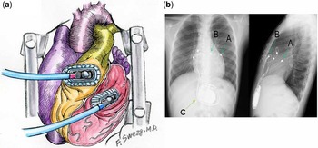
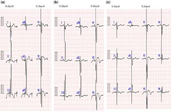
A median sternotomy with a limited skin incision was made under general anaesthesia. A pair of bipolar steroid-eluting ventricular pacing leads (Model 4968; Medtronic, Minneapolis, Minnesota, United States of America) was implanted. The negative pole of the pacing lead was implanted into the lateral wall of the left ventricle using a Tentacles device (Sumitomo Bakelite, Tokyo, Japan), which was used to expose the lateral wall of the left ventricle without haemodynamic compromise (Fig 1a).Reference Arai, Mizuno and Yoshizaki2 The indifferent electrode was implanted into the right ventricular outflow tract, which was assessed to be a well-synchronised site of the left ventricle, on the diagonal line and away from the left ventricular lead based on transesophageal echocardiography (Fig 1b). A bipolar epicardial lead was placed in the right atrium, and two pairs of pacing leads were connected to a Medtronic pacemaker (Enpulse E2DR01; Medtronic, Minneapolis) The lead parameters (threshold, sensing, and impedance) were confirmed. The post-operative course was uneventful. The post-operative QRS duration was 106 milliseconds after 1 week, 114 milliseconds after 1 year, and 110 milliseconds after 2 years in patient 1; 119 milliseconds after 1 week, 112 milliseconds after 2 years, and 114 milliseconds after 3.5 years in patient 2; and 102 milliseconds after 1 week and 106 milliseconds after 2 months in patient 3 (Fig 2). Post-operative left ventricular function in the two patients was estimated by echocardiography (Vivid 7 echocardiographic system; GE Healthcare, Wauwatosa, Wisconsin, United States of America). Mechanical left ventricular synchrony was measured by tissue Doppler imagingReference Suffoletto, Dohi, Cannnesso, Saba and Gorcsan3 and two-dimensional speckle tracking imaging.Reference Anderson, Miyazaki, Sutherland and Oh4 The septal–lateral delay in the time-to-peak myocardial systolic velocity using tissue Doppler imaging was measured as Bax’s index.Reference Bax, Bleeker and Marwick5 The two-dimensional speckle tracking imaging-radial dyssynchrony index was defined as the time difference of the radial strain between the anteroseptal and posterolateral regions.Reference Anderson, Miyazaki, Sutherland and Oh4 In patient 1, Bax’s indices before the procedure, after the procedure, and follow-up periods – 2 years after procedure – were 45, 30, and 40 milliseconds, respectively. In addition, the two-dimensional speckle tracking radial dyssynchrony indices were 70, 60, and 60 milliseconds, respectively. In patient 2, Bax’s indices at follow-up periods – 1, 2, and 3.5 years after the procedure – were 65, 19, and 10 milliseconds, respectively. The two-dimensional speckle tracking radial dyssynchrony indices were 43, 43, and 39 milliseconds, respectively.
(a) Intraoperative schema; left ventricular pacing lead was implanted into the lateral wall of the left ventricle using a Tentacles device without haemodynamic compromise. (b) Position of the pacing leads from anteroposterior (left) and lateral (right) projections. Positioning of the ventricular epicardial pacing leads on the basal lateral wall of the left ventricle (arrow A) and right ventricular outflow tract (arrow B) was directed by transesophageal echocardiography. A usual dual chamber device was used (arrow C).

Post-operative 12-lead electrocardiogram shows short QRS duration in patient 1 (a, 114 milliseconds), patient 2 (b, 112 milliseconds), and patient 3 (c, 106 milliseconds).

Discussion
Chronic right ventricular pacing in patients with complete atrioventricular block is associated with left ventricular dyssynchrony causing deleterious left ventricular remodelling and decreased exercise capacity.Reference Thambo, Bordachar and Garrigue6 In order to prevent dyssynchrony of the left ventricle induced by pacing, Rumeau et alReference Rumeau, Dulac and Coulier7 reported primary implantation of a biventricular pacemaker in children with complete atrioventricular block.Reference Rumeau, Dulac and Coulier7 Our method of biventricular pacing could minimise the adverse effects of conventional right ventricular pacing, such as QRS prolongation and inducible left ventricular dyssynchrony. Biventricular pacing by conventional methods, however, requires greater pacemaker battery usage and a larger epicardial space for implantation, which is considered unsuitable for young children; moreover, primary biventricular pacemaker implantation is not allowed in Japan for a diagnosis of complete atrioventricular block. This method might be planned in situations in which a conventional biventricular pacing device is not available.
Recently, off-pump cardiac operating techniques, such as off-pump coronary artery bypass grafting, have made it possible to implant left as well as right ventricular epicardial leads with minimal surgical insult.Reference Arai, Mizuno and Yoshizaki2 Transesophageal echocardiography is useful not only in securing the position of the left ventricular lead but also in evaluating left ventricular wall motion and mitral valve performance after implantation of a left ventricular pacemaker.
To assess left ventricular synchrony in children who undergo cardiac resynchronization therapy, the QRS duration and septal–posterior wall-motion delay using tissue Doppler imaging are commonly analysed.Reference Takabayashi, Shimpo, Mitani and Komada8 Recently, two-dimensional speckle tracking has emerged as a modality for assessing left ventricular dyssynchrony in adult patients with dilated cardiomyopathy.Reference Anderson, Miyazaki, Sutherland and Oh4 This method has an advantage over tissue Doppler imaging because of its capability to evaluate regional function independent of angle correction, and this provides greater information regarding left ventricular synchrony compared with conventional methods.Reference Marwick, Yu and Sun9 Therefore, we used this method in patients with complete atrioventricular block to assess left ventricular regional radial function and synchrony after biventricular pacing.
In conclusion, biventricular pacing in children with complete atrioventricular block may be safe and effective, and would result in the clinical improvement of cardiac function with minimal mechanical left ventricular dyssynchrony and a short QRS duration. The assessment of left ventricular synchrony after biventricular pacemaker implantation revealed that no left ventricular dysfunction due to ventricular dyssynchrony occurred in the mid-term follow-up period. However, small children with complete atrioventricular block would typically require long-term pacemaker support.
Acknowledgements
The authors thank Hiroshi Imagawa, MD, for surgical assistance and Katsuji Inoue, MD, for technical assistance with data. We also thank Fuminaga Suetsugu, MD, for a beautiful schema of the procedure.