Case description
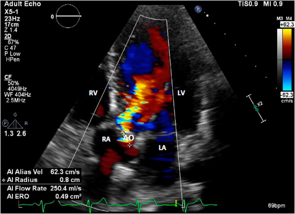
A 26-year-old woman – gravida 1, para 0 – with a history of severe aortic stenosis secondary to congenital bicuspid aortic valve disease and asthma presented to the emergency room at 22 weeks gestation with chest pain and pre-syncopal symptoms. She had previously undergone an aortic valve repair at day of life 4. On admission, echocardiogram (Fig 1) revealed an ejection fraction of 60–65% and a bicuspid aortic valve with severe stenosis, with a valve area of 0.56 cm2 and mean gradient of 65 mmHg. Patient also had episodes of non-sustained ventricular tachycardia and hypotension [blood pressure range: systolic blood pressure (72–99) and diastolic blood pressure (37–63)] on presentation. The patient was extensively counselled about the risk of maternal and fetal mortality associated with her condition; however, she strongly desired to continue the pregnancy.
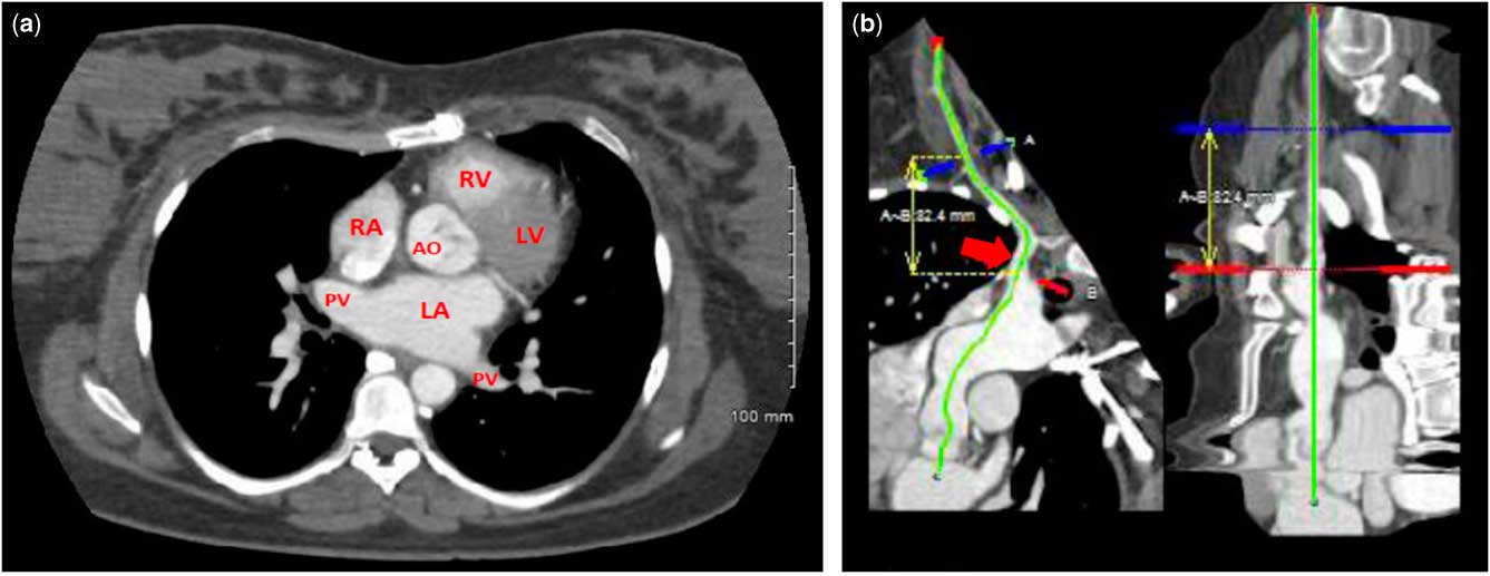
Following evaluation with CT angiogram of chest and cardiac catheterisation (Fig 2), she was found to be a poor candidate for balloon aortic valvuloplasty or transcatheter aortic valve replacement. This case was discussed multiple times at the weekly multi-disciplinary cardiac valve conference with representation from Cardiology, Interventional Cardiology, Cardiothoracic Surgery, Obstetric and Cardiac Anaesthesiology, and Maternal Fetal Medicine. Several options were discussed including scheduled open-heart valve replacement during the pregnancy; initiation of dobutamine infusion or extracorporeal membrane oxygenation to maintain cardiac output up to 28 weeks of gestation and then delivery followed by open-heart valve replacement, if development of sustained arrhythmia and/or worsening hypotension; and planned caesarean delivery followed by open-heart valve replacement at 28 weeks if development of frequent arrhythmias and/or worsening hypotension.
Transthoracic echocardiography at the time of presentation. Five-chamber view, including the aorta, showing severe aortic stenosis and regurgitation. AO=bicuspid aortic valve; LA=left atrium; LV=left ventricle; RA=right atrium; RV=right ventricle.

CT scan angiogram chest with and without contrast with three-dimensional reconstruction. ( a ) Bicuspid aorta (AO) is noted. There are no calcifications noted at the AO valve leaflets. There are no calcific or non-calcific atherosclerotic plaques of the thoracic AO and its branching vessels. ( b ) Owing to narrowing of the femoral artery, the right subclavian artery was evaluated for an alternative access site for transcatheter AO valve replacement. There is common origin of right subclavian and left common carotid artery. Right subclavian artery (red arrow) measurements (minimal diameters): proximal: 7×8 mm, mid: 5.5×6 mm, and distal: 5.5×5.1 mm (at the level of origin of lateral thoracic artery). LA=left atrium; LV=left ventricle; RA=right atrium; RV=right ventricle; PV=pulmonary vein.

Patient’s prognosis remained guarded with frequent episodes of non-sustained ventricular tachycardia and persistent hypotension until 28 weeks, at which time she underwent a caesarean section followed by a sternotomy with mechanical aortic valve replacement and pericardial enlargement of the sinotubular junction. There was delivery of a female neonate weighing 1360 g with Apgar scores of 8 and 9 at 1 and 5 minutes. The infant was transferred to the neonatal ICU where she was intubated but extubated a day later and subsequently discharged 5 weeks after delivery without significant complications. On postoperative day 4, the patient was discharged home from the cardiac ICU in stable condition.
Discussion
The most common cause of aortic stenosis among women of childbearing age is congenital bicuspid aortic valve disease, which accounts for 2% of CHDs.Reference Silversides, Colman, Sermer, Farine and Siu 1 – Reference Fedak, Verma, David, Leask, Weisel and Butany 5 On the basis of WHO classification of maternal cardiovascular risk by cardiac condition, severe symptomatic aortic stenosis in pregnancy is classified under WHO class IV. These patients are considered extremely high risk for maternal morbidity and mortality. Data quantifying maternal and fetal risk of pregnancy in women specifically with these high-risk features of aortic stenosis are limited. A systematic search of peer-reviewed literature between 1985 and 2007 on pregnancy in women with structural CHD reported rates of maternal risks including arrhythmias (2.4%), heart failure (7%), and cardiovascular events such as myocardial infarction, stroke, and cardiovascular mortality (2.5%).Reference Drenthen, Pieper and Roos-Hesselink 6 Although this study reported no difference in neonatal outcomes when compared with the general population, Hameed et alReference Hameed, Karaalp and Tummala 7 reported adverse fetal outcomes such as preterm delivery and intrauterine growth restriction in patients with severe aortic stenosis.
In severe aortic stenosis, there is elevation of ventricular systolic and diastolic filling pressures owing to a fixed stroke volume and limited increase in cardiac output. Patients with aortic stenosis become very sensitive to acute changes in preload and afterload, in which ventricular arrhythmias and heart failure can occur. Symptomatic patients may benefit from admission in a tertiary referral hospital with a specialised unit for close maternal cardiac surveillance, blood pressure control, and heart rate regulation. In our institution, we also keep a STAT caesarean section cart and an infant warmer bedside in case an emergent delivery is necessary and mobilisation of patient to the operating room will cause loss of valuable time. Initiation of antepartum fetal surveillance including monthly growth ultrasounds should be considered during admission, starting at viability, to assess fetal well-being and growth. During the course of our patient’s hospitalisation, she received two full courses of antenatal corticosteroids for promotion of fetal lung maturity and prevention of prematurity-associated complications.
A conservative management strategy was deemed high risk for both the mother and fetus owing to complaint of chest pain associated with intermittent arrhythmias and hypotensive episodes noted on presentation. Treatment options were discussed by a multi-disciplinary team with special attention paid to maternal and fetal risks and feasibility of each option. In addition to the diagnosis of severe aortic stenosis, our patient was found to have severe aortic regurgitation, which might have played a role in her presentation and course. The American College of Cardiology and the American Heart Association have recommended that pregnant patients with severe aortic regurgitation should only be considered for a valve operation if they have refractory NYHA class IV heart failure symptoms.Reference Nishimura, Otto and Bonow 8 Overall, aortic regurgitation is better tolerated in pregnancy than aortic stenosis. However, when both conditions present simultaneously in one individual, it might make the management challenging and possibly worsen the prognosis. Information is scarce on the management of pregnant patients with a combination of both aortic stenosis and aortic regurgitation.
Valvular surgery has been performed successfully during pregnancy for many years. However, aortic valve replacement during pregnancy does pose an increased risk of mortality to the mother and fetus (3–15% and 16–33%, respectively).Reference John, Gurley and Schaff 9 , Reference Chambers and Clark 10 Balloon aortic valvuloplasty has an overall lower associated risk of complication; however, it has been reported to increase or worsen existing aortic regurgitation. Although there are sparse data on transcatheter aortic valve replacement during pregnancy, the existing data suggest that transcatheter aortic valve replacement performed during pregnancy would likely be successful with a lower risk to the fetus and acceptable maternal risk compared with surgery.Reference Hodson, Kirker, Swanson, Walsh, Korngold and Ramelli 11 Following evaluation, our patient was not an ideal candidate for balloon aortic valvuloplasty owing to already existing severe aortic regurgitation. Additionally, our patient was not a candidate for transcatheter aortic valve replacement owing to significant narrowing of the femoral artery and the absence of calcification on the aortic valve annulus, which would enable anchoring of the prosthetic valve.
The total blood volume rises steadily during the first trimester and is increased by almost 50% by the 30th week, remaining constant thereafter in late pregnancy. Furthermore, the enlarged uterus partially impedes venous return by compressing the inferior caval vein, accounting for lower cardiac output. Ultimately, a decision was made to proceed with delivery and surgical aortic valve replacement at 28 weeks’ gestational age owing to increased frequency of non-sustained ventricular tachycardia and persistent hypotension. However, at 28 weeks there is increased survival rate of the fetus, and beyond this gestational age increased cardiac performance might increase maternal mortality. Of note, the decision to deliver at 28 weeks does not come without risk of morbidity to the neonate, including risk of respiratory distress syndrome, neonatal ICU admission, intraventricular haemorrhage, and necrotising enterocolitis. Nonetheless, when compared with the risk of mortality associated with sudden cardiac arrest or decompensation that may arise in the patient, delivery is a reasonable option.
During delivery, care is best provided through the cooperative efforts of a multi-disciplinary team involving Obstetrics/Maternal Fetal Medicine, Neonatology, Cardiothoracic surgery, and Obstetric Anaesthesia. Decision to proceed with a caesarean section was based on the concern for significant demands on the cardiovascular system during the first and second stages of labour, placing an acute load on the diseased heart.
Conclusion
Pregnancy in women with severe bicuspid aortic stenosis is associated with marked increase in maternal morbidity and unfavourable effect on fetal outcome, which are related to severity of disease. This risk is further exacerbated when there is coexistence of another valvular pathology, in this case, severe aortic regurgitation. Choice of treatment option can be challenging especially when trying to balance the potential risks of each option to mother and fetus. A collaborative team of physicians with experience in managing haemodynamic changes in pregnancy can help alleviate some of these challenges.
Acknowledgement
The authors thank Dr Robert Hooker for the excellent care he provided to the patient, and for reviewing the manuscript.
Financial Support
This research received no specific grant from any funding agency, commercial or not-for-profit sectors.
Conflicts of Interest
None