Pulmonary atresia with intact ventricular septum is a cardiac malformation encompassing a wide spectrum. The incidence is ~4.5–5.4/100,000 live births.Reference Calder, Peebles and Occleshaw 1 Fistulous connections between the right ventricle and the coronaries are common, with an incidence estimated to range from 31 to 68%.Reference Calder, Peebles and Occleshaw 1 – Reference Giglia, Mandell, Connor, Mayer and Lock 3
A unique feature of this disease is the concept of right ventricle-dependent coronary circulation – that is, dependence of myocardial perfusion on connections from the right ventricle caused by proximal coronary obstruction and inadequate anterograde aortic flow through the coronaries such that decreasing right ventricular pressure would compromise the myocardium. This can occur in as many as one-third of patients with fistulae.Reference Cheung, Richmond, Turner, Bacha and Torres 4 Determining the coronary anatomy and myocardial dependence is an important step in the evaluation and management of these patients.
Classically, the presence of coronary fistulae is thought to be dependent on a hypertensive right ventricle. We describe for the first time a case of pulmonary atresia with intact ventricular septum and coronary fistulae with a subsystemic right ventricle.
Case presentation
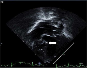
At 28 weeks of gestation, fetal echocardiography demonstrated pulmonary atresia with an intact ventricular septum with a severely hypoplastic right ventricle and suggestion of coronary fistulae. It also noted that the basal ventricular septum bowed into the left ventricle. The child was born at 38 weeks of gestation with a birth weight of 2.56 kg. Prostaglandin E1 was subsequently initiated and the neonate was transported to the cardiac intensive care unit. The postnatal echocardiogram confirmed the diagnosis and also demonstrated right ventricular hypoplasia with a small (z score −1.8) and dysplastic tricuspid valve with moderate regurgitation. The ventricular septum was noted to be thin and dyskinetic (Fig 1). The first postnatal electrocardiogram was notable for T-wave inversions in the inferior leads without signs of ischaemia.
Apical four-chamber view from the first postnatal echocardiogram demonstrating the hypoplastic right ventricle, the dysplastic tricuspid valve, and the thin interventricular septum (arrow) that bows into the left ventricle.

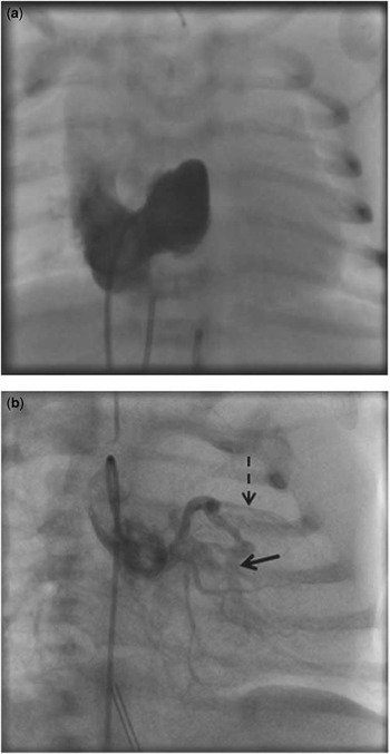
Catheterisation was performed before planned surgical shunt placement. The right ventricular pressure was 26/6 when the aorta was 54/26 mmHg. During the right ventriculogram (Fig 2a), the coronaries did not opacify. With selective left coronary angiography (Fig 2b), the right ventricular cavity opacified via multiple fistulous connections into the apex and outflow tract, and a tripartite right ventricle was demonstrated with a heavily hypertrophied apical portion. The left anterior descending artery was seen, and the distal branches beyond the fistulae were diminutive. The left circumflex and right coronary arteries were seen to fill normally from the aorta. The patient subsequently had surgical placement of a 3.5-mm modified Blalock–Taussig shunt without cardiopulmonary bypass. The post-operative electrocardiogram, in comparison with the first electrocardiogram, was notable for T-wave inversions being more prominent in the anterior leads but without significant ST changes. After an uneventful recovery, the patient was discharged home at 15 days of age. The patient’s clinical status was consistent with well-balanced circulation. An echocardiogram before discharge showed mild regurgitation through a dysplastic tricuspid valve with continued dyskinesia and bowing of the thinned ventricular septum and moderately diminished right ventricular function. The estimated right ventricular pressure remained low.
( a ) During the right ventriculogram, the coronaries did not opacify with the contrast. There is at least moderate tricuspid regurgitation. ( b ) With a selective injection into the left coronary artery, the right ventricular (RV) cavity opacified (arrow) with contrast via fistulous connections and a tripartite, but hypoplastic, RV was demonstrated. The left anterior descending artery was seen and the distal branches beyond the fistulae were diminutive (dashed arrow).

Discussion
The wide spectrum of pulmonary atresia with intact ventricular septum is due to variability in size and morphology of the right ventricle.Reference Daubeney, Delany and Anderson 5 The presence of coronary cameral fistulae with or without right ventricular dependency adds another level of complexity. Current management typically includes a selective strategy involving a combination of catheterisation and surgical interventions.Reference Alwi 6 , Reference Ashburn, Blackstone and Wells 7 The endpoint for palliation can range from salvaging a two-ventricle circulation to a single-ventricle strategy culminating in a Fontan circulation. Regardless of the endpoint, careful assessment of the size and morphology of the tricuspid valve, the right ventricle, and the coronary arteries is necessary. Our institution has employed a strategy of performing a cardiac catheterisation on all neonates for delineation of coronary anatomy before catheter-based or surgical palliation. A very small subset is known to have coronary ostial atresia and is at high risk of sudden death, and some centres have listed these patients for primary cardiac transplantation.Reference Cheung, Richmond, Turner, Bacha and Torres 4 , Reference Selamet, Hsu, Thaker and Gersony 8
We report a unique case of pulmonary atresia with intact ventricular septum with a subsystemic right ventricle. The precise aetiology of coronary cameral fistulae is unknown, but it is believed that a lack of egress from the right ventricle with severe hypertension of the chamber plays an important role in the development of these abnormal connections. The connections are usually found in patients with severe right ventricular hypertension, tricuspid valvar stenosis, or hypoplasia and right ventricular hypoplasia.Reference Freedom, Anderson and Perrin 2
Our case did not have significant stenoses in the ventricle–coronary connections, and in the context of a subsystemic right ventricle there was no evidence for right ventricle-dependent coronary circulation. In essence, the patient already had a “decompressed” right ventricle. To our knowledge, there has been no specific report of this lesion presenting with a subsystemic right ventricle without intervention in the neonatal period. We selected a strategy of surgical shunting with longer-term plans for further single ventricle palliations due to hypoplasia of the tricuspid valve and right ventricle along with significant dysplasia and regurgitation of the tricuspid valve. These factors, along with the presumed antenatal ischaemic injury to the right ventricle, made it very unlikely, in our estimation, for the right ventricle to be a meaningful contributor to pulmonary blood flow. Although the right ventricle was “decompressed”, we did not elect to open up the right ventricular outflow tract because of concerns about potentially deleterious consequences of pulmonary insufficiency and tricuspid regurgitation.
Owing to the low incidence, evidence-based guidelines for management are lacking and reports have been limited to small case series, individual-centre retrospective chart reviews, and the prospective Congenital Heart Surgeons Society study. The overall survival of this lesion has been estimated to be about 80% from the Congenital Heart Surgeons Society study,Reference Ashburn, Blackstone and Wells 7 but the role of abnormal coronary artery connections in the prognosis of these patients remains to be determined. Studies at specific centres have reported that the right ventricle-dependent coronary circulation is associated with higher mortality but this has not been replicated in larger studies pooling data from multiple institutions.Reference Freedom, Anderson and Perrin 2 , Reference Cheung, Richmond, Turner, Bacha and Torres 4 , Reference Ashburn, Blackstone and Wells 7 , Reference Guleserian, Armsby, Thiagarajan, del Nido and Mayer 9
The finding of a subsystemic right ventricle is an unusual finding and could represent a high-risk subset of this lesion. The presence of coronary cameral fistulae in our patient suggests that the right ventricle was hypertensive at some point during fetal development, thus allowing for the formation of these abnormal connections. It is unknown as to what led the right ventricle to become subsystemic, but an ischaemic event in the right ventricle or worsening tricuspid insufficiency could potentially be inciting factors. It is impossible to know precisely when this inciting event took place but it could have occurred in the late fetal period after the development of abnormal coronary connections, which then persisted. The presence of a thin-walled dyskinetic ventricular septum in utero lends some support for this hypothesis, despite the lack of typical findings for myocardial infarction in the serial electrocardiograms. In the absence of stenoses in the coronary cameral fistulae, there is a theoretical risk of ongoing ischaemia due to coronary run-off from the higher pressured aorta into the subsystemic right ventricle. In fact, it is tempting to speculate that run-off through the fistula accounts for the diminutive left anterior descending artery distal to the fistulous connection. For this reason, our patient had a modified Blalock–Taussig shunt placed without cardiopulmonary bypass out of concern for difficulty protecting the myocardium. At this point, we can only speculate about the aetiology of decompression of the right ventricle in our patient. We continue to believe that all patients with this disease deserve early cardiac catheterisation for definition of coronary artery anatomy and flow distribution with recognition of the possibility for co-existing coronary cameral fistulae and a subsystemic right ventricle.
Acknowledgements
None.
Financial Support
None.
Conflicts of Interest
None.