Introduction
Helminth infections remain a persistent public health problem in resource-poor areas of the world (Hotez et al., Reference Hotez, Brindley, Bethony, King, Pearce and Jacobson2008b). Three helminth pathogens, the liver flukes Opisthorchis viverrini and Clonorchis sinensis and the blood fluke Schistosoma haematobium, are categorized as Group I carcinogens by the World Health Organization's International Agency for Research on Cancer (IARC) (Bouvard et al., Reference Bouvard, Baan, Straif, Grosse, Secretan, El Ghissassi, Benbrahim-Tallaa, Guha, Freeman, Galichet and Cogliano2009; IARC, 2012). Opisthorchiasis is endemic in the lower Mekong Basin countries of Thailand, Lao PDR, Cambodia and Vietnam (Sripa et al., Reference Sripa, Bethony, Sithithaworn, Kaewkes, Mairiang, Loukas, Mulvenna, Laha, Hotez and Brindley2011). About 9 million people in Thailand alone are infected with O. viverrini, with many progressing to cholangiocarcinoma (Sripa et al., Reference Sripa, Brindley, Mulvenna, Laha, Smout, Mairiang, Bethony and Loukas2012). The geographical distribution of opisthorchiasis reflects the distribution of its intermediate hosts, both Bithynia spp. snails and cyprinid fish, as well as the dietary habits of the people resident in these regions (Smout et al., Reference Smout, Sripa, Laha, Mulvenna, Gasser, Young, Bethony, Brindley and Loukas2011; Sithithaworn et al., Reference Sithithaworn, Andrews, Nguyen, Wongsaroj, Sinuon, Odermatt, Nawa, Liang, Brindley and Sripa2012). Infection with O. viverrini is caused by consumption of undercooked fish, in which metacercariae (MC), the infective stage, of O. viverrini have encysted. Metacercariae excyst in the host duodenum and migrate into the intrahepatic biliary system, where they develop into adult flukes (Sripa et al., Reference Sripa, Bethony, Sithithaworn, Kaewkes, Mairiang, Loukas, Mulvenna, Laha, Hotez and Brindley2011).
Residents of endemic regions are often infected for a lifetime. Opisthorchiasis induces inflammation of the epithelia lining the intrahepatic bile ducts, with subsequent periductal fibrosis. Chronic opisthorchiasis can in turn progress to severe hepatobiliary disease, including cholangitis, obstructive jaundice, hepatomegaly, cholecystitis, cholelithiasis and cholangiocarcinoma (CCA), bile duct cancer (Mairiang et al., Reference Mairiang, Laha, Bethony, Thinkhamrop, Kaewkes, Sithithaworn, Tesana, Loukas, Brindley and Sripa2012). The mechanisms by which the parasite causes CCA are likely multi-factorial and include chronic immunopathology, mechanical damage from the worms grazing the epithelium lining the bile ducts, elevated dietary nitrosamines, and the secretion by the parasite of growth factors and other proteins that stimulate cell proliferation and interfere with homeostatic apoptosis (Smout et al., Reference Smout, Sripa, Laha, Mulvenna, Gasser, Young, Bethony, Brindley and Loukas2011). Despite the humoral and cell-mediated immune response elicited in both humans and rodents, it is unclear whether a protective immune response is acquired to O. viverrini (Wongratanacheewin et al., Reference Wongratanacheewin, Sermswan and Sirisinha2003). Due to the chronic nature of opisthorchiasis and its carcinogenicity, control measures to combat this infection are a public health priority, especially in Thailand and Lao PDR (Prichard et al., Reference Prichard, Basanez, Boatin, McCarthy, Garcia, Yang, Sripa and Lustigman2012). Praziquantel and, recently, tribendimidine are effective anthelmintics to treat opisthorchiasis (Soukhathammavong et al., Reference Soukhathammavong, Odermatt, Sayasone, Vonghachack, Vounatsou, Hatz, Akkhavong and Keiser2011; Keiser et al., Reference Keiser, Adelfio, Vargas, Odermatt and Tesana2013). However, successful clearance of the infection does not lead to protection against reinfection, and there is concern of increased risk of CCA from repeated rounds of praziquantel for reinfection (Sripa et al., Reference Sripa, Brindley, Mulvenna, Laha, Smout, Mairiang, Bethony and Loukas2012).
Despite the pernicious impact of opisthorchiasis and the inability of drug treatment to induce lasting protection, there are few reports on attempts to develop a vaccine against this neglected tropical disease. Syrian golden hamsters (Mesocricetus auratus) can be reliably infected with MC of O. viverrini, leading to patent infection (Flavell & Lucas, Reference Flavell and Lucas1983; Thamavit et al., Reference Thamavit, Kongkanuntn, Tiwawech and Moore1987). Notwithstanding the clinical relevance of this model, it has been under-studied as a model for the development of vaccines. This is despite the outcome of passive transfer to naïve hamsters of spleen cells and/or sera from infected hamsters that results in reductions in fecundity compared to control hamsters, implying that both humoral and cellular factors contribute to this protective outcome (Flavell et al., Reference Flavell, Pattanapanyasat and Flavell1980). In addition, immunization with parasite extracts has also induced modest reductions in fluke burdens (Sirisinha & Wongratanacheewin, Reference Sirisinha and Wongratanacheewin1986). However, a key step in vaccine development is proof of concept that protective immunity can indeed be induced. Many successful viral and bacterial vaccines are attenuated versions of the microbe. Whereas diverse forms of attenuation have shown utility, e.g. serial passage, attenuation by irradiation of the infective stages of helminth parasites has been used widely. Examples include irradiation of the third-stage larvae of Ancylostoma caninum, which provided the platform for industrial production of a vaccine for hookworm disease of dogs (Fujiwara et al., Reference Fujiwara, Loukas, Mendez, Williamson, Bueno, Wang, Samuel, Zhan, Bottazzi, Hotez and Bethony2006) and the irradiated cercariae model of schistosomes (Yole et al., Reference Yole, Pemberton, Reid and Wilson1996). This report presents the initial steps towards proof of concept that attenuation of O. viverrini by gamma irradiation of metacercariae can induce a protective immune response as a basis for vaccine development. We anticipate that this model will facilitate screening of vaccine candidates and formulations for consideration in human clinical trials against this carcinogenic liver fluke.
Materials and methods
Irradiation and viability of metacercariae
Metacercariae of O. viverrini (Ov-MC) were obtained from naturally infected cyprinid fish (Sithithaworn et al., Reference Sithithaworn, Pipitgool, Srisawangwong, Elkins and Haswell-Elkins1997). These larvae were dispensed into a glass block (4 × 4 × 1 cm) containing 0.85% NaCl (1 ml) and exposed to gamma (γ) radiation at increasing doses. The γ-rays were discharged from a 60Co linear accelerator (Theratron 780C, Theratronics, Ottawa, Ontario, Canada) at a rate of 112 rad/min, with target Ov-MC located 80 cm from the 60Co source, in a radiation field of 400 cm2 (20 × 20 cm).
To monitor the effect of γ-irradiation on viability of Ov-MC in vitro, 20 Ov-MC were irradiated with 0, 5, 10, 12, 20, 25 or 50 Gray (Gy). Thereafter, the irradiated Ov-MCs were induced to excyst in vitro for 20 min in 1 ml 0.25% trypsin in phosphate-buffered saline (PBS) containing 0.2% EDTA (excystation buffer). Newly excysted juveniles were washed several times in saline (0.85% NaCl), then examined and quantified for activity/viability by light microscopy.
Experimental infection and vaccination
One day before infection with irradiated Ov-MC, blood samples were collected from the retro-orbital vein of hamsters. To determine the effect of radiation on infectivity of irradiated Ov-MC, groups of five hamsters were infected with 50 Ov-MC that had been irradiated with increasing doses of γ-radiation – 3, 5, 10, 12, 20, 25 and 50 Gy. Infection of hamsters with Ov-MC was accomplished by oro-gastric gavage (Bhamarapravati et al., Reference Bhamarapravati, Thammavit and Vajrasthira1978; Sripa & Kaewkes, Reference Sripa and Kaewkes2002). Hamsters infected with non-irradiated (intact/wild-type) Ov-MC were included as controls. Six weeks after infection, hamsters were euthanized, after which livers, blood and faeces were collected.
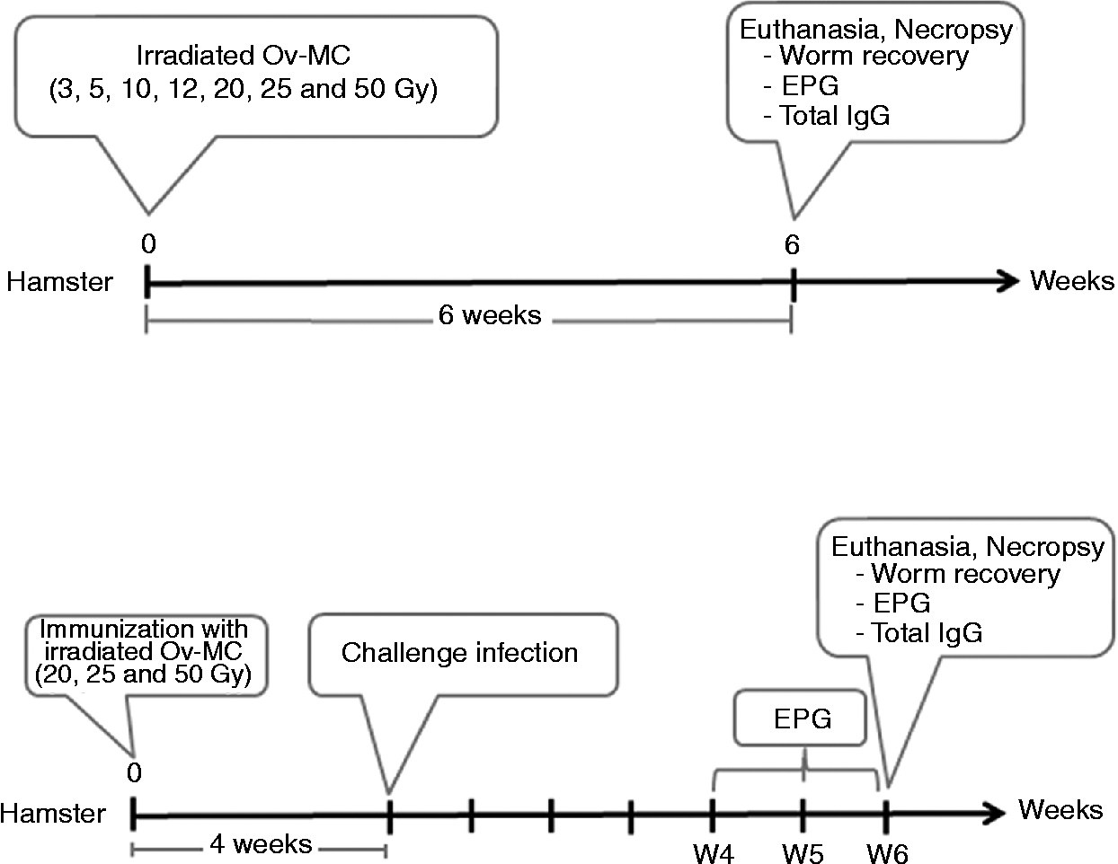
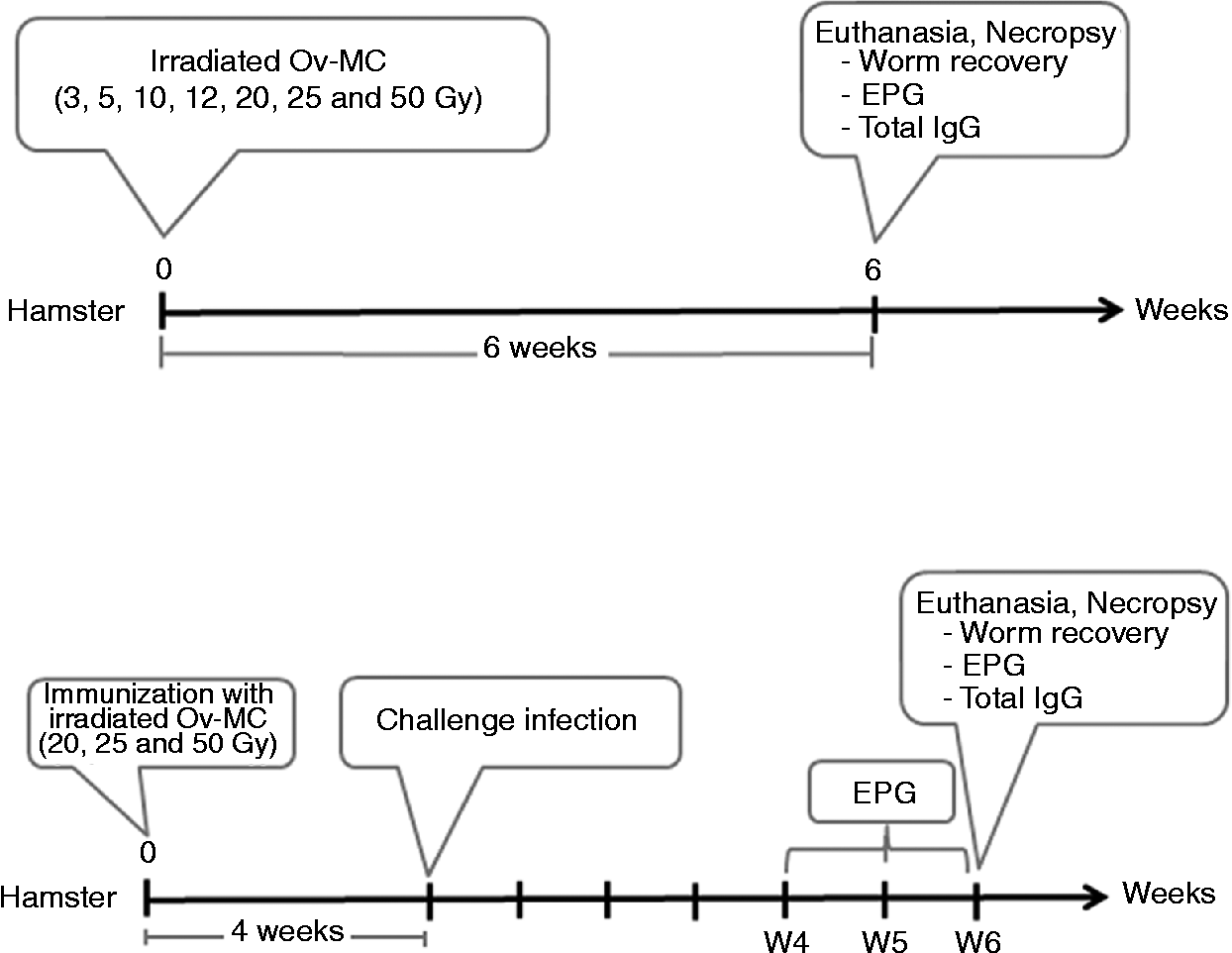
A putatively protective dose of irradiated Ov-MC for vaccination was predicted to be that which resulted in impaired parasite development, as ascertained by reduced adult fluke numbers in the biliary tree, but which also stimulated a strong antibody response to O. viverrini fluke antigens. Immunization was carried out using Ov-MC subjected to increasing doses of radiation. Six treatment groups (n= 5 per group) were included: group 1, control hamsters infected with 50 intact/wild-type (non-irradiated) Ov-MC (0 Gy); groups 2, 3 and 4 immunized with a single dose of 50 Ov-MC that had received 20, 25 and 50 Gy γ-radiation, respectively. Hamsters were challenged with 50 intact Ov-MC 4 weeks after exposure to irradiated Ov-MC. Faeces of the hamsters were collected 4, 5 and 6 weeks later to ascertain numbers of fluke eggs per gram of faeces. Figure 1 presents a schematic of the experimental design and timetable for immunization with irradiated Ov-MC, challenge infection, euthanasia and sample collection.
Experimental design and the timelines of immunization with irradiated O. viverrini metacercariae, challenge infection and necropsy. Upper panel: infection of hamsters with radiation-attenuated metacercariae. Lower panel: vaccine study involving immunization with radiation-attenuated metacercariae, followed by challenge with intact parasites. Ov-MC, Opisthorchis viverrini metacercariae; Gy, Gray; EPG, worm eggs per gram of hamster faeces; W, weeks.
Faecal eggs, hamster sera, recovery of adult flukes
Faeces from each group of hamsters were pooled and the eggs of O. viverrini counted as eggs per gram (EPG) of faeces using a modified formalin–ethyl acetate technique (Elkins et al., Reference Elkins, Haswell-Elkins and Anderson1986). Food was not provided to the hamsters for 12 h prior to euthanasia. After euthanasia, blood was recovered from the heart and allowed to clot at 4°C for 30 min, centrifuged at 2100 g for 10 min at 4°C, and serum separated from blood cells and stored at − 20°C. Adult worms of O. viverrini were recovered by dissecting the liver, with the number of worms determined by examination under light microscopy (Flavell et al., Reference Flavell, Flavell and Field1983).
Preparation of O. viverrini antigen
Worms of O. viverrini were recovered from bile ducts of hamsters at necropsy. Worms were washed in phosphate-buffered saline, pH 7.2 (PBS) and resuspended in PBS containing a 1 × protease cocktail (GE Healthcare Life Sciences, Pittsburg, Philadelphia, USA). Worms were homogenized in a hand-held tissue grinder on ice and subsequently the lysate was subjected to ultrasonic sonication with amplitude 20% for 2-s bursts with 5-s intervals for 20 cycles (Vibra-Cell, Sonics & Materials Inc, Newtown, Connecticut, USA). The lysate was clarified by centrifugation at 18,000 g for 30 min at 4°C, the supernatant collected, and the protein concentration determined using the bicinchoninic acid assay (Amresco, Solon, Ohio, USA). The soluble lysate of adult liver flukes, for use as antigen in serological assays (below), was divided into aliquots and stored at − 20°C.
Serology
To detect O. viverrini-specific immunoglobulin (Ig) G, flat-bottom 96-well microtitre plates (Maxi-Sorp Immunoplates, Nunc, Denmark) were coated with 1 μg/ml of the soluble lysate of adult worms at 100 μl per well (Saichua et al., Reference Saichua, Sithithaworn, Jariwala, Deimert, Sithithaworn, Sripa, Laha, Mairiang, Pairojkul, Periago, Khuntikeo, Mulvenna and Bethony2013) in 15 mm Na2CO3, 35 mm NaHCO3, pH 9.6, and incubated at 4°C overnight. Wells were washed with 300 μl of 154 mm NaCl, 0.05% Tween 20 (wash solution) five times before being blocked with 200 μl of 5% skimmed milk powder buffer. Plates were incubated at 37°C for 2 h and then washed three times with wash solution (above). Sera were diluted 1:1000 in incubation buffer (2% skimmed milk powder in 137 mm NaCl, 9 mm Na2HPO4.2H2O, 0.8 mm NaH2PO4.2H2O, 0.05% Tween-20), added to plates at 100 μl per well, with the plates incubated for 2 h at 37°C. The washing process was repeated as above, after which 100 μl goat anti-hamster IgG–horseradish peroxidase (HRP) (Invitrogen, Grand Island, New York, USA) diluted 1:2500 in PBS was added (100 μl per well) and incubated for 1 h at 37°C. Plates were washed four times with wash solution (above), followed by a final wash with 1 × PBS. O-Phenylenediamine (Life Technologies/Invitrogen) was added (100 μl/well) and the plates incubated at 37°C for 30 min. Finally, substrate development was halted by addition to wells of 100 μl 0.5 m H2SO4. Optical densities (OD) at 490 nm were determined (Spectramax, Molecular Devices, Sunnydale, California, USA), with data collected and stored using SoftMax Pro (Molecular Devices). Duplicate wells without control or experimental sera, but with the above reagents, were included to determine the extent of non-specific binding of the antibodies.
SDS-PAGE, Western blot analysis
Fifty microgram of PBS-soluble adult worm antigen extract were separated by 15% sodium dodecyl sulphate polyacrylamide gel electrophoresis (SDS-PAGE) using the mini-PROTEIN electrophoresis system (Biorad, Hercules, California, USA) and transferred to nitrocellulose using a rapid semi-dry blotting system, Fastblot B43/B44 (Biometra, Göttingen, Germany). The nitrocellulose membrane was cut into 14 strips (~3.5 μg soluble adult worm antigen extract per strip) and blocked with 5% skimmed milk powder in PBS-T (0.1% Tween-20 in PBS) for 2 h at room temperature. Membranes were briefly rinsed twice with PBS-T, before incubating in pooled hamster sera diluted 1:100 in 2% skimmed milk powder in PBS-T for 2 h at room temperature. After washing three times for 10 min each with PBS-T, membranes were incubated with goat anti-hamster IgG-HRP (Invitrogen) diluted in 2% skimmed milk powder in PBS-T for 1 h. Membranes were washed twice for 10 min each with PBS-T, followed by a final 10-min rinse with PBS. The bands were visualized using a chemiluminescence detection system (ECL PlusTM, GE Healthcare).
Data analysis
Protection levels were calculated by comparing worm recovery of control and vaccinated groups. Data were analysed by one-way analysis of variance using Prism 5 (GraphPad, La Jolla California, USA); P values of ≤ 0.05 were considered to be statistically significant.
Results
Gamma-radiation attenuated metacercariae of O. viverrini
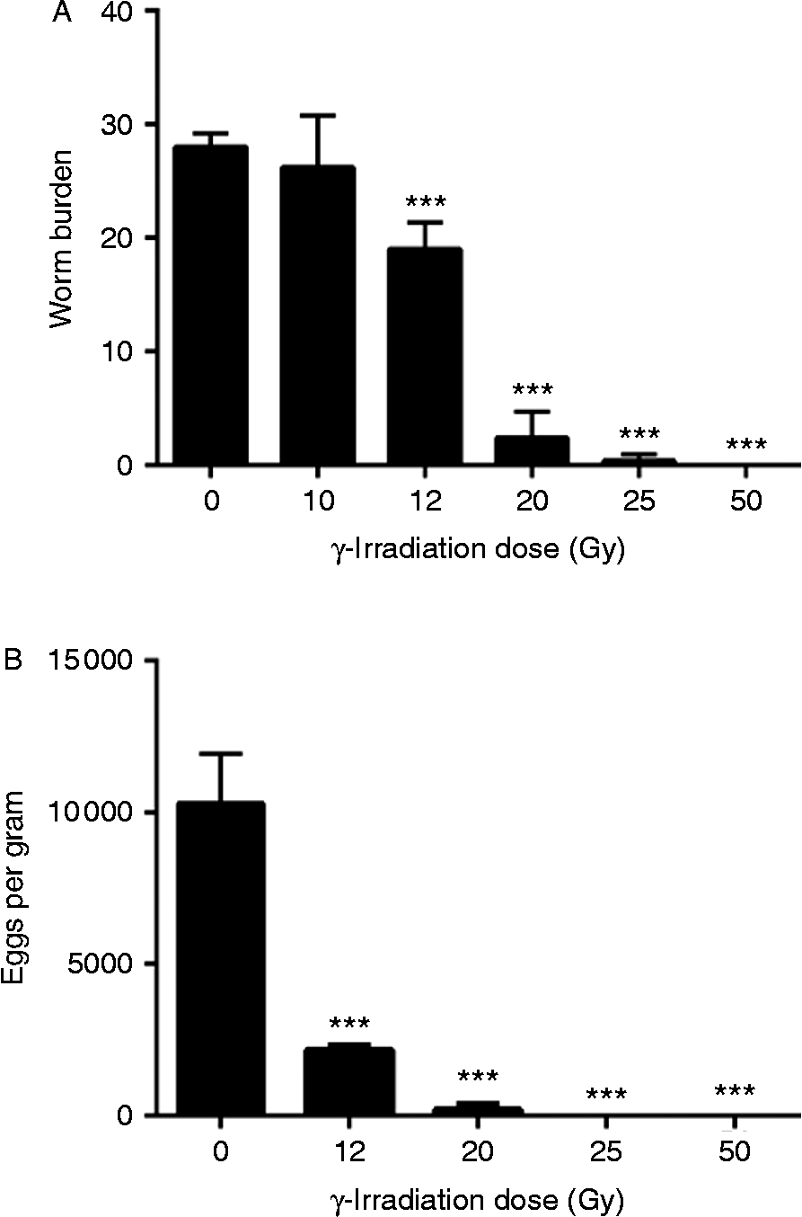
Metacercariae of O. viverrini exposed to 5, 10, 12, 20, 25 and 50 Gy of γ-radiation from 60Co exhibited 85–100% active excystation in vitro. Excystation rates of MC that had been γ-irradiated did not differ from that of controls, i.e. non-irradiated Ov-MC (not shown). To address the in vivo effect of γ-radiation on the establishment of irradiated Ov-MC in hamsters, as well as to determine the optimal dose to attenuate irradiated the Ov-MC to elicit a protective immune response, hamsters were infected with Ov-MC that had been exposed to increasing doses of radiation. Radiation doses greater than 12 Gy resulted in progressively negative impacts on the ability of the Ov-MC to develop into adult worms. Whereas similar numbers of adult worms were recovered from hamsters infected with Ov-MC irradiated at 10 Gy and intact MC, significantly fewer adult worms were recovered after infection with 12 Gy Ov-MC. Less than 10% survived at 25 Gy, and none survived to the adult stage when Ov-MC were exposed to 50 Gy. Overall, the number of adult worms recovered from the hamster biliary tract ranged from 32 to 0 (fig. 2A). The numbers of eggs in faeces of infected hamsters reflected the numbers of adult worms (fig. 2B), where egg output was significantly decreased in groups of hamsters infected with Ov-MC irradiated with 12, 20, 25 and 50 Gy, compared to hamsters infected with control, intact (non-irradiated) MC (P≤ 0.001) which shed faeces containing ~10,000 EPG.
The number of adult worms of O. viverrini in the bile ducts, and eggs recovered in pooled faeces, from hamsters infected with O. viverrini metacercariae subjected to γ-radiation. (A) The number of worms recovered 6 weeks after infection with 50 irradiated Ov-MC subjected to different doses of γ-radiation. (B) Faecal egg counts of hamsters infected with irradiated and wild-type metacercariae (0 Gy). *** Denotes significant differences between worms treated compared with control groups infected with wild-type parasites (P≤ 0.001).

Immunoglobulin responses to soluble lysate of adult O. viverrini
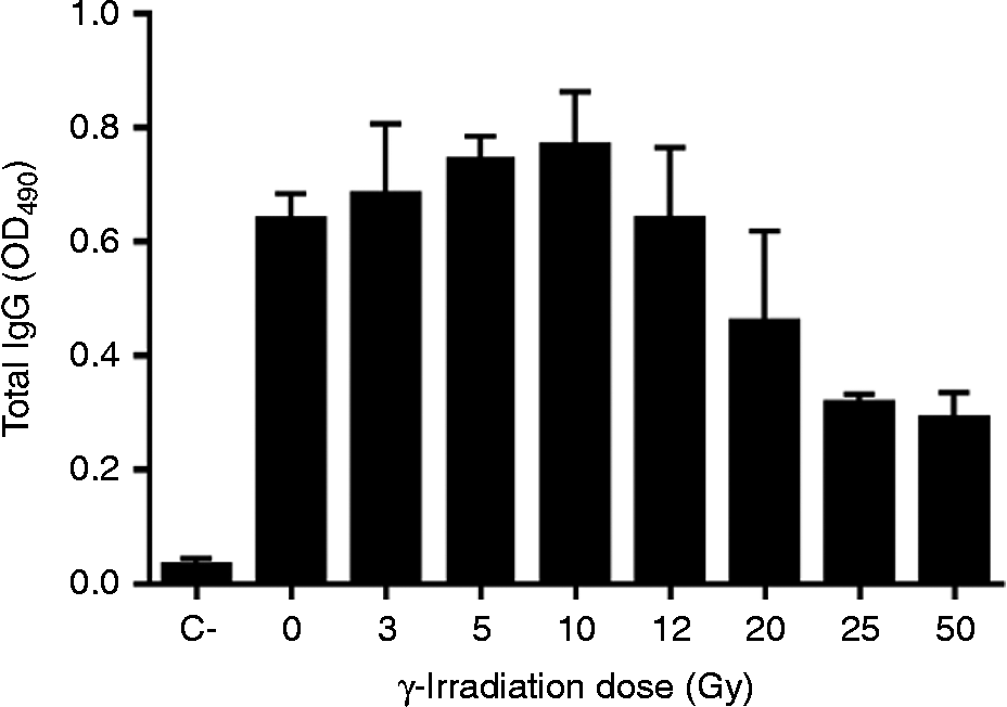
Immunoglobulin G (IgG) responses against the soluble lysate of adult O. viverrini worms were detected in infected, but not uninfected, hamsters after the primary exposure to MC (fig. 3). Titres of IgG greater than OD490= 0.6 against soluble lysate of adult O. viverrini were elicited in hamsters exposed to Ov-MC exposed to 0, 3, 5, 10 and 12 Gy. Exposure to MC exposed to greater doses of radiation (Ov-MC at 20, 25 and 50 Gy) also elicited strong IgG responses against soluble lysate of adult O. viverrini, although not as markedly elevated as evident in sera from rodents receiving MC exposed to less radiation. As noted, an appropriate immunizing dose of γ-radiation would require the impairment of parasite development upon reaching the small intestine/biliary system as well as eliciting a strong anti-adult O. viverrini-IgG response. A radiation dose of 20 Gy induced a robust antibody response against adult soluble lysate, along with significant reductions in adult worms and faecal eggs (fig. 2). Accordingly, a dose of 20 Gy, as well as the higher 25 Gy and 50 Gy doses of radiation were utilized in the immunization study (below).
IgG responses to a lysate of adult O. viverrini worms in the sera of hamsters 6 weeks after infection with irradiated metacercariae. C: uninfected hamsters.
Anti-worm and anti-fecundity effects against challenge infection
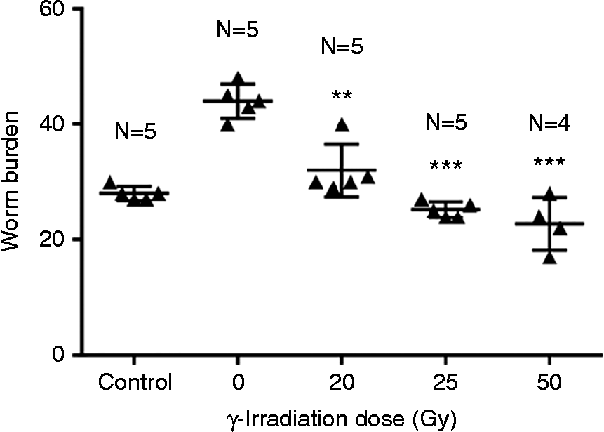
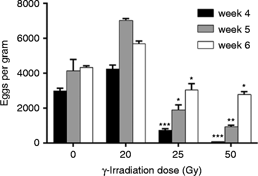
Metacercariae of O. viverrini irradiated with 20, 25 and 50 Gy were used to immunize hamsters before challenge infection with intact MC (fig. 1). Immunized groups were compared with non-immunized control groups, which were challenged with 50 intact Ov-MC. Significantly fewer worms were recovered from hamsters immunized with Ov-MC irradiated at 20, 25 and 50 Gy than from the control hamsters infected with intact Ov-MC or 0 Gy (fig. 4). Similarly, faecal egg numbers were decreased significantly in hamsters immunized with 25 Gy and 50 Gy Ov-MC, although not with the 20 Gy Ov-MC immunization dose. Reductions in faecal egg counts were marked at weeks 4 and 5. Although the reductions waned by week 6, significantly fewer eggs were still apparent in comparison to the control hamsters challenged with wild-type metacercariae (fig. 5).
The number of adult worms of Opisthorchis viverrini recovered from hamsters after immunization with irradiated metacercariae and subsequent challenge with 50 wild-type metacercariae. Control: hamsters infected with 50 intact Ov-MC. Levels of significant differences: ** P≤ 0.01 and *** P≤ 0.001.
The number of eggs per gram of faeces (EPG) in hamsters exposed to irradiated metacercariae at 4, 5 and 6 weeks after infection and challenged with 50 wild-type metacercariae. Levels of significant differences: * P≤ 0.05, ** P≤ 0.01 and *** P≤ 0.001.
IgG levels following challenge infection
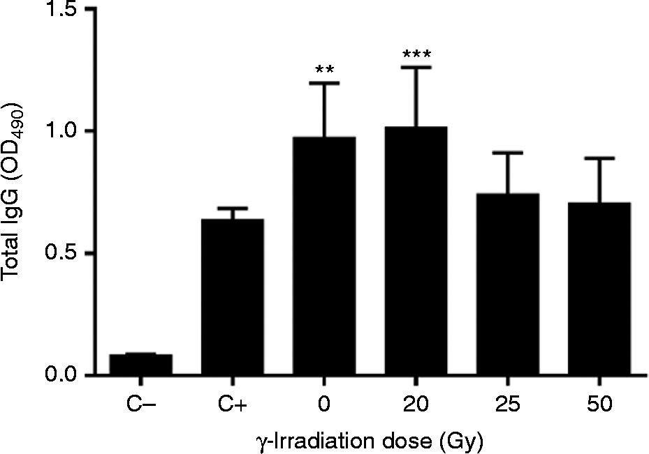
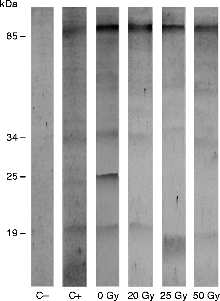
The serum IgG antibody profiles in hamsters immunized with irradiated Ov-MC were determined by enzyme-linked immunosorbent assay (ELISA) (fig. 6). IgG responses against O. viverrini were induced in all hamsters infected with irradiated Ov-MC, with significantly increased IgG responses against soluble lysate of adult O. viverrini in groups that received 0 and 20 Gy irradiation compared to non-vaccinated but challenged rodents (fig. 6). Immunoblot analysis showed that serum IgG from the group of hamsters vaccinated with non-irradiated MC (0 Gy) reacted to the soluble lysate of O. viverrini worms with bands of molecular masses of ~89, 34, 25 and 20 kDa. In hamsters that received irradiated Ov-MC at 25 Gy and 50 Gy, proteins of 89, 34 and 20 kDa were recognized; the 25 kDa protein was not recognized (fig. 7). The most prominent band recognized by vaccinated animals was at 89 kDa.
IgG responses against soluble lysate of adult O. viverrini as determined by ELISA in sera of hamsters 6 weeks after challenge infection with wild-type O. viverrini metacercariae following immunization with radiation-attenuated metacercariae at 20, 25 and 50 Gy. The term ‘ − ve control’ (C − ) refers to uninfected hamsters whereas ‘control’ (C+) refers to non-vaccinated but infected hamsters. Levels of significant differences: ** P≤ 0.01 and *** P≤ 0.001.
Immunoblot analysis of IgG in hamster sera against a soluble lysate of adult O. viverrini worms. The terms 20, 25 and 50 Gy refer to hamsters immunized with radiation-attenuated O. viverrini metacercariae that had been exposed to 20, 25 and 50 Gy. The term 0 Gy refers to hamsters vaccinated with non-irradiated metacercariae and infected with wild-type metacercariae; C − refers to a pool of pre-immunized serum. C+ refers to non-vaccinated hamsters infected with 50 wild-type metacercariae.
Discussion
The energy from ionizing radiation, such as γ-rays, X-rays and ultraviolet (UV) light, damages cellular nucleic acids, proteins and lipids, leading to physiological changes or cell death (Spitz et al., Reference Spitz, Azzam, Li and Gius2004). These types of radiation have been used to attenuate diverse species of helminths for subsequent administration, as proof of concept that vaccines can be developed against these pathogens (Bain, Reference Bain1999; Bickle, Reference Bickle2009). Several efficacious vaccines currently in use, including some against helminths, are based on immunization with irradiated microbes. The use of ionizing radiation to attenuate helminth parasites forms the basis of a number of commercial vaccines for companion animals and livestock (Bain, Reference Bain1999; Bickle, Reference Bickle2009). In a pioneering report from half a century ago, it was demonstrated that irradiated larvae of the cattle lungworm Dictyocaulus viviparous fail to develop to adulthood, yet still stimulate a protective response, an outcome that propelled commercial development of a vaccine for this infection (Jarrett et al., Reference Jarrett, Jennings, McIntyre, Mulligan and Urquhart1958).
In Schistosoma mansoni, radiation-attenuated cercariae induce protective immunity in mammals, including non-human primates (Bickle, Reference Bickle2009). With vaccines for schistosomes, the protection obtained with cercariae that have been attenuated with ionizing radiation is considered to be the ‘gold’ standard against which other vaccine candidates are compared for clinical development (McManus & Dalton, Reference McManus and Dalton2006; Hotez et al., Reference Hotez, Bethony, Oliveira, Brindley and Loukas2008a; McManus & Loukas, Reference McManus and Loukas2008; Loukas et al., Reference Loukas, Gaze, Mulvenna, Gasser, Brindley, Doolan, Bethony, Jones, Gobert, Driguez, McManus and Hotez2011). Elevated levels of protection against Schistosoma japonicum can be induced by exposure to X-irradiated cercariae in macaques (Hsu et al., Reference Hsu, Hsu and Osborn1969) and cattle (Hsu et al., Reference Hsu, Hsu, Xu, Shi, He, Clarke and Johnson1983), by exposure to γ-irradiated cercariae in bovines (Hsu et al., Reference Hsu, Xu, He, Shi, Shen, Hsu, Osborne and Clarke1984), and UV-attenuated cercariae in pigs (Tian et al., Reference Tian, Lin, Wu, Gao, Zhang, Ji and Wu2010). Vaccine efficacies are variable, however, as exemplified by immunization of mice with γ-irradiated cercariae of S. japonicum, which elicits only modest resistance to reinfection (Zhang et al., Reference Zhang, Taylor, Bickle, Wang and Ge1999). Whereas it would be impractical to use γ-radiation of fish to block transmission of O. viverrini, we examined radiation-attenuated MCs here with the goal of obtaining a baseline level of protection (either strong or limited protection against challenge) against which to compare future vaccine preparations.
At the outset, we determined that exposure to γ-radiation reduced the infectivity of metacercariae of O. viverrini; i.e. doses >10 Gy reduced infectivity, with, for example, >50% reduction caused by 12 Gy. At 50 Gy, flukes failed to develop from irradiated larvae. Similar findings have been described with radiation sensitivity of MC of the minute intestinal fluke Metagonimus yokagawi (Chai et al., Reference Chai, Kim, Kook and Lee1995). Metacercariae of the closely related liver fluke C. sinensis may be even more sensitive to damage from γ-radiation than O. viverrini, with only ~25% of worms exposed to 5 Gy developing to the adult stage in rats (Park & Yong, Reference Park and Yong2003; Quan et al., Reference Quan, Lee, Bae, Ohwatari, Min and Yang2005). We also observed that immunization of hamsters with γ-irradiated MC of O. viverrini resulted in decreased infection and fecundity upon challenge infection with intact/wild-type MC. Passive transfer of immune spleen cells and serum to protect against O. viverrini has been examined, but failed to induce resistance to reinfection in hamsters (Flavell et al., Reference Flavell, Pattanapanyasat and Flavell1980). The immunization of hamsters with a soluble lysate of adult worms before challenge infection afforded modest protection, a 30% reduction of worms (Sirisinha & Wongratanacheewin, Reference Sirisinha and Wongratanacheewin1986). Vaccination with radiation-attenuated MC of related liver flukes has also been reported, e.g. a single dose of γ-irradiated (12 Gy) MC induced resistance in rats to challenge infection with C. sinensis (Quan et al., Reference Quan, Lee, Bae, Ohwatari, Min and Yang2005).
When helminth parasites are exposed to ionizing radiation, they can suffer growth retardation, vacuolization of the interstitial tissues, elevation of the tegument, malformation of reproductive organs, including vitellaria, the uterus and numbers of eggs in utero, failure of reproduction and attenuation of pathogenicity (Bickle et al., Reference Bickle, Dobinson and James1979; Chai et al., Reference Chai, Kim, Kook and Lee1995). Irradiation of MC of O. viverrini led to malformation of reproductive organs and impairment of, and/or failure to, produce eggs. Moreover, an abnormal morphology, particularly of the reproductive organs, was apparent in worms exposed to 25 Gy. A dose of 50 Gy was lethal, with no adult worms surviving. The number of eggs released in the faeces of hamsters that were immunized with 25 Gy was significantly reduced in the fourth and fifth weeks of infection compared to hamsters infected with wild-type parasites. We selected this dose of γ-radiation for vaccine development because 25 Gy Ov-MC induced robust IgG responses yet attenuated adult flukes sufficiently to arrest their development and ensure failure of normal reproduction. Metacercariae of O. viverrini exposed to 25 Gy were capable of inducing a robust IgG response to soluble lysate of adult O. viverrini, which we conjecture is the vaccine-induced protective response that negatively influenced fecundity of the challenge cohort of wild-type flukes.
In summary, vaccination of hamsters with irradiated Ov-MC induced significant levels of protection, as evidenced by reduced worm numbers and reduced fecundity of the parasites. The methods and findings provided proof of concept for assessment of performance of vaccines for human opisthorchiasis in the context of informative vaccine endpoints – titre of IgG antibody to antigens of O. viverrini, the numbers of flukes resident in the biliary system after immunization, and the number of fluke eggs in hamster faeces, i.e. fecundity effects. Hamsters provide an informative model host for development of a vaccine against infection with O. viverrini because not only is this rodent a permissive, definitive host, but also because the hamster will develop O. viverrini-induced CCA (Bhamarapravati et al., Reference Bhamarapravati, Thammavit and Vajrasthira1978; Smout et al., Reference Smout, Sripa, Laha, Mulvenna, Gasser, Young, Bethony, Brindley and Loukas2011; Lvova et al., Reference Lvova, Tangkawattana, Balthaisong, Katokhin, Mordvinov and Sripa2012; Sripa et al., Reference Sripa, Brindley, Mulvenna, Laha, Smout, Mairiang, Bethony and Loukas2012). Efforts toward developing a vaccine for opisthorchiasis might now focus on refining this model involving infection with radiation-attenuated metacercariae. The iterative process might include multiple immunizations, size of the inoculum of irradiated Ov-MC, duration between immunization and challenge, and analysis of cellular and cytokine responses. With advances in immuno-proteomics, and using mass spectrometry-based approaches, sera from hamsters vaccinated with irradiated MC could be used to identify the protective antigens. It would also be informative to investigate the role of the immunization on biliary tree and intestinal microbiota, which are modified by opisthorchiasis (Plieskatt et al., Reference Plieskatt, Deenonpoe, Mulvenna, Krause, Sripa, Bethony and Brindley2013). Progress toward a vaccine that would not only protect against opisthorchiasis but also against opisthorchiasis-induced bile duct cancer would be welcomed, given that these are among the most problematic neglected tropical diseases and infection-induced cancers of East Asia.
Financial support
These studies were supported by The Higher Education Research Promotion and National Research University Project of Thailand, Office of the Higher Education Commission through the Health Cluster (SHeP-GMS) Khon Kaen University to A.P. and T.L. In addition, B.S., T.L., J.M.B. and P.J.B. were supported by a Tropical Medicine Research Center grant (P50AI098639) from the National Institute of Allergy and Infectious Diseases (NIAID), National Institutes of Health (NIH). The content of this report is solely the responsibility of the authors and does not necessarily represent the official views of the NIH.
Conflict of interest
None.
Ethics statement
Male Syrian (golden) hamsters (Mesocricetus auratas) were reared at the animal facilities of the Faculty of Medicine, Khon Kaen University. Protocols for the experiments were approved by the Animal Ethics Committee of Khon Kaen University, approval number AEKKU25/2554, according to the Ethics of Animal Experimentation of the National Research Council of Thailand.