Case report
A 13-year-old boy born with bicuspid aortic valve was referred at our Centre as part of the routine follow-up. Echocardiography examination incidentally reported an anomalous coronary artery. The patient was asymptomatic. The physical examination was unremarkable. Electrocardiography was normal. Echocardiography showed also normal biventricular function with competent atrioventricular valve and bicuspid aortic valve. Bicuspid aortic valve was found in the setting of trisinuate root with appropriate separation of the right and left coronary aortic sinuses with one raphe between right and left coronary valvular leaflets associated with trivial central aortic regurgitation.
A coronary CT scan confirmed anomalous right coronary artery arising from the opposite sinus with inter-arterial course. As described in previous publications, Reference Harris, Whitehead, Shin, Keller, Weinberg and Fogel1,Reference Cheezum, Liberthson and Shah2 the coronary CT scan protocol allows us to assess number of ostia and ostial morphology; asymmetry index, defined as the ratio between the maximal and minimal diameter the level of commissure between the right and left coronary leaflets; proximal vessel stenosis compared to normal distal reference and length of the stenosis; takeoff angle; and takeoff level of the anomalous coronary artery. The coronary CT examination was acquired with prospective ECG gating in a diastolic phase.
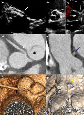
The radiological findings of the patient are shown in Figure 1. The anomalous right coronary artery had a separated origin. The ostia of the right coronary artery was not characterised by an elevated ratio of the vertical-to-horizontal measurements and therefore appeared round. Cross-sectional area of the anomalous coronary artery obtained in double oblique view at the level of the anterior aortic commissure had an asymmetry index of 1, as in normal patients. Proximal vessel stenosis of the anomalous coronary artery has not been found. The anomalous coronary artery had an acute takeoff angle (define <45°) with a takeoff level above the aortic valve commissure (high origin). The orifice of the right coronary artery was to the left of the raphe immediately above the septal commissure.
( a ) Echocardiographic parasternal short axis provides longitudinal views of the proximal portion of the coronary arteries. The right coronary artery (white arrow) has anomalous origin from the opposite coronary sinus. ( b ) Echocardiographic parasternal short-axis view provides a short-axis view of the bicuspid aortic valve. The aortic valve has one raphe between the right and left coronary leaflets. ( c ) Colour Doppler flow revealed a trivial central aortic regurgitation. ( d ) Coronary CT identified morphological features of the anomalous coronary artery. Multiplanar axial CT reconstruction at the level of the coronary artery origin demonstrates the right coronary artery (white arrow) travelling between the aorta and the pulmonary artery (asterisk). The anomalous coronary artery has a separate vessel origin. ( e ) Lumen cross-sectional area of the anomalous coronary artery obtained in double oblique view at the level of the anterior aortic commissure. This view is known as the “Angelini/Cheong cut.” In the anomalous coronary artery, a normal ratio between the maximal and minimal diameter was found. ( f ) Vessel’s takeoff level (above the commissure) is shown in three dimensional reformatted images. The white arrow points to the origin of the right coronary artery. ( g ) Virtual angioscopy post-processing technique is used to visualise the coronary ostia.

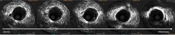
Intravascular ultrasonography was performed starting from three cm distal to the coronary ostium. Intravascular ultrasonography assessment reported no evidence of lateral compression in the proximal anomalous coronary artery. As described in recent publications, Reference Angelini, Uribe, Monge, Tobis, Elayda and Willerson3 intravascular ultrasonography was used to measure systolic proximal vessel cross-sectional area percentage narrowing compared to normal distal reference (%); and ratio between the maximal and minimal diameter at maximal proximal narrowing point. Results showed that there was no narrowing of the proximal tract of the anomalous coronary artery (Fig 2). The highest ratio between the maximal and minimal diameter in the proximal portion of the anomalous coronary artery was 1.05. The absence of elliptic shape and proximal coronary stenosis was considered consistent with the absence of associated intramural course. A stress echocardiography did not detect signs of myocardial ischaemia. Accordingly, recent ESC Guideline for the management of adult congenital heart disease surgery is not recommended for anomalous right coronary artery from the opposite coronary sinus in asymptomatic patient without myocardiac ischaemia and without intramural course and orifice anomalies. Reference Lerer and Edwards4 The patient was advised regular follow-up, with no medical treatment.
Intravascular ultrasound images from the patient with right anomalous coronary artery arising from the opposite sinus. The inner edges of the cross-sectional lumen from the distal to the proximal portion of the anomalous coronary artery were discerned. This segment of the artery is round and has a 37.5 mm uniform diameter, with an area of 12.7 cmq.

Discussion
This report illustrates the case of a patient with aortic bicuspid valve and an anomalous right coronary artery arising from the opposite sinus with inter-arterial course without evidence of intramural course. Absence of intramural course in patient with anomalous right coronary artery from the opposite coronary sinus and intra-arterial course is an unusual finding. Angelini et al described the universal presence of intramural course in patients with the origin of the right coronary artery from the opposite sinus of Valsalva at the intravascular ultrasonography evaluation. Reference Angelini, Uribe, Monge, Tobis, Elayda and Willerson3 In humans, however, little is known about the relationships between aortic valve morphology and anomalous coronary arteries. Subject with bicuspid aortic valve had a significantly higher frequency of the left dominance coronary arterial system in comparison with normal heart. In patients with congenitally bicuspid aortic valves, the left coronary ostium seems to rise above the aortic sinotubular junction more frequently compared than the normal population. Reference Lerer and Edwards4 High incidence of coronary anomalies has been found in the congenital heart defects involving maldevelopment of the outlet portion (conotruncus) of the developing heart. Reference Burch and Sahn5 Outlet portion is highly dependent upon neural crest cell involvement for normal development, and neural crest seems to play an important role in determining the aortic origin site and the proximal course of the coronary arteries. In the animal model, the frequency of relevant coronary artery anomalies significantly increases when the aortic valve is bicuspid. Reference Sans-Coma, Arqué, Durán, Cardo and Fernández6 In Syrian hamster, the probability of occurrence of anomalous coronary artery patterns increases continuously according to the deviation degree of the aortic valve from its normal (tricuspid) morphology. Reference Fernández, Durán and Real7 The development of cardiac arterial valves and coronary arteries has not been studied in human. We suggest that the occurrence of bicuspid aortic valve and absence of intramural course may be investigated further. The bicuspid aortic valve may attenuate the presence of the intramural course in the anomalous coronary artery. The presence of one raphe between right and left coronary leaflets and absence of a normally developed aortic valve commissure between the left and right aortic leaflets may be a potential biological mechanism which explains this association.
Conclusion
We report on a patient with bicuspid aortic valve and anomalous right coronary artery from the opposite sinus without evidence of intramural course. Experts support that anomalous right coronary artery from the opposite sinus in normal heart is constantly associated with some degree of intramural stenosis. Although further investigation is required, we suggest that such unusual finding might be correlated to the contemporary occurrence of bicuspid valve and anomalous origin of the right coronary artery.
Acknowledgements
None.
Financial support
This research received no specific grant from any funding agency, commercial, or not-for-profit sectors.
Conflicts of interest
None.