The implantable vascular access device, also known as Port-a-Cath or implantable chemoport, is a central venous catheter connected to a small reservoir implanted in the subcutaneous tissue.Reference Aparna, Ramesh and Appaji1 The most widely used port system was first introduced by Niederhuber et al. in 1982.Reference Niederhuber, Ensminger, Gyves, Liepman, Doan and Cozzi2 Chemoport provides a method to ensure prolonged central venous access for patients with chronic diseases requiring prolonged parenteral treatment. One advantage of the chemoport is that the puncturing needle can be removed following each infusion. Another advantage is that the skin covering its reservoir offers a natural protection against infection.Reference Aparna, Ramesh and Appaji1
Similar to other subcutaneous devices, prolonged use of chemoport has been associated with early and late complications.Reference Freytes, Reid and Smith3 If left underdiagnosed and untreated, these complications can lead to catastrophic sequelae. Late complications include drug extravasation, venous thrombosis, infection, sepsis, mechanical dysfunction, catheter disconnection, and embolisation.Reference Poorter, Lauw, Bemelman, Bakker, Taat and Veenhof4–Reference Robinson and Halverson8 Dislodgement and migration of chemoport catheter is a rare complication with potentially serious consequences.Reference Wang, Tsai and Hou9 The complication is uncommon, and its prevalence is scarce in the literature.
In this article, we are presenting a 15-year experience regarding chemoport catheter migration at the American University of Beirut Medical Center (AUBMC) which is a tertiary medical centre in the Middle east adjoining two specialised centers; the Children’s Heart Center (CHC) and the Children’s Cancer Center of Lebanon (CCCL). CCCL is a non-profit leading research, medical, and teaching institution in Lebanon and the Middle East and North Africa region. Most of the patients getting treatment in our CCCL required implantable chemoport for long-term chemotherapy administration.
In this study, we investigated the incidence and the clinical presentation of this complication in our paediatric population diagnosed with cancer.
Methods
This study has received the approval of the Institutional Review Board. Data from the past 15 years have been retrospectively analysed ranging from 1 April, 2002 until 31 March, 2018. All patients who were under the age of 18 at the time of chemoport insertion were eligible for enrollment in our study. We analysed the following variables: age, sex, type of cancer diagnosed, site of chemoport insertion, and frequency of migration or fracture.
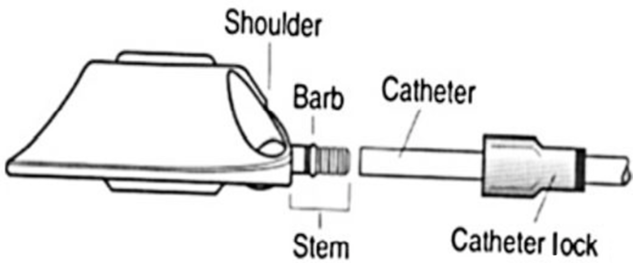
As per our centre’s protocol, unless general anaesthesia is contraindicated, all paediatric cancer patients requiring prolonged IV chemotherapy undergo chemoport insertion. A peripherally inserted central catheter is an alternative to chemoport if general anaesthesia is contraindicated or in selected patients who receive their treatment mostly as outpatient over a duration of 6 months or less (as in patients with Hodgkins lymphoma), are not too young or mentally disabled, and whose care givers are expected to provide proper care for the peripherally inserted central catheter line at home. Surgery is done by a trained paediatric surgeon. The surgical procedure for chemoport insertion starts by accessing a central vein (subclavian vein, internal or external jugular vein) through a neck incision. A subclavicular incision is made, and the chemoport chamber is inserted and fixed to the pectoralis fascia. The chemoport is introduced from the chest into the neck incision. A venotomy is done in the central vein, and the catheter is introduced under fluoroscopic guidance all the way centrally to the level of superior vena cava and right atrial region. The catheter is then connected to the port chamber, and a catheter lock is advanced over the catheter to secure the system (Fig 1).Reference Faraj, Zaghal, El-Beyrouthy and Kutoubi10
The port chamber with the catheter and catheter lock, Courtesy of Faraj et al.Reference Faraj, Zaghal, El-Beyrouthy and Kutoubi10.

According to the Center’s policy for implantable venous access devices care, chemoport care and handling are only done by certified nurses. Accessed chemoports for more than 24 hours are flushed with heparin twice daily using a pulsated push-pause method. Blood return shall be checked prior to administration of IV therapy. Forceful flushing is contraindicated in case of non-functioning chemoport. Nonfunctioning is defined as failure to aspirate or infuse fluids in the chemoport.
The median, maximum, minimum, and interquartile ranges were reported for continuous variables. Incidence of the event of Port-a-Cath fracture and migration in relation to 1222 patients was calculated. Cases requiring non-surgical intervention were assigned to paediatric cardiology team or interventional radiology team according to availability and scheduling.
Results
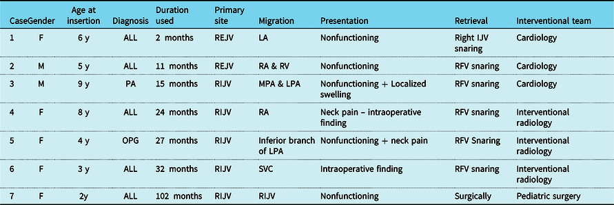
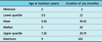
The total number of patients who underwent chemoport insertion in the allocated time period was 1222. Only 7 out of the 1222 suffered from chemoport catheter dislodgement (0.57%). The data set for the seven cases with catheter dislodgement is summarized in Tables 1 and 2. There were four girls and three boys. Their ages at Port-a-Cath insertion ranged from 2 years to 9 years. Totally, five patients had acute lymphoblastic leukemia, one had pilocytic astrocytoma, and one had optic pathway glioma. The duration of chemoport use before the event of dislodgement varied from 2 to 102 months. The primary site for insertion in five patients was the right internal jugular vein, whereas it was the right external jugular vein in the other two patients. Clinical presentation varied with non-functioning chemoport being the most common presentation, occurring in four cases. Two patients reported neck pain at the time of the dislodgement, and one patient reported localised swelling due to fluids extravasation. The dislodgement was discovered intraoperatively during removal after end of chemotherapy in two patients. One of these two cases presented with neck pain during chemotherapy injection, and a fluoroscopy study showed extravasation at the right base of the neck consistent with catheter break. The chemoport was removed surgically and replaced with a new one on the left side. Part of the old catheter was retained during removal and was retrieved by one of our interventional radiologists. The other case was asymptomatic, and fracture was incidentally discovered during chemoport removal after the end of treatment. The surgeon was able to retrieve the fragmented part from proximal part of internal jugular vein.
Clinical data set for seven patients with catheter dislodgement

Abbreviations: ALL: acute lymphoblastic leukemia; F: female; LA: left atrium; LPA: left pulmonary artery; M: male; MPA: main pulmonary artery; PA: pilocytic astrocytoma; OPG: optic pathway glioma; REJV: right external jugular vein; RA: right atrium; RFV: right femoral vein; RIJV: right internal jugular vein; RV: right ventricle; SVC: superior caval vein
Descriptive analysis of continuous variables

The different migration sites included the right internal jugular vein, the superior caval vein, the right atrium, the right ventricle, the main pulmonary artery, and the left pulmonary artery. Catheter migration to the left atrium occurred in one case. This patient presented with non-functioning chemoport 2 months after insertion. A fluoroscopic study showed the disconnected catheter crossing from the right atrium through the Foramen Ovale and terminating in the region of the left atrium. Retrieval was done via right femoral vein snaring in five patients. Right internal jugular vein snaring was performed in another patient. In the last patient, the fragment was tracked by the surgeon and retrieved from the internal jugular vein intraoperatively during chemoport removal. Snaring was done by a paediatric cardiologist in three cases and by an interventional radiologist in another three cases.
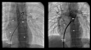
From a cardiological standpoint, we performed the procedure for retrieval of the dislodged catheter under fluoro-imaging in the cardiac catheterisation laboratory. All patients had venous access via the right femoral or internal jugular routes utilising six French vascular sheaths under local anaesthesia with 2% lidocaine. We advanced a five French Judkins catheter towards the site of the migrated fragment. The Judkins catheter was exchanged for a snare guiding catheter, and a snare was advanced through the snare guiding catheter towards the lodged site. Most dislodged catheters could be snared directly or following repositioning by a five French pigtail catheter. In some cases, we applied mild force to release the catheter from the migrated site. We utilised two kinds of snares, the triple loop 6 French/12–20 mm EN Snare® endovascular snare system and the 6 French/15 mm ONE Snare® Endovascular system. The choice of the device was made based on the operator’s personal preference. The catheter fragment and snare were carefully pulled back and introduced into the venous sheath and were extracted gently from the skin. The venous access site was compressed manually. There were no complications during the procedure (Fig 2).
Fluoroscopic pictures during retrieval of a migrated catheter from the LPA. Right femoral vein is accessed percutaneously. A sheath is introduced into the central vein then a catheter is advanced from inferior caval vein towards the LPA. An exchange wire is then placed distally (Left). The angiography catheter is then exchanged with a delivery sheath (Right). IVC, inferior caval vein; LPA, left pulmonary artery; LV, left ventricle; MPA, main pulmonary artery; RA, right atrium; RPA, right pulmonary artery; RV, right ventricle; SVC, superior caval vein.

Discussion
Patients with chemoport catheter dislodgement and migration may present with a spectrum of symptoms. Most patients are asymptomatic.Reference Çilingiroğlu and Akkuş11,Reference Cheng, Tsai, Yang and Han12 When there are symptoms, patients present with non-flushing catheter, absence of blood return, pain, or extravasation during infusion.Reference Ribeiro, Monteiro, Menezes, Schettini and Vianna13 Other uncommon presentations include palpitations due to ventricular tachycardia, thoracic pain, cough, and dyspnoea.Reference Çilingiroğlu and Akkuş11,Reference Wassef, Kass, Parmar and Ravandi14,Reference Gowda, Gowda and Khan15 Endocarditis, cardiac tamponade, heart and vessel perforation, and cardiac arrest have also been reported.Reference Ho, Chou, Chang, Jan, Lin and Fu16 In our series, non-functioning catheter was the most common presentation occurring in five patients (Table 1).
Catheter migration is a rare complication of chemoport devices that is reported in 0.2–8% of the cases in the literature.Reference Ballarini, Intra and Ceretti17–Reference Vlasveld, Rodenhuis and Rutgers20 In the paediatric population, it is reported to reach 1.4% to 4.1%.Reference Ho, Chou, Chang, Jan, Lin and Fu16,Reference Babu and Spicer21,Reference Dillon and Foglia22 The incidence at our institution in the past 15 years is 0.57% which, we believe, is the lowest reported incidence in the paediatric population in the literature to the best of our knowledge. Catheter migration seems to be less likely with catheters placed at the internal jugular vein compared with those placed at the subclavian vein.Reference Plumhans, Mahnken and Ocklenburg23,Reference Wu, Yeh, Huang, Lau and Yeh24 The most frequent location for catheter dislodgement is between the right atrium and inferior caval vein.Reference Tsai, Han and Lin25 A less frequent migration site is the left pulmonary artery which occurred in one case in our series. Migration to the coronary sinus was reported by Karam et al. in an adult patient.Reference Karam, Hourani and Al-Kutoubi26 In our series, migration to the left atrium through a patent Foramen Ovale occurred in one case. This has not been reported before in the literature to the best of our knowledge.
In terms of retrieval, Cheng et al. reported a success rate of 97.8% for percutaneous retrieval of dislodged catheter fragments with a 3.3% rate of complications.Reference Cheng, Tsai, Yang and Han12 Therefore, the endovascular approach is considered the first choice for retrieval of dislodged chemoport catheters.Reference Wang, Tsai and Hou9
Catheter fracture and migration could be a result of many surgical and non-surgical factors; however, it is usually difficult to identify the exact aetiology.Reference Dillon and Foglia27 Surgical factors include poor connection of the catheter to the port, damage of the catheter during assembly at the site of anastomosis,Reference Ho, Chou, Chang, Jan, Lin and Fu16,Reference Dillon and Foglia27,Reference Kirvelä and Satokari28 and improper positioning of the catheter.Reference Çilingiroğlu and Akkuş11,Reference Liu, Tseng, Chen, Chern and Chang29–Reference D’Angelo, Ramacciato and Caramitti31 Non-surgical factors include forced flushing, vigorous movements of upper extremities, neck flexion, congestive heart failure, increasing in thoracic pressure with coughing and vomiting, and catheter damage by chemotherapeutic drugs.Reference Vlasveld, Rodenhuis and Rutgers20,Reference Wu, Yeh, Huang, Lau and Yeh24,Reference Yedlicka, Carlson, Hunter, Castañeda-Zúñiga and Amplatz32 We believe that we achieved a very low rate of catheter dislodgement at our centre by taking these surgical and non-surgical factors into consideration. Chemoport insertion is done by trained paediatric surgeons. Our paediatric surgery team consists of three paediatric surgery attendings and a rotating senior surgery resident. Surgery is done by the attending paediatric surgeon with the assistance of the senior resident. As mentioned above, chemoport accessing, handling, and care are only done by certified trained nurses according to a clear written policy. It might be worth empathising that out of 1222 patients included in our study, none of the patients who had their ports inserted after the age of 9 had a fracture.
Pinch-off syndrome is a specific phenomenon for catheters placed at the subclavian vein. It occurs when a subclavian catheter is compressed or kinked during arm motion as it passes through a small anatomic space between the first rib and the clavicle, which causes its fracture.Reference Biffi, De Braud and Orsi18,Reference Charvat, Linke, Horaekova and Prausova19,Reference Dillon and Foglia27,Reference Behrend, Paboura and Raab33 This phenomenon was first described by Aitken and Minton in 1984.Reference Aitken and Minton34 They stated that the pinch-off sign seen as the catheter passed under the clavicle signified potential stress on the catheter which could result in fracture and migration of the catheter. The pinch-off syndrome is not reported in the jugular vein ports which we use at our centre.
Conclusion
Dislodgement and migration of chemoport catheter is a rare complication of uncertain aetiology and with potentially serious consequences. It is likely related to many surgical and non-surgical factors. Institutions should have a clear policy for chemoport care that takes these factors into consideration. Percutaneous retrieval, done by experienced cardiologist or interventional radiologist, is the first choice for management as it is considered a safe and effective approach.
Limitation
Risk factors could not be fully assessed due to the rarity of this complication.
Acknowledgements
The authors thank Dr. Ahmed Zaghal, MD, from the paediatric surgery department at our institution for his contribution to this work by sharing his experience and review over this topic. The authors are also grateful to Dr. Abdulrahman Dardeer, from the Anesthesia department at Hamad Medical Corporation, Doha, Qatar, for his valuable contribution.
Conflicts of interest
None.