




No CrossRef data available.
Article contents
Human infectivity trait in Trypanosoma brucei: stability, heritability and relationship to sra expression
Published online by Cambridge University Press: 14 September 2004
Abstract
Some Trypanosoma brucei lines infect humans whereas others do not because the parasites are lysed by human serum. We have developed a robust, quantitative in vitro assay based on differential uptake of fluorescent dyes by live and dead trypanosomes to quantify the extent and kinetics of killing by human serum. This method has been used to discriminate between 3 classes of human serum resistance; sensitive, resistant and intermediate. TREU 927/4, the parasite used for the T. brucei genome project, is intermediate. The phenotype is expressed in both bloodstream and metacyclic forms, is stably expressed during chronic infections and on cyclical transmission through tsetse flies. Trypanosomes of intermediate phenotype are distinguished from sensitive populations of cells by the slower rate of lysis and by the potential to become fully resistant to killing by human serum as a result of selection or long-term serial passaging in mice, and to pass on full resistance phenotype to its progeny in a genetic cross. The sra gene has been shown previously to determine human serum resistance in T. brucei but screening for the presence and expression of this gene indicated that it is not responsible for the human serum resistance phenotype in the trypanosome lines that we have examined, indicating that an alternative mechanism for HSR exists in these stocks. Examination of the inheritance of the phenotype in F1 hybrids for both bloodstream and metacyclic stages from 2 genetic crosses demonstrated that the phenotype is co-inherited in both life-cycle stages in a manner consistent with being a Mendelian trait, determined by only one or a few genes.
Information
- Type
- Research Article
- Information
- Copyright
- 2004 Cambridge University Press
INTRODUCTION
Trypanosoma brucei has been divided into 3 subspecies, T. b. brucei, T. b. rhodesiense and T. b. gambiense based on a combination of geographical distribution, host specificity and the course of infection in humans (chronic or acute). T. b. brucei is solely infective to animals including domestic livestock and game, T. b. rhodesiense infects humans and animals in East and Southern Africa, while T. b. gambiense infects humans and domestic animals in West and Central Africa (Hoare, 1972). An important aspect of research on this group of parasites has been to develop methods to distinguish these 3 morphologically identical subspecies so as to be able to identify the human infective subspecies in reservoir hosts and in the tsetse vector. Extensive analyses using a range of biochemical and molecular markers (Tait et al. 1985; Godfrey et al. 1990; Matthieu-Daude et al. 1995; Gibson, 2002) have largely been unable to identify consistent differences between the 3 subspecies with 2 exceptions; firstly, using both iso-enzyme and molecular markers, 2 distinct clades of T. b. gambiense could be defined (type 1 and 2) with type 2 being more closely related to West African T. b. brucei (Tait, Babiker & Le Ray, 1984; Gibson, 1986). Second, in the Busoga focus in Uganda, it has been possible to define T. b. rhodesiense using multilocus genotypes as the sole criteria (Hide et al. 1994; MacLeod et al. 2000).
The early observation that non-human infective isolates (T. b. brucei) are lysed by human serum and human infective isolates (T. b. gambiense and T. b. rhodesiense) are not, led to the development of the blood incubation infectivity test (BIIT), which can identify human infective isolates based on their ability to resist the lytic effects of human serum (Rickman & Robson, 1970a). These authors showed that, in addition to fully resistant and fully sensitive trypanosomes, a class of ‘subresistant’ parasite isolates could be defined (Rickman & Robson, 1970b). However, given that these authors were not using cloned lines of trypanosomes, it was unclear whether this phenotype was the result of mixtures of resistant and sensitive trypanosomes or a distinct phenotype.
The identification of the sra gene as exclusively expressed by human serum resistant (HSR) trypanosome lines and the demonstration that the gene can confer HSR when transfected into the bloodstream expression site (ES) of sensitive trypanosomes, has provided a further potentially valuable marker for the definition of human infective trypanosomes (Xong et al. 1998; Milner & Hajduk, 1999). The identification of sra as a determinant of resistance/sensitivity also solved the conundrum that human serum resistance is an unstable phenotype in at least some trypanosome stocks with different subclones being either sensitive or resistant. This variability of expression arises because the sra gene is expressed from an ES and thus expression of sra is linked to antigenic variation. There are multiple expression sites in the genome but the parasite uses only 1 ES at a time and switches use between ESs; which may or may not contain sra (Xong et al. 1998).
A PCR-based method for the detection of the sra gene has been developed and the presence of the gene has been shown to correlate strongly with the human serum-resistant (HSR) phenotype in the Busoga focus, and so this method has been heralded as a means of defining human infective trypanosomes (Welburn et al. 2001). However, while it is clear that the sra gene is responsible for human infectivity in a large number of T. b. rhodesiense isolates (Welburn et al. 2001; Gibson et al. 2002), there are exceptions to this in trypanosome lines from East Africa (Rifkin et al. 1994; Agbo et al. 2003) and, critically, this gene is not present in West African T. b. gambiense isolates (De Greef & Hamers, 1994; Radwanska et al. 2002). Thus, there must be alternative mechanisms determining human infectivity/human serum resistance other than those based on the sra gene.
The question arises as to whether these other mechanisms are likely to determine a phenotype that is a stable and heritable trait. Given the experience with sra these issues should not be assumed; sra expression is unstable and, although the gene is inherited, its position in the genome is potentially variable by virtue of location in an ES. In this paper we report on the stability and heritability of the HSR phenotype in trypanosome stocks where it is not determined by sra.
MATERIALS AND METHODS
Parasite stocks and lines
Trypanosomes of 5 different genotypes were used in these studies as indicated in Table 1. Using the classical criteria of Hoare (1972), STIB 247 and TREU 927 were classified as T. b. brucei, ELIANE and STIB 386 are type 1 and type 2 T. b. gambiense respectively (Gibson, 1986), and the isogenic lines Etats 1.2 and 1.10 are T. b. rhodesiense clones derived from the stock EATRO 3 (Xong et al. 1998). STIB 247, STIB 386, TREU 927 and EATRO 3 were transmitted separately through tsetse flies and metacyclic forms of TREU 927 were cloned as previously described (Turner et al. 1990). STIB 386 and TREU 927 were also rapidly syringe-passaged between mice for 49 passages before recloning to generate lines that were human serum sensitive (HSS) and HSR, 386S and GUTat 10.1, respectively (Turner et al. 1991; van Deursen et al. 2001). A subline of TREU 927 was selected for human serum resistance by 2 rounds of incubating cells in 25% human serum for 4 h and inoculation of mice with 3×10⁷ cells – TREU 927R. We also used 74 F1 hybrid progeny from 2 genetic crosses, STIB 247×TREU 927 and STIB 247×STIB 386 described previously (Turner et al. 1990; MacLeod et al., manuscript in preparation). Six of the progeny were transmitted separately through tsetse flies to generate metacyclic forms.
Table 1. Human serum resistance phenotypes and sra expression and genotypes of trypanosome lines (STIB 386S is a subclone of STIB 386, GUTat 10.1 a subclone of TREU 927 and Etats 1.2 and 1.10 are subclones of EATRO 3. Human serum resistant clones are defined here as Trypanosoma brucei rhodesiense and human serum sensitive clones as T. b. brucei. A, wild animal; H, human; T, tsetse; S, sensitive; I, intermediate; R, resistant. ‘?’ indicates equivocal subspecies definition as discussed in the text.)

In vitro human serum resistance assay
In a series of preliminary experiments the following potential variables were investigated: serum from different individuals, the use of heat-inactivated/non heat-inactivated serum, incubation temperature (25 or 37 °C), serum concentration, trypanosome density, source of control serum, duration of incubation, parasite viability staining methodology (data not shown).
In our optimized assay, trypanosomes were isolated by differential centrifugation (Ghiotto et al. 1979) rather than using anion-exchange chromatography, as the latter method always generated a ‘background’ of 5–20% ‘dead’ cells. The cells were resuspended in a volume of 500 μl of either 25% human or guinea-pig serum (Serotec) in PBS at a concentration of 1×10⁷/ml and incubated at 37 °C for 0, 2 and 4 h. The human serum was from a single individual after overnight fasting. The same donor was used for all assays. On completion of the incubation the suspension was centrifuged at 900 g for 5 min at 4 °C, the pellet resuspended in 200 μl of nucleic acid staining solution (1 in 500 dilution of Syto 10 and Dead Red dyes in PBS; Molecular Probes) and incubated in the dark at room temperature for 15 min, followed by centrifugation at 900 g for 5 min. The cell pellet was resuspended in 50 μl of PBS followed by the addition of 400 μl of 4% glutaraldehyde and fixation for at least 15 min. At least 200 cells were scored as green or red using a fluorescence microscope and Zeiss filter set 9. These dyes stain the nuclei of viable cells green and those of dead cells red when they are viewed by fluorescence microscopy, thus allowing the number of viable and non viable cells to be measured. No double-labelled cells were detected and unlabelled cells comprised <1% of the numbers counted in all assays.
In vivo human serum resistance assay
The assay we used was a minor variant of the Blood Incubation Infectivity Test (BIIT) Rickman & Robson, 1970a). Trypanosomes were separated from blood as described above and diluted in MEM/Earles salts with 25% human serum from a single human donor to a final concentration of 1×10⁷ cells/ml. After incubation for 2–4 h at 37 °C, 100 cells, diluted in PBS, were inoculated intraperitoneally into each of 2 ICR mice, which had been immunosuppressed by i.p. inoculation with 150 mg/kg cyclophosphamide 24 h previously. Mice were screened for patent parasitaemias 6 days a week for at least 2 weeks. A third mouse (positive control) in each experiment was inoculated with an equivalent number of parasites after incubation in 25% guinea-pig serum under the same experimental conditions.
DNA preparation and PCR
DNA was purified from bloodstream-stage parasites using standard methods (Lanham & Godfrey, 1970; Sambrook, Fritsch & Maniatis, 1989). A PCR test of genomic DNA, to verify the quality of the DNA stocks under investigation, was carried out using primers TIM-C and TIM-D as described by MacLeod, Turner & Tait (1997). The PCR amplification of the sra gene was undertaken using the conditions and primers as described by Radwanska et al. (2002), Gibson et al. (2002) and Welburn et al. (2001). The products were separated on agarose gels and visualized under UV light. The correct sized product for sra from STIB 247 was excised from the gel under UV illumination then loaded onto a Spin-X column (Costar) and centrifuged at 13000 g for 5 min. One microlitre of a 1/20 dilution of the eluted DNA was amplified in a semi-nested reaction using primers SP-A, 5′ ACAGCAACATCTCAGCGCTTTATGC and 538 (Welburn et al. 2001). The PCR conditions were as follows: 95 °C for 50 sec, 58 °C for 50 sec and 64 °C for 1 min for 30 cycles, using the primer concentrations and the PCR buffer described elsewhere (MacLeod, Turner & Tait, 1999). The products were cloned using the TA cloning kit (Invitrogen) and sequence determination carried out using the same primers and an ABI sequencer.
Northern analysis
Total RNA was prepared using the Trizol TM reagent (Gibco-BRL) according to the manufacturer's instructions. The isolated RNA was treated with amplification grade DNAse 1 to remove contaminating DNA. For Northern blots, 4 μg total RNA was denatured in formaldehyde and separated by agarose gel electrophoresis (Sambrook et al. 1989). After separation, the RNA was transferred to Hybond nylon membrane using standard protocols, pre-hybridized in 7% SDS, 0·5 M sodium phosphate, pH 7·2, 1 mM EDTA, at 65 °C for 2 h, followed by overnight hybridization with a random primed probe (Sambrook et al. 1989). The probe for the sra gene was prepared by PCR amplification of genomic DNA from Etat 1.10 using the primers and conditions described by Welburn et al. (2001). In order to generate a tubulin probe, a 630 bp fragment was amplified from DNA using the primers TUB 3′ GGGAATTCTTTCGCATCGAACATCTGCTGC and TUB 5′ GGGAATTCCCCCGACAACTTCATCTTTGGA, under the following conditions: 95 °C for 50 sec, 55 °C for 50 sec and 64 °C for 1 min for 30 cycles.
RESULTS
HSR phenotypes using in vivo assays of bloodstream forms
An in vivo human serum resistance assay was used to determine the serum resistance profile of 3 stocks used as parents in genetic crosses. Using an inoculum of 100 parasites per mouse, STIB 247, previously reported as human serum sensitive (Brun & Jenni, 1987) failed to infect any of 10 mice and STIB 386 infected all 10 mice as expected. TREU 927, however, infected only 2 out of 8 mice, an outcome previously described for other field stocks and termed ‘subresistant’ (Rickman & Robson, 1970b; Hawking, 1977). At a higher inoculum (1000 cells), STIB 247 still failed to infect any of 8 mice whereas TREU 927 now infected all 8 mice. Equal numbers of trypanosomes were incubated with guinea-pig serum prior to inoculation in TO mice, as controls, which resulted in all mice becoming infected. The results are summarized in Table 1.
HSR phenotypes using in vitro assays
We first analysed the serum resistance profile of the stocks STIB 386, TREU 927 and STIB 247 in this assay to compare with the in vivo data. The fluorescent staining of these stocks is shown in Fig. 1, and the percentage death depicted graphically in Fig. 2A. To verify this new assay we also tested a number of reference lines. The HSR stock, ELIANE and 3 isogenic lines of T. b. rodesiense, EATRO 3, Etat 1.2 and Etat 1.10, the latter 2 having previously been described as being HSS and HSR, respectively (De Greef & Hamers, 1994; Rifkin et al. 1994; Xong et al. 1998). These results are summarized in Table 1. Lines Etat 1.2 and STIB 247 were examples of the HSS phenotype class, with a substantial loss of viability (>90%), while ELIANE and Etat 1.10 were resistant to human serum with only ~18% loss of viability over a 4 h incubation period. However, TREU 927 and EATRO 3 both showed intermediate kinetics of lysis over the 4 h time-course. These data are consistent with those obtained with the in vivo assay and confirm that intermediate resistance is a definable phenotype, distinct from either resistance or sensitivity. To address the formal possibility that the intermediate phenotype could be due to a mixture of cells either expressing or not expressing human serum resistance, a 1[ratio ]1 mixture of HSR (STIB 386) and HSS (STIB 247) trypanosomes was assayed as before and resulted in ~50% lysis and different lysis kinetics compared with those obtained for the intermediate phenotype (Fig. 2A). Based on these results, we conclude that, in addition to the phenotypes of human serum sensitivity and resistance, a third phenotype of intermediate resistance can be defined by both in vivo and in vitro assays.

Fig. 1. Three stocks of Trypanosoma brucei parasites incubated for 4 h in human serum and stained using Syto 10/Dead Red. (A) STIB 247 trypanosomes were killed by human serum and stained red; (B) STIB 386 cells were largely resistant to human serum and stained green and (C) TREU 927 cells gave an intermediate result.

Fig. 2. Quantitative analysis of human serum resistance/sensitivity in lines of Trypanosoma brucei. (A) The kinetics of killing of bloodstream TREU 927 ([utrif ]) is intermediate between that of STIB 247 (□, T. b. brucei) and STIB 386 ([dtrif ], T. b. gambiense type 2) and different from the kinetics of killing of a 1[ratio ]1 mixture of resistant and sensitive bloodstream trypanosomes ([bull ]). (B) The kinetics of killing of metacyclic forms of STIB 247 (□), STIB 386 ([dtrif ]), TREU 927 ([utrif ]) and EATRO 3 ([bull ]). Average values are shown, n=3, 2S.E.<10% in all cases. Lysis in parallel incubations in guinea-pig serum as negative controls <10% in all cases.
To determine if the human serum resistance phenotypes are stable through the course of chronic infections, we initiated infections of STIB 247, STIB 386 and TREU 927 in groups of mice and at weekly intervals assayed populations of trypanosomes. Over a 5-week period for STIB 247 and a 3-week period for each of the other two stocks, the HSR phenotypes were unchanged for each stock (data not shown).
Human serum resistance in metacyclic trypanosomes
To determine whether the human serum resistance phenotype was the same in metacyclic forms as in bloodstream forms, we transmitted EATRO 3, STIB 247, STIB 386 and TREU 927 through tsetse flies, isolated metacyclic forms directly from the salivary glands and determined their human serum resistance profiles using the in vitro assay. The results, presented in Fig. 2B and summarized in Table 1, are directly comparable to those in Fig. 2A and show that the metacyclic forms of each stock had the same resistance phenotype as the bloodstream stages. EATRO 3 and TREU 927 were intermediate, STIB 386 was resistant and STIB 247 was sensitive to lysis by human serum. In all cases, the percentage lysis was slightly higher for metacyclic than for bloodstream forms. This can be attributed to the method of isolating metacyclic forms as they will be contaminated with pre-metacyclic trypanosomes which would be lysed by the alternative pathway of complement.
To further investigate the intermediate phenotype of stock TREU 927, single metacyclic forms of the parasites were optically isolated from the salivary glands of tsetse flies and amplified in mice. The resultant subcloned bloodstream populations were analysed for human serum resistance using the in vitro assay and all had an intermediate phenotype equivalent to that observed for TREU 927 before fly transmission (data not shown). These results show that the intermediate phenotype is a genuine third phenotypic state which is stable after fly transmission and subsequent expansion in mice.
Stability of human serum resistance
The instability of the human serum resistance phenotype on passaging in mice has been previously described for some T. b. rhodesiense stocks, including EARTO 3 (De Greef & Hamers, 1994; Hajduk et al. 1989; Rifkin et al. 1994; Xong et al. 1998). The resistance phenotypes of STIB 247, TREU 927 and STIB 386 are stable through the natural life-cycle of the trypanosomes i.e. in chronic infections and on fly transmission, but it is possible that their phenotype could change on serial bloodstream syringe passage. To investigate human serum resistance stability in these stocks we examined their ability to firstly, change phenotype on long-term passaging, followed by subcloning and second, under selection for resistance. The line GUTat 10.1, which is being used for the T. brucei genome sequencing project, changed HSR phenotype from intermediate to resistant (Table 1). Also, just 2 rounds of selection of TREU 927 for 4 h in human serum caused an equivalent change in phenotype, to generate the line TREU 927R. We attempted to select HSR STIB 247 lines in the same manner, but without success. Long-term passaging of STIB 386 and subcloning led to identification of a subclone that was sensitive to lysis by human serum (STIB 386S, Table 1). These results indicate that the HSR phenotype is a variable trait in the intermediate and resistant stocks, but is invariant in the sensitive stock, STIB 247. To investigate this last result further, 11 different lines which were sensitive to human serum, as determined by the in vitro assay, were selected in human serum in an attempt to generate resistant lines. No resistant trypanosomes were isolated (data not shown).
Presence/absence of the sra gene
In order to test if the sra gene is present in the stocks under investigation in this study, we attempted to amplify this gene by PCR, using the PCR test described by Welburn et al. (2001). A sequence homologous to the sra gene, which has been identified in TREU 927 on chromosome II and shown to be a VSG pseudogene (Campillo & Carrington, 2003), was amplified from all stocks using this test. Since this generates a larger sized product than the sra gene it acts as an internal control for PCR amplification (Fig. 3A). The 743 bp product derived from the sra gene was amplified from Etat 1.10 and Etat 1.2 as expected. The PCR assay failed to amplify sra from TREU 927 and STIB 386, although a series of products was amplified from STIB 247. To identify if any of these products was from the sra gene, semi-nested PCR was performed on gel-extracted DNA using primers SP-A and 538. A single product of the expected size (576 bp) was obtained. To establish the relationship between the amplified products and the sra gene sequence, this amplified fragment was cloned and sequenced. The fragment obtained from STIB 247 showed 99·5% identity to the published sequence (data not shown). This level of homology suggests that the PCR product from STIB 247 is the sra gene. Further attempts to amplify the sra gene from TREU 927 and STIB 386 using alternative primer combinations (Gibson, Backhouse & Griffiths, 2002; Radwanska et al. 2002) failed to produce a product, although amplification of the expected product in the positive controls was observed (Table 1). As a positive control to demonstrate that the DNA samples contained amplifyable DNA, the tim gene was amplified by PCR from each stock under the conditions described previously (MacLeod et al. 1997). The expected sized product was obtained for all samples (data not shown). These data suggest that TREU 927/GUTat 10.1 and STIB 386 do not contain the sra gene despite being HSR.

Fig. 3. (A) PCR amplification of the sra gene using primers and conditions described by Welburn et al. (2001). PCR products from the lines STIB 247, Etat 1.10, Etat 1.2, TREU 927, TREU 927R, STIB 386R and STIB 386S were separated on a 1% Seakem agarose gel and visualized by eithidium bromide staining. The 1085 bp product is the related VSG pseudogene and the 743 bp band is the sra gene (Campillo & Carrington, 2003). (B) (i) The sra gene probe was hybridized with a Northern blot of total RNA (4 μg per track). Exposure time was 4 h. (ii) Ethidium-stained gel of the RNA prior to Northern blotting. (iii) The Northern blot was reprobed with a tubulin probe as a positive control.
Expression of the sra gene in bloodstream forms
In order to test whether the sra gene in STIB 247 was expressed, Northern blot analysis was undertaken. Etat 1.10 and Etat 1.2 were used as positive and negative controls (Xong et al. 1998). TREU 927, TREU 927R, STIB 386 and STIB 386S were also included in this analysis. The probe was a 575 bp fragment of the sra gene amplified from Etat 1.10 genomic DNA, which was sequenced to confirm it was part of the sra gene (data not shown). The results, shown in Fig. 3B(i), revealed a strong hybridization signal for Etat 1.10 but no signal for ETAT 1.2 or from STIB 247. Similarly, no signal was observed with either STIB 386 (either resistant or sensitive) or TREU 927 (either resistant or intermediate), suggesting that the human serum resistance expressed by these stocks is not determined by the sra gene. A tubulin probe was used as a control for loading (Fig. 3B(ii)). The results are summarized in Table 1.
Inheritance of HSR
An investigation into the inheritance of the phenotype was undertaken to determine if the ability to resist the lytic effects of human serum is a trait that is inherited in a Mendelian fashion. The HSR phenotypes of a series of F1 hybrids (bloodstream forms) from 2 genetic crosses (previously described by Tait et al. (2002) were analysed using the in vitro assay. Some of the results are presented in Fig. 4A and indicate that all the progeny from the STIB 247×STIB 386 cross and the STIB 247×TREU 927 cross fall into 1 of 3 discrete categories either sensitive, resistant or intermediate, and not a range of different levels of resistance; thus the phenotype segregates in the cross. For the STIB 247 (sensitive)×STIB 386 (resistant) cross, 8 were HSR, 14 were HSS and 12 were intermediate and for the STIB 247 (sensitive)×TREU 927 (intermediate) cross, 10 progeny were HSR, 11 were HSS and 17 were intermediate in phenotype. It is clear, therefore, that the phenotype is inherited in a manner consistent with being a Mendelian trait, and that only one or a few genes determine the phenotype. As STIB 247 is invariably sensitive to human serum, the resistant hybrids presumably inherited the ability to resist human serum from the other parent, either TREU 927 or STIB 386. Therefore, TREU 927 can confer full resistance as well as intermediate resistance to its progeny and STIB 386 can pass on intermediate resistance as well as full resistance to its progeny.
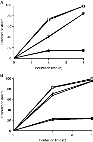
Fig. 4. (A) The kinetics of killing of bloodstream form F1 hybrids from a STIB 247×TREU 927 cross. (B) The kinetics of killing of metacyclic forms of the same hybrids. F532/63bcl3 ([lozf ]) and 5 (□), F532/72mcl1([squf ]), 4([utrif ]), 5([dtrif ]) and 9 (○). Average values are shown, n=3, 2S.E.<10% in all cases. Lysis in parallel incubations in guinea-pig serum as negative controls <10% in all cases.
In order to determine if the same phenotype is expressed in metacyclic-form 6 hybrids from the STIB 247×TREU 927 cross were transmitted through tsetse flies and metacyclics tested. The human serum resistance profiles were the same for metacyclic forms as for bloodstream forms, indicating that the inherited phenotypes are stable on fly transmission and that the same gene(s) are likely to be responsible for both bloodstream and metacyclic HSR (Fig. 4B).
DISCUSSION
The BIIT assay (Rickman & Robson, 1970a) was the first technique which could differentiate T. b. brucei from T. b. rhodesiense, by assaying the ability of trypanosomes to survive in human serum and subsequently infect mice. A number of in vitro assays have been developed since then that discriminate trypanosome lines as HSR or HSS (cf. Brun & Jenni, 1987; Hajduk et al. 1989; Tomlinson et al. 1995). In this paper, we describe a simple, robust quantitative method which directly measures the survival of trypanosomes in human serum and that discriminates the response to human serum into 3 phenotype classes – sensitive, resistant and intermediate. The intermediate phenotype is defined by a slower rate of parasite killing than that observed with the sensitive phenotype. The comparison of our in vitro and in vivo data suggests that the intermediate phenotype may correspond to the ‘subresistant’ class previously described using the BIIT (Rickman & Robson, 1970a; Hawking, 1977).
What is the biological significance of the intermediate phenotype? At first glance, the similarity between the intermediate and HSS phenotypes is such that the intermediate phenotype could be considered a slower form of HSS. However, 3 lines of evidence demonstrate that the distinction between sensitive and intermediate is biologically important. Trypanosomes with intermediate sensitivity were capable of becoming fully resistant by (1) rapid selection, (2) by long-term passaging and recloning and (3) can pass on a resistant phenotype to progeny in a genetic cross between parents of intermediate and sensitive phenotypes.
Our discrimination of 3 phenotype classes and the demonstration in this paper and by others (Hajduk et al. 1989; De Greef & Hamers, 1994) that lines of trypanosomes can be developed that differ in phenotype from that of the stocks from which they originated, calls into question the biological validity of the subspecies definitions used for T. brucei. This is because the classical criterion to discriminate T. b. brucei from T. b. rhodesiense and T. b. gambiense is the ability to infect humans (Hoare, 1972). In light of this ambiguity we denote some of the subspecies definitions in Table 1 with question marks. How the subspecies definition issue might be resolved is important but falls outside the scope of this current paper.
We have shown that the serum resistance phenotype is stable through the course of chronic infections, after cyclical transmission through tsetse flies and is the same in both bloodstream and metacyclic forms. The only occasions in which we have found the trait to be variable is under selection pressure and in the artificial laboratory situation of long-term serial passaging between mice (for intermediate and resistant, but not sensitive, lines). This variability has been noted before (Hajduk et al. 1989; De Greef & Hamers, 1994) and indeed has been exploited extremely successfully to create isogenic pairs of sensitive and resistant lines, comparison of which has led to the identification of a gene, sra, that can determine resistance/sensitivity (De Greef & Hamers, 1994; Xong et al. 1998; Milner & Hajduk, 1999). It is possible that the mechanism of variability is common to many stocks irrespective of whether a sra or non-sra mode of action is employed to determine human serum resistance.
The identification of sra as a determinant of resistance in T. b. rhodesiense has resulted in the use of a simple PCR assay to distinguish between human serum sensitive and resistant trypanosomes (Welburn et al. 2001; Gibson et al. 2002; Radwanska et al. 2002), as opposed to the lengthy and costly BIIT (Rickman & Robson, 1970a). This PCR test is based on the assumption that human serum resistant trypanosomes have the sra gene and non-human infective trypanosomes do not which appears to hold true for the majority of isolates tested from east and southern Africa (Welburn et al. 2001; Gibson et al. 2002; Agbo et al. 2003), but not for west and central Africa (Berberof, Perez-Morga & Pays, 2001; Radwanska et al. 2002).
In order to determine the relevance of the sra gene to human serum resistance phenotypes described here, the PCR tests of Gibson et al. (2002) and Welburn et al. (2001) were used to determine if the sra gene is present in the stocks which were identified as HSR, HSS and intermediate. The HSS stock, STIB 247, tested positive for the sra gene (confirmed by sequence analysis) whereas the HSR and intermediate stocks, STIB 386 and TREU 927 were both negative. The sequence homologous to the sra gene, which has been identified in TREU 927 and is present in all isolates tested here, is a pseudogene it cannot be responsible for the HSR phenotype (Campillo & Carrington, 2003). This clearly indicates that a test for the presence/absence of the sra gene, while being a good approximation, is not an absolute test to predict the phenotype of HSR and that alternative mechanisms for HSR exist not only in West African stocks (Berberof et al. 2001; Radwanska et al. 2002) but also in the East African TREU 927 isolate. The lack of correlation between human serum resistance and expression of the sra gene was further confirmed by Northern analysis, which demonstrated that neither STIB 386 nor TREU 927 lines expressed the sra gene or a related sra homologue. The HSR phenotype of these stocks is also expressed in the metacyclic stage which suggests that the gene determining resistance is unlikely to be expressed from a bloodstream expression site, as is the case for the sra gene, as bloodstream expression sites are not thought to be transcribed at the metacyclic stage (Graham et al. 1990). It is formally possible that low-level transcription of the sra gene could occur to the extent that it confers HSR but is not detectable by Northern analysis. However, the fact that no sra gene was detected by 3 independent PCR tests would suggest that the gene is not present in these stocks and so cannot be expressed or is so divergent that it cannot be amplified by a PCR test or detected by hybridization. It is possible that both the sra mechanism and the alternative mechanism of HSR are present and functioning in the same organism. Indeed, the clone EATRO 3 has an intermediate phenotype and so, presumably, is expressing resistance from the alternative mechanism (as expression of the sra gene confers full resistance), while its derivative, Etat 1.10 is fully resistant by expressing sra.
These results presented here clearly demonstrate that the human serum resistance phenotype of the stocks under study is not due to the expression of the sra gene but due to an alternative mechanism. To determine the genetic basis of this mechanism, the inheritance pattern of resistance was investigated by the analysis of a series of F1 hybrids from 2 genetic crosses, STIB 247×TREU 927 and STIB 247×STIB 386. It would have been interesting to investigate inheritance in the cross TREU 927×STIB 386 but unfortunately there are too few F1 hybrids available to be informative (Turner et al. 1990). For both crosses, the F1 hybrids fell into 1 of 3 phenotypes, fully resistant, fully sensitive or intermediate, demonstrating that this phenotype segregates in the hybrids and is potentially amenable to linkage analysis. A genetic map of TREU 927 is currently being generated with the first 2 chromosomes, I and II, having been completed (El Sayed et al. 2003; Hall et al. 2003). Thus it should be possible to identify markers in the genetic map which cosegregate with the phenotype, and so the region of the genome harbouring the gene which determines human serum resistance can be located. A similar analysis of a cross STIB 386×STIB 247 should allow us to map and identify the gene(s) responsible for the HSR phenotype in West African human infective trypanosomes.
This work was supported by a grant from the Wellcome Trust to A.T. and C.M.R.T. We acknowledge with gratitude the gift of Etat 1.2 and Etat 1.10 from Etienne Pays.
References
REFERENCES
AGBO, E. C.,
CLAUSEN, P-H.,
BUSCHER, P.,
MAJIWA, P. A. O.,
CLAAASSEN, E. &
TE PAS, M. F. W. (2003).
Population genetic structure and cladistic analysis of Trypanosoma brucei isolates.
Infection, Genetics and Evolution 3,
165–174.CrossRefGoogle Scholar
BERBEROF, M.,
PEREZ-MORGA, D. &
PAYS, E. (2001).
A receptor-like flagellar pocket glycoprotein specific to Trypanosoma brucei gambiense.
Molecular and Biochemical Parasitology 113,
127–138.CrossRefGoogle Scholar
BRUN, R. &
JENNI, L. (1987).
Human serum resistance of metacyclic forms of Trypanosoma brucei brucei, T. brucei rhodesiense and T. brucei gambiense.
Parasitology Research 73,
218–223.Google Scholar
CAMPILLO, N. &
CARRINGTON, M. (2003).
The origin of the human serum resistance associated (SRA) gene and a model of the structure of the SRA polypeptide from Trypanosoma brucei rhodesiense.
Molecular and Biochemical Parasitology 127,
79–84.CrossRefGoogle Scholar
DE GREEF, C. &
HAMERS, R. (1994).
The serum-resistance associated (SRA) gene of Trypanosoma brucei rhodesiense encodes a VSG-like protein.
Molecular and Biochemical Parasitology 68,
277–284.CrossRefGoogle Scholar
EL-SAYED, N. M. A.,
GHEDIN, E.,
SONG, J.,
MacLEOD, A.,
BRINGAUD, F.,
LARKIN, C.,
WANLESS, D.,
PETERSON, J.,
HOU, L.,
TAYLOR, S.,
TWEEDIE, A.,
BITEAU, N.,
KHALAK, H. G.,
LIN, X.,
MASON, T.,
HANNICK, L.,
CALER, E.,
BLANDIN, G.,
BARTHOLOMEU, D.,
SIMPSON, A. J.,
KAUL, S.,
ZHAO, H.,
PAI, G.,
VAN AKEN, S.,
UTTERBACK, T.,
HAAS, B.,
KOO, H. L.,
UMAYAM, L.,
SUH, B.,
GERRARD, C.,
LEECH, V.,
QI, R.,
SCHWARTZ, D.,
FELDBLYUM, T.,
SALZBERG, S.,
TAIT, A.,
TURNER, C. M. R.,
ULLU, E.,
WHITE, O.,
MELVILLE, S.,
ADAMS, M. D.,
FRASER, C. M. &
DONELSON, J. E. (2003).
The sequence and analysis of Trypanosoma brucei chromosome II.
Nucleic Acids Research 31,
4856–4863.CrossRefGoogle Scholar
GHIOTTO, V.,
BRUN, R.,
JENNI, L. &
HECKER, H. (1979).
Trypanosoma brucei: morphometric changes and loss of infectivity during transformation of bloodstream forms to procyclic culture forms in vitro.
Experimental Parasitology 48,
447–456.CrossRefGoogle Scholar
GIBSON, W. C. (1986).
Will the real Trypanosoma b. gambiense please stand up.
Parasitology Today 2,
255–257.CrossRefGoogle Scholar
GIBSON, W. (2002).
Will the real Trypanosoma brucei rhodesiense please step forward?
Trends in Parasitology 18,
486–490.Google Scholar
GIBSON, W.,
BACKHOUSE, T. &
GRIFFITHS, A. (2002).
The human serum-resistance associated gene is ubiquitous and conserved in Trypanosoma brucei rhodesiense throughout East Africa.
Infection, Genetics and Evolution 1,
207–214.CrossRefGoogle Scholar
GODFREY, D. G.,
BAKER, R. D.,
RICKMAN, L. R. &
MEHLITZ, D. (1990).
The distribution, relationships and identification of enzymatic variants within the subgenus Trypanozoon.
Advances in Parasitology 29,
1–74.Google Scholar
GRAHAM, S. V.,
MATTHEWS, K. R.,
SHIELS, P. G. &
BARRY, J. D. (1990).
Distinct, developmental stage-specific activation mechanisms of trypanosome VSG genes.
Parasitology 101,
361–367.CrossRefGoogle Scholar
HAJDUK, S. L.,
MOORE, D. R.,
VASUDEVACHARYA, J.,
SIQUEIRA, H.,
TORRI, A. F.,
TYTLER, E. M. &
ESKO, J. D. (1989).
Lysis of Trypanosoma brucei by a toxic subspecies of human high density lipoprotein.
Journal of Biological Chemistry 264,
5210–5217.Google Scholar
HALL, N.,
BERRIMAN, M.,
LENNARD, N. J.,
HARRIS, B. R.,
BART-DELABESSE, E. N.,
HERTZ-FOWLER, C.,
GERRARD, C. S.,
ATKIN, R. J.,
BARRON, A. J.,
BOWMAN, S.,
BRAY-ALLEN, S. P.,
BRINGAUD, F.,
CLARK, L. N.,
CORTON, C. H.,
CRONIN, A.,
DAVIES, R.,
DOGGETT, J.,
FRASER, A.,
GRUTER, E.,
HALL, S.,
HARPER, A. D.,
KAY, M. P.,
LEECH, V.,
MAYES, R.,
PRICE, C.,
QUAIL, M. A.,
RABBINOWITSCH, E.,
REITTER, C.,
RUTHERFORD, K.,
SASSE, J.,
SHARP, S.,
SHOWNKEEN, R.,
MacLEOD, A.,
TAYLOR, S.,
TWEEDIE, A.,
TURNER, C. M. R.,
TAIT, A.,
GULL, K.,
BARRELL, B. &
MELVILLE, S. (2003).
The DNA sequence of chromosome I of an African trypanosome: gene content, chromosome organisation, recombination and polymorphism.
Nucleic Acids Research 31,
4864–4873.CrossRefGoogle Scholar
HAWKING, F. (1977).
The resistance to human plasma of Trypanosoma brucei, T. rhodesiense and T. gambiense. 1. Analysis of the composition of trypansome strains.
Transactions of the Royal Society of Tropical Medicine and Hygiene 70,
504–512.Google Scholar
HIDE, G.,
WELBURN, S. C.,
TAIT, A. &
MAUDLIN, I. (1994).
Epidemiological relationships of Trypanosoma brucei stocks from South East Uganda: evidence for different population structures in human infective and non-human infective isolates.
Parasitology 109,
95–111.CrossRefGoogle Scholar
HOARE, C. A. (1972).
The Trypanosomes of Mammals.
Blackwell Scientific Publications,
Oxford.
LANHAM, S. M. &
GODFREY, D. G. (1970).
Isolation of salivarian trypanosomes from man and other mammals using DEAE-cellulose.
Experimental Parasitology 28,
521–534.CrossRefGoogle Scholar
MacLEOD, A.,
TURNER, C. M. R. &
TAIT, A. (1997).
Detection of single copy gene sequences from single trypanosomes.
Molecular and Biochemical Parasitology 84,
267–270.CrossRefGoogle Scholar
MacLEOD, A.,
TURNER, C. M. R. &
TAIT, A. (1999).
A high level of mixed Trypanosoma brucei infections in tsetse flies detected by three hypervariable minisatellites.
Molecular and Biochemical Parasitology 102,
237–248.CrossRefGoogle Scholar
MacLEOD, A.,
TWEEDIE, A.,
WELBURN, S. C.,
MAUDLIN, S. C.,
MAUDLIN, I.,
TURNER, C. M. R. &
TAIT, A. (2000).
Minisatellite marker analysis of Trypanosoma brucei: reconciliation of clonal, panmictic, and epidemic population genetic structures.
Proceedings of the National Academy of Sciences, USA 97,
13442–13447.CrossRefGoogle Scholar
MATHIEU-DAUDE, F.,
STEVENS, J.,
WELSH, J.,
TIBAYRENC, M. &
McCLELLAND, M. (1995).
Genetic diversity and population structure of Trypanosoma brucei: clonality versus sexuality.
Molecular and Biochemical Parasitology 72,
89–101.CrossRefGoogle Scholar
MILNER, J. D. &
HAJDUK, S. L. (1999).
Expression and localization of serum resistance associated protein in Trypanosoma brucei rhodesiense.
Molecular and Biochemical Parasitology 30,
271–283.CrossRefGoogle Scholar
RADWANSKA, M.,
CHAMEKH, M.,
VANHAMME, L.,
CLAES, F.,
MAGEZ, S.,
MAGNUS, E.,
BAETSELIER, P.,
BUSCHER, P. &
PAYS, E. (2002).
The serum resistance-associated gene as a diagnostic tool for the detection of Trypanosoma brucei rhodesiense.
American Journal of Tropical Medicine and Hygiene 67,
684–690.CrossRefGoogle Scholar
RICKMAN, L. R. &
ROBSON, J. (1970 a).
The blood incubation infectivity tests: a simple test which may serve to distinguish Trypanosoma brucei from T. rhodesiense.
Bulletin of the World Health Organization 42,
650–651.Google Scholar
RICKMAN, L. R. &
ROBSON, J. (1970 b).
The testing of proven Trypanosoma brucei and T. rhodesiense strains by the Blood Incubation Test.
Bulletin of the World Health Organization 42,
911–916.Google Scholar
RIFKIN, M. R.,
DE GREEF, C.,
JIWA, A.,
LANDSBERGER, F. R. &
SHAPIRO, S. Z. (1994).
Human serum-sensitive Trypanosoma brucei rhodesiense: a comparison with serologically identical human serum-resistant clones.
Molecular and Biochemical Parasitology 66,
211–220.CrossRefGoogle Scholar
SAMBROOK, J.,
FRITSCH, E. F. &
MANIATIS, T. (1989).
Molecular Cloning: A Laboratory Manual, 2nd Edn.
Cold Spring Harbor Laboratory Press,
Cold Spring Harbor, New York.
TAIT, A.,
BABIKER, E. A. &
LERAY, D. (1984).
Enzyme variation in T. brucei spp. I. Evidence for the sub-speciation of T. b. gambiense.
Parasitology 89,
311–326.Google Scholar
TAIT, A.,
BARRY, J. D.,
WINK, R.,
SANDERSON, A. &
CROWE, J. S. (1985).
Enzyme variation in Trypanosome brucei spp. II. Evidence for T. b. rhodesiense being a set of variants of T. b. brucei.
Parasitology 90,
89–100.Google Scholar
TAIT, A.,
MASIGA, D.,
OUMA, J.,
MacLEOD, A.,
SASSE, J.,
MELVILLE, S.,
LINDEGARD, G.,
McINTOSH, A. &
TURNER, C. M. R. (2002).
Genetic analysis of phenotype in Trypanosoma brucei: a classical approach to potentially complex traits. Phil.
Transactions of the Royal Society of London B 357,
89–99.Google Scholar
TOMLINSON, S.,
JANSEN, A-M.,
KOUDINOV, A.,
GHISO, J. A.,
MIURA, N-H. C.,
RIFKIN, M. R.,
OHTAKI, S. &
NUSSENZWEIG, V. (1995).
High-density-lipoprotein-independent killing of Trypanosoma brucei by human serum.
Molecular and Biochemical Parasitology 70,
131–138.CrossRefGoogle Scholar
TURNER, C. M. R.,
ASLAM, N.,
SMITH, E.,
BUCHANAN, N. &
TAIT, A. (1991).
The effects of genetic exchange on variable antigen expression in Trypanosoma brucei.
Parasitology 103,
379–386.CrossRefGoogle Scholar
TURNER, C. M. R.,
STERNBERG, J.,
BUCHANAN, N.,
SMITH, E.,
HIDE, G. &
TAIT, A. (1990).
Evidence that the mechanism of gene exchange in Trypanosoma brucei involves meiosis and syngamy.
Parasitology 101,
377–386.CrossRefGoogle Scholar
VAN DEURSEN, F. J.,
SHAHI, S. K.,
TURNER, C. M. R.,
HARTMANN, C.,
GUERRA-GIRALDEZ, C.,
MATTHEWS, K. R. &
CLAYTON, C. E. (2001).
Characterization of the growth and differentiation in vivo and in vitro of bloodstream-form Trypanosoma brucei TREU 927.
Molecular and Biochemical Parasitology 112,
163–171.CrossRefGoogle Scholar
WELBURN, S. C.,
PICOZZI, K.,
FEVRE, E. M.,
COLEMAN, P. G.,
ODIIT, M.,
CARRINGTON, M. &
MAUDLIN, I. (2001).
Identification of human-infective trypanosomes in animal reservoir of sleeping sickness in Uganda by means of serum-resistance-associated (SRA) gene.
Lancet 358,
2017–2019.CrossRefGoogle Scholar
XONG, H. V.,
VANHAMME, L.,
CHAMEKH, M.,
CHIMFWEMBE, C. E.,
VAN DEN ABBEELE, J.,
PAYS, A.,
VAN MEIRVENNE, N.,
HAMERS, R.,
DE BAETSELIER, P. &
PAYS, E. (1998).
A VSG expression site-associated gene confers resistance to human serum in Trypanosoma rhodesiense.
Cell 95,
839–846.CrossRefGoogle Scholar

Table 1. Human serum resistance phenotypes and sra expression and genotypes of trypanosome lines
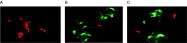
Fig. 1. Three stocks of Trypanosoma brucei parasites incubated for 4 h in human serum and stained using Syto 10/Dead Red. (A) STIB 247 trypanosomes were killed by human serum and stained red; (B) STIB 386 cells were largely resistant to human serum and stained green and (C) TREU 927 cells gave an intermediate result.

Fig. 2. Quantitative analysis of human serum resistance/sensitivity in lines of Trypanosoma brucei. (A) The kinetics of killing of bloodstream TREU 927 ([utrif ]) is intermediate between that of STIB 247 (□, T. b. brucei) and STIB 386 ([dtrif ], T. b. gambiense type 2) and different from the kinetics of killing of a 1[ratio ]1 mixture of resistant and sensitive bloodstream trypanosomes ([bull ]). (B) The kinetics of killing of metacyclic forms of STIB 247 (□), STIB 386 ([dtrif ]), TREU 927 ([utrif ]) and EATRO 3 ([bull ]). Average values are shown, n=3, 2S.E.<10% in all cases. Lysis in parallel incubations in guinea-pig serum as negative controls <10% in all cases.
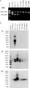
Fig. 3. (A) PCR amplification of the sra gene using primers and conditions described by Welburn et al. (2001). PCR products from the lines STIB 247, Etat 1.10, Etat 1.2, TREU 927, TREU 927R, STIB 386R and STIB 386S were separated on a 1% Seakem agarose gel and visualized by eithidium bromide staining. The 1085 bp product is the related VSG pseudogene and the 743 bp band is the sra gene (Campillo & Carrington, 2003). (B) (i) The sra gene probe was hybridized with a Northern blot of total RNA (4 μg per track). Exposure time was 4 h. (ii) Ethidium-stained gel of the RNA prior to Northern blotting. (iii) The Northern blot was reprobed with a tubulin probe as a positive control.

Fig. 4. (A) The kinetics of killing of bloodstream form F1 hybrids from a STIB 247×TREU 927 cross. (B) The kinetics of killing of metacyclic forms of the same hybrids. F532/63bcl3 ([lozf ]) and 5 (□), F532/72mcl1([squf ]), 4([utrif ]), 5([dtrif ]) and 9 (○). Average values are shown, n=3, 2S.E.<10% in all cases. Lysis in parallel incubations in guinea-pig serum as negative controls <10% in all cases.
 You have
Access
- 29
- Cited by