Introduction
The Antarctic Pearlwort, Colobanthus quitensis (Kunth.) Bartl. (Caryophyllaceae) is one of the two native vascular plant species that have successfully colonized the maritime Antarctic during the Holocene (Smith Reference Smith and Laws1984). Within the Antarctic biome, it is only found on the Antarctic Peninsula and on islands in the maritime Antarctic (Smith Reference Smith and Laws1984). Its distribution also includes South Georgia (sub-Antarctic), the Falkland Islands, and sites along the Andes, reaching c. 10°N in Mexico (Moore Reference Moore1970). Moore (Reference Moore1970) observed that C. quitensis is self-compatible and appears to be largely, if not entirely, self-pollinated. Convey (Reference Convey1996), found that maritime Antarctic C. quitensis showed lower allocation to seed production than plants from sub-Antarctic sites.
Ecophysiological studies have shown that C. quitensis is sensitive to the rapid climate warming trend and to the biologically damaging high UV-B radiation in spring (Day et al. Reference Day, Ruhland, Grobe and Xiong1999). Edwards & Smith (Reference Edwards and Smith1988), suggested that the achievement of about 30% of its maximum photosynthetic rate at 8°C is the most important feature for survival of C. quitensis in Antarctica. However, cold resistance mechanisms and biochemical adaptations to it (Bravo et al. Reference Bravo, Ulloa, Zúñiga, Casanova, Corcuera and Alberdi2001, Alberdi et al. Reference Alberdi, Bravo, Gutiérrez, Gidekel and Corcuera2002), are no less important in C. quitensis. The establishment of this plant under controlled environmental conditions is an important tool for research on adaptations of terrestrial plants to the prevailing Antarctic conditions. Here we describe a simple method for the rapid and recurrent propagation of C. quitensis for experimental use.
Materials and methods
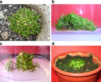
Individuals of C. quitensis were collected at the northern end of Admiralty Bay, South Shetland Islands (62°05′S; 58°23′W) where the mean air temperature in the summer (December–February) ranges from -3°C to +1°C, with an annual mean of -2.7°C (Smith Reference Smith and Van Der Maael1993). Soil is generally an oligotrophic lithosol, with low nutrient availability to plants (Smith Reference Smith, Siegfried, Condy and Laws1985). Plants of C. quitensis have a cushion growth form, comprising an agregation of individual shoots (Fig. 1a). Groups of four shoots were propagated vegetatively in soil:peat mixture (3:1) at 16 ± 2°C in a growth chamber with photon flux density of 180 µmol m-2 s-1 and 16/8 h light/dark period for two months.
Different stages of in vitro micropropagation of C. quitensis. (a) Mother plant collected in the Antarctic (b) Development of multiple shoots from nodal shoot segments on MS medium (c) One month in vitro plants growing in growth chamber (d) In vitro C. quitensis plants grown in soil in a growth chamber.

Shoots were thoroughly washed and surface sterilized for 15 min in Erlenmeyer flasks with 300 ml of an aqueous suspension of 1.5 g l-1 Captan 80W (BASF Chile-SA), under constant stirring, and then rinsed three times with sterile distilled water. The plants were transferred to 100 ml of 10% sodium hypochlorite and 0.01% Tween 20 for 10 min, and then rinsed three times with sterile distilled water. The explants (5 mm) from the sterile plants were used to initiate cultures into culture vessels (250 ml) containing MS (Murashige & Skoog Reference Murashige and Skoog1962) basal medium. Medium was prepared using MS salts, vitamins, 4% sucrose and this basal medium was also used as the control. Plant growth regulators tested included kinetin (K) and 2,4 Dichlorophenoxyacetic acid (2,4 D) at concentrations of 0, 1.0, or 2.0, mg l-1 (Table I). The medium pH was adjusted to 4.5 or 5.6 prior to addition of agar and autoclaved at 120°C for 20 min. Cultures were incubated at 16 ± 2°C under 16-h photoperiod using cool, white fluorescent light irradiance of 40 µmol m-2 s-1. Explants were sub-cultured to fresh medium every four weeks.
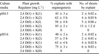
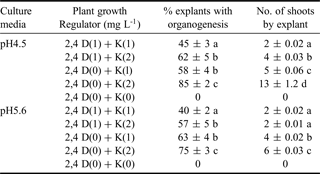
Effect of pH and hormone treatment on shoot induction in Colobanthus quitensis. Percentage of explants exhibiting organogenesis and number of shoots by explant was determined after four weeks on induction media MS pH 4.5 and 5.6 with different hormones, compared to the total number of initial explants on treatment. Data were subjected to analyses of variance, and means were separated by an LSD test at P = 0.05
Well-developed plantlets were removed from culture vessels and were transplanted to plastic cups containing a mixture of autoclaved soil:sand (1:1), and were maintained in growth chambers at 14 ± 2°C, relative humidity 80%, irradiance of 100 μmol m-2 s-1 for 16-h photoperiod. The plants were irrigated weekly with Hoagland solution. After four weeks, hardened plants were transferred to pots containing soil:sand (1:1) and maintained in the same conditions described before.
A completely randomized design was used for all treatments. One shoot explant was used per test vessel, with twelve replicates for each treatment. The experiments were repeated twice. Percentage of responding explants and number of shoots developed per responding explant were recorded weekly. Results were subjected to analyses of variance, and means were separated by an LSD test at the 5% level of significance using Statgraphics Plus 5.1 (Manugistics Inc, Rockville, MD, USA).
Results and discussion
After two weeks, the first sign of organogenesis induction was the swelling of the explants. Organogenesis appeared to occur directly from the explants even when 2,4-D was included in the culture medium. In very few cases (< 5%), a small amount of callus was initiated from the explants. MS medium supplemented only with K (2 mg l-1) at pH 4.5 produced the largest shooting response (P < 0.05) with the maximal number of shots per explant 13 ± 1.2. The same treatment at pH 5.6 resulted in the induction of less than half the number of shoots induced (Table I). MS medium without hormones resulted in no shoot induction at pH 4.5 or 5.6 (Table I). In plants growing in MS medium with K (2 mg l-1) at pH 4.5, shoot extension and subsequent root formation occurred within 4–5 weeks after transferring to media without growth regulators. Root formation was visible 3–4 weeks after shoot elongation. Separate plantlets were transferred to fresh medium to continue micropropagation (Fig. 1). In addition, some in vitro plants (about 10%) generated flowers (Fig. 1c). However, these flowers did not produce viable seeds.
It became apparent that the hardening-off stage was of crucial importance to a successful micropropagation protocol. Colobanthus quitensis was found to be very sensitive to high moisture content. Plantlets transferred to autoclaved soil and tightly covered with plastic bags, rotted and died within 10–14 days. In contrast, 80% survival was recorded for plantlets subjected to a twice-daily mist spray for 2–3 weeks after planting out. Plant growth stopped when the temperature was above 20°C, and 100% of survival was recorded when temperature was ≤ 14°C. The regenerated plants had similar morphological characteristics to the source plants (colour, leaf morphology and growth in a cushion form) (Fig. 1a & d) and 100% survived after transplantation to soil (Fig. 1d). This in vitro method yields an increase of approximately four to five fold of the initial number of plants within two months. In addition, vegetative plant production is three fold higher by micropropagation.
This procedure offers a potential system for mass propagation of C. quitensis with conservation implications for the collection of field material for experimentation.
Acknowledgements
We appreciated the technical assistance of Cristina Vargas and acknowledge Instituto Antartico Chileno (INACH) for logistic support.